Cells and Tissues Chapter 4 A Tour of

- Slides: 127
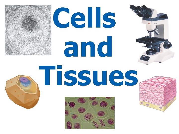
Cells and Tissues
Chapter 4 A Tour of the Cell Power. Point Lectures for Biology: Concepts & Connections, Sixth Edition Campbell, Reece, Taylor, Simon, and Dickey Copyright © 2009 Pearson Education, Inc. Lecture by Richard L. Myers Translated by Nabih Baeshen
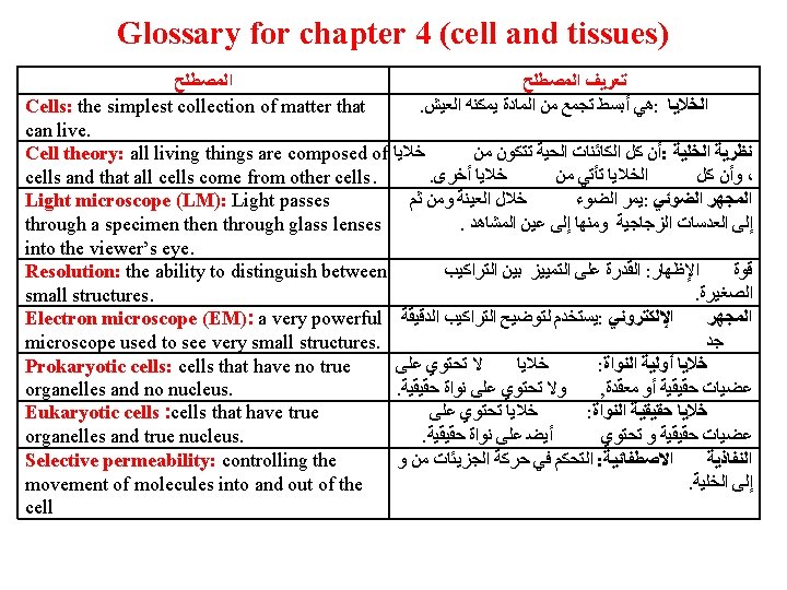
Introduction: Cells on the Move § Cells, the simplest collection of matter that can live, were first observed by Robert Hooke in 1665 § Antoni van Leeuwenhoek later described cells that could move – He viewed bacteria with his own handcrafted microscopes Copyright © 2009 Pearson Education, Inc.
Introduction: Cells on the Move Paramecium § The early microscopes provided data to establish the cell theory – That is, all living things are composed of cells and that all cells come from other cells Copyright © 2009 Pearson Education, Inc.
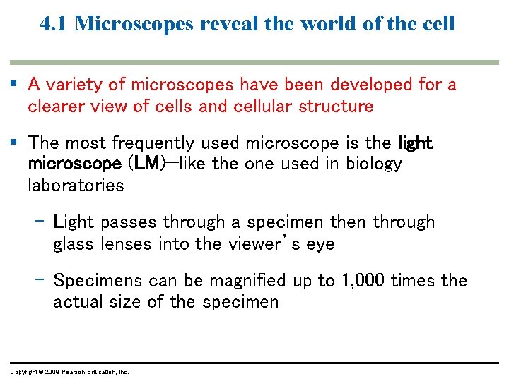
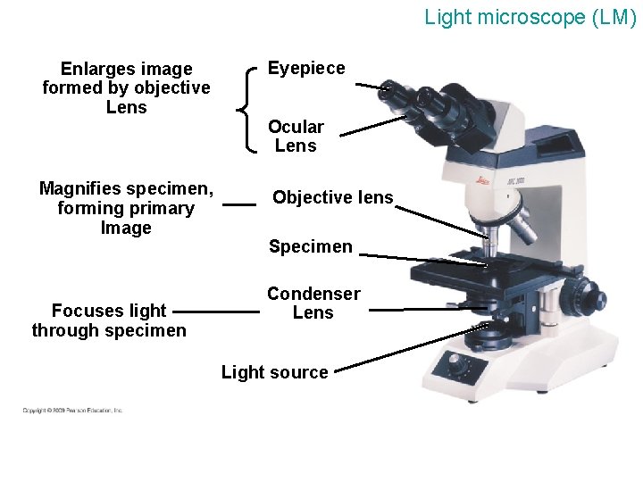
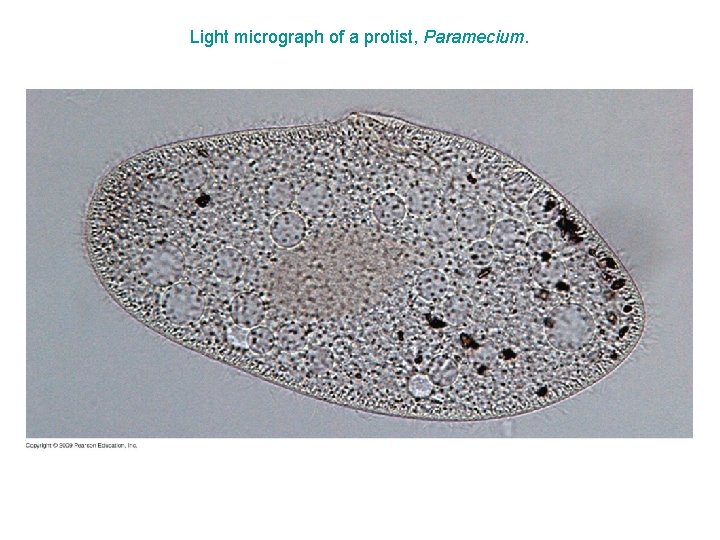
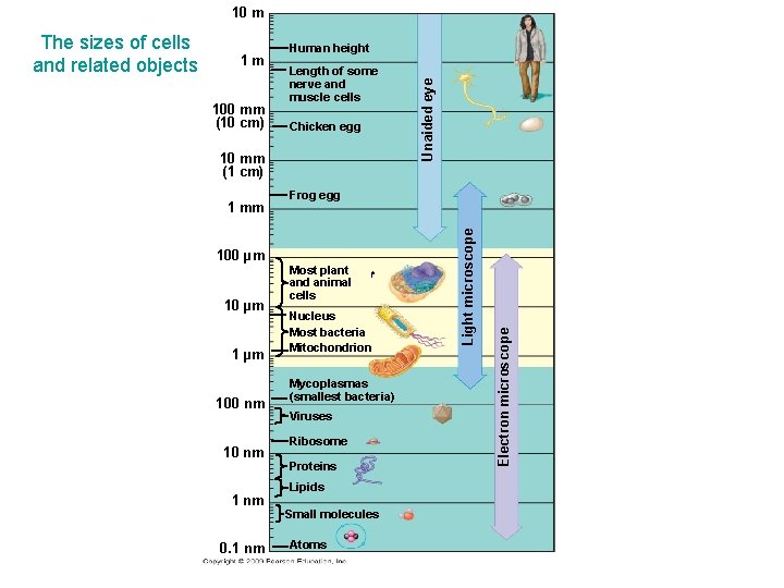
4. 1 Microscopes reveal the world of the cell § A variety of microscopes have been developed for a clearer view of cells and cellular structure § The most frequently used microscope is the light microscope (LM)—like the one used in biology laboratories – Light passes through a specimen through glass lenses into the viewer’s eye – Specimens can be magnified up to 1, 000 times the actual size of the specimen Copyright © 2009 Pearson Education, Inc.
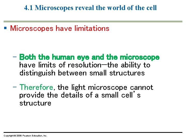
4. 1 Microscopes reveal the world of the cell § Microscopes have limitations – Both the human eye and the microscope have limits of resolution—the ability to distinguish between small structures – Therefore, the light microscope cannot provide the details of a small cell’s structure Copyright © 2009 Pearson Education, Inc.
Light microscope (LM) Enlarges image formed by objective Lens Magnifies specimen, forming primary Image Focuses light through specimen Eyepiece Ocular Lens Objective lens Specimen Condenser Lens Light source
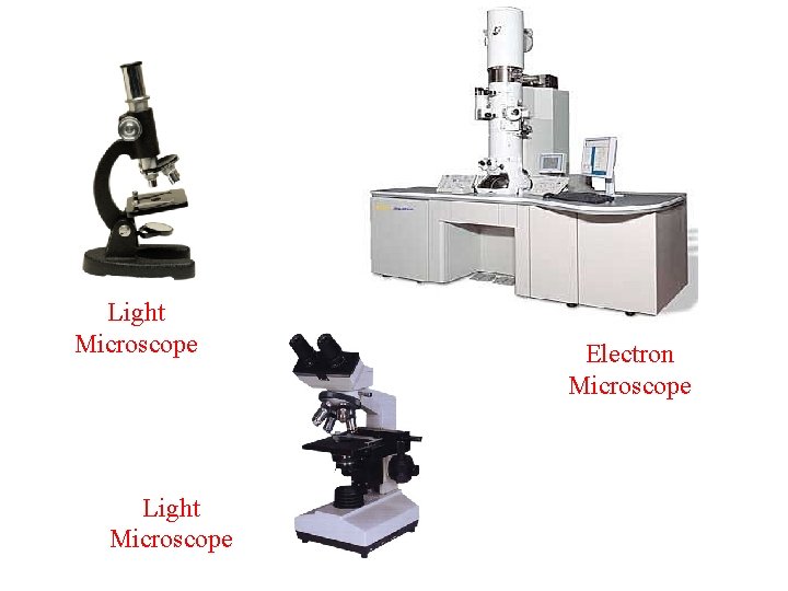
Light Microscope Electron Microscope
Light micrograph of a protist, Paramecium.
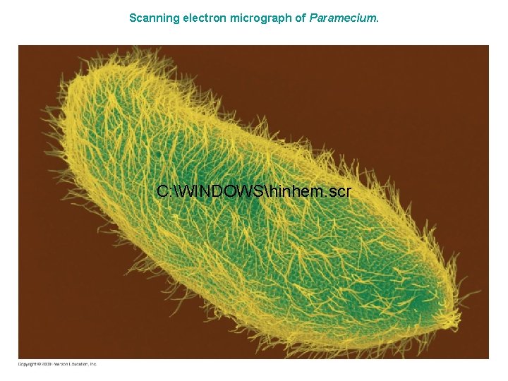
4. 1 Microscopes reveal the world of the cell § Biologists often use a very powerful microscope called the electron microscope (EM) to view the ultrastructure of cells – It can resolve biological structures as small as 2 nanometers and can magnify up to 100, 000 times – Instead of light, the EM uses a beam of electrons Copyright © 2009 Pearson Education, Inc.
Scanning electron micrograph of Paramecium. C: WINDOWShinhem. scr
10 m Length of some nerve and muscle cells Chicken egg 10 mm (1 cm) 1 mm Frog egg 100 µm 100 nm 1 nm 0. 1 nm Most plant and animal cells ﻡ Nucleus Most bacteria Mitochondrion Mycoplasmas (smallest bacteria) Viruses Ribosome Proteins Lipids Small molecules Atoms Electron microscope 100 mm (10 cm) Human height Light microscope 1 m Unaided eye The sizes of cells and related objects
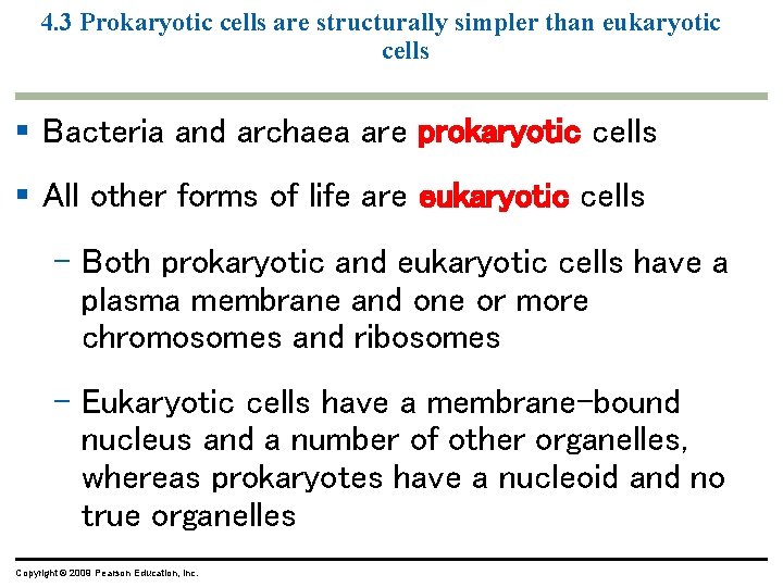
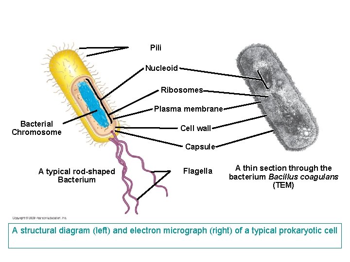
4. 3 Prokaryotic cells are structurally simpler than eukaryotic cells § Bacteria and archaea are prokaryotic cells § All other forms of life are eukaryotic cells – Both prokaryotic and eukaryotic cells have a plasma membrane and one or more chromosomes and ribosomes – Eukaryotic cells have a membrane-bound nucleus and a number of other organelles, whereas prokaryotes have a nucleoid and no true organelles Copyright © 2009 Pearson Education, Inc.
Pili Nucleoid Ribosomes Plasma membrane Bacterial Chromosome Cell wall Capsule A typical rod-shaped Bacterium Flagella A thin section through the bacterium Bacillus coagulans (TEM) A structural diagram (left) and electron micrograph (right) of a typical prokaryotic cell
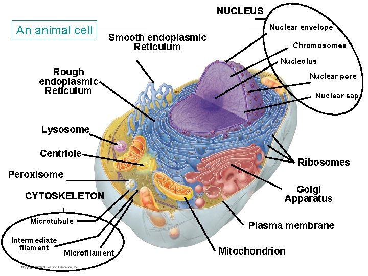
4. 4 Eukaryotic cells are partitioned into functional compartments § There are four life processes in eukaryotic cells that depend upon structures and organelles – Manufacturing – Breakdown of molecules – Energy processing – Structural support, movement, and communication Copyright © 2009 Pearson Education, Inc.
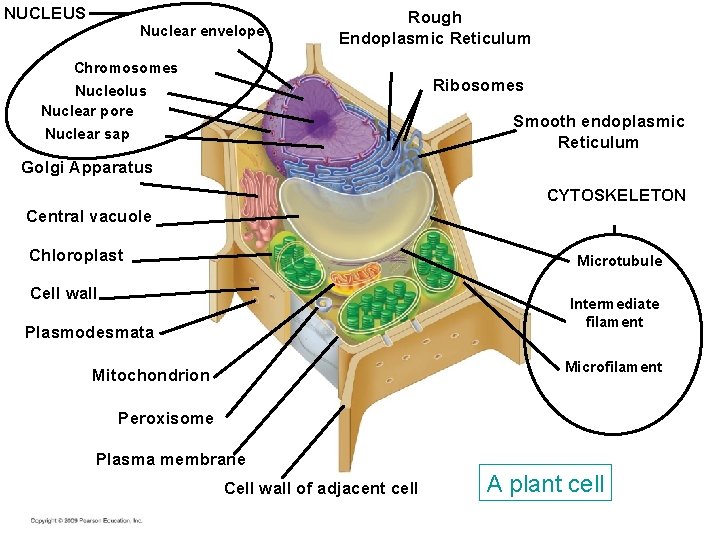
NUCLEUS Nuclear envelope Rough Endoplasmic Reticulum Chromosomes Ribosomes Nucleolus Nuclear pore Smooth endoplasmic Reticulum Nuclear sap Golgi Apparatus CYTOSKELETON Central vacuole Chloroplast Microtubule Cell wall Intermediate filament Plasmodesmata Microfilament Mitochondrion Peroxisome Plasma membrane Cell wall of adjacent cell A plant cell
NUCLEUS An animal cell Nuclear envelope Smooth endoplasmic Reticulum Chromosomes Nucleolus Rough endoplasmic Reticulum Nuclear pore Nuclear sap Lysosome Centriole Ribosomes Peroxisome CYTOSKELETON Microtubule Intermediate filament Microfilament Golgi Apparatus Plasma membrane Mitochondrion
4. 4 Eukaryotic cells are partitioned into functional compartments § Although there are many similarities between animal and plant cells, differences exist – Lysosomes and centrioles are not found in plant cells – Plant cells have a rigid cell wall, chloroplasts, and a central vacuole not found in animal cells Copyright © 2009 Pearson Education, Inc.
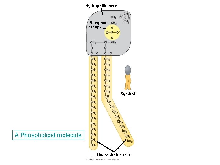
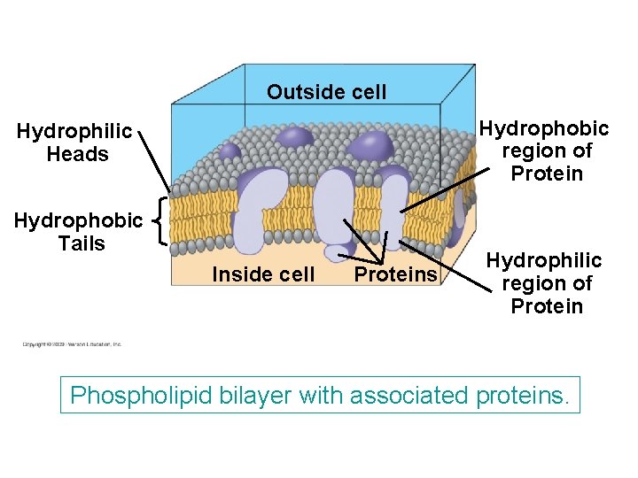
4. 5 The structure of membranes correlates with their functions § The plasma membrane controls the movement of molecules into and out of the cell, a trait called selective permeability – The structure of the membrane with its component molecules is responsible for this characteristic – Membranes are made of lipids, proteins, and some carbohydrate, but the most abundant lipids are phospholipids Copyright © 2009 Pearson Education, Inc.
Hydrophilic head Phosphate group Symbol A Phospholipid molecule Hydrophobic tails
Outside cell Hydrophobic region of Protein Hydrophilic Heads Hydrophobic Tails Inside cell Proteins Hydrophilic region of Protein Phospholipid bilayer with associated proteins.
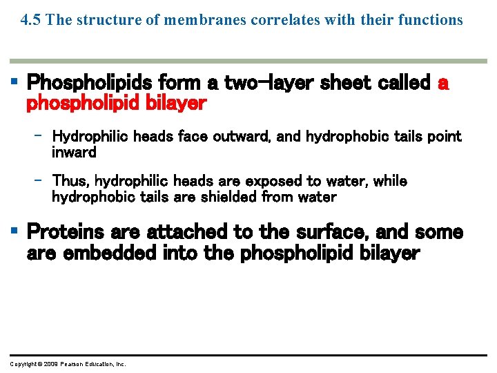
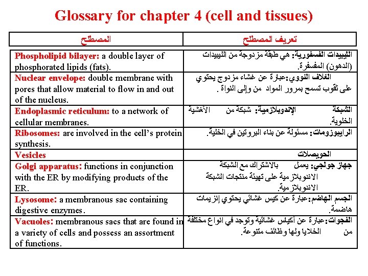
4. 5 The structure of membranes correlates with their functions § Phospholipids form a two-layer sheet called a phospholipid bilayer – Hydrophilic heads face outward, and hydrophobic tails point inward – Thus, hydrophilic heads are exposed to water, while hydrophobic tails are shielded from water § Proteins are attached to the surface, and some are embedded into the phospholipid bilayer Copyright © 2009 Pearson Education, Inc.
CELL STRUCTURES INVOLVED IN MANUFACTURING AND BREAKDOWN Copyright © 2009 Pearson Education, Inc.
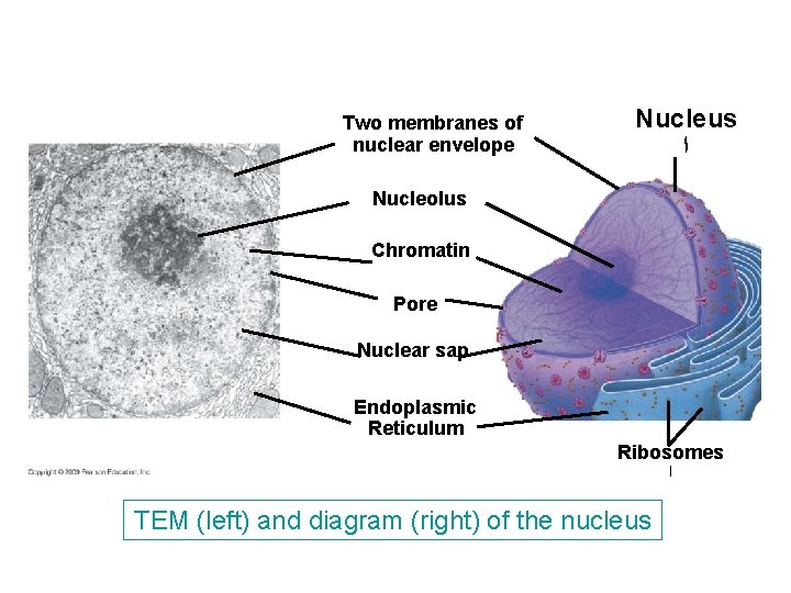
Two membranes of nuclear envelope Nucleus ﺍ Nucleolus Chromatin Pore Nuclear sap Endoplasmic Reticulum Ribosomes ﺍ TEM (left) and diagram (right) of the nucleus
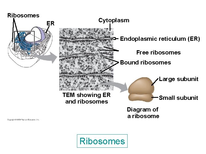
Ribosomes ER Cytoplasm Endoplasmic reticulum (ER) Free ribosomes Bound ribosomes Large subunit TEM showing ER and ribosomes Small subunit Diagram of a ribosome Ribosomes
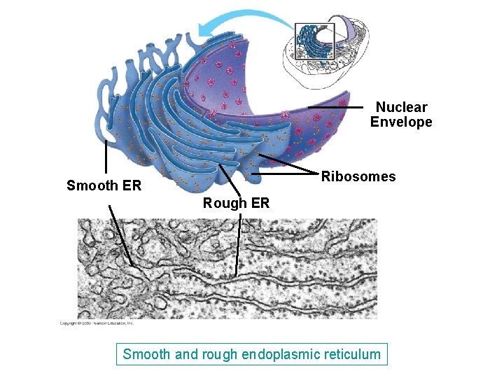
Nuclear Envelope Ribosomes Smooth ER Rough ER Smooth and rough endoplasmic reticulum
4. 6 The nucleus is the cell’s genetic control center § The nucleus controls the cell’s activities and is responsible for inheritance – Inside is a complex of proteins and DNA called chromatin, which makes up the cell’s chromosomes – DNA is copied within the nucleus prior to cell division Copyright © 2009 Pearson Education, Inc.
4. 6 The nucleus is the cell’s genetic control center § The nuclear envelope is a double membrane with pores that allow material to flow in and out of the nucleus – It is attached to a network of cellular membranes called the endoplasmic reticulum Copyright © 2009 Pearson Education, Inc.
4. 7 Ribosomes make proteins for use in the cell and export § Ribosomes are involved in the cell’s protein synthesis – Ribosomes are synthesized in the nucleolus, which is found in the nucleus – Cells that must synthesize large amounts of protein have a large number of ribosomes Copyright © 2009 Pearson Education, Inc.
4. 7 Ribosomes make proteins for use in the cell and export § Some ribosomes are free ribosomes; others are bound – Free ribosomes are suspended in the cytoplasm – Bound ribosomes are attached to the endoplasmic reticulum (ER) associated with the nuclear envelope Copyright © 2009 Pearson Education, Inc.
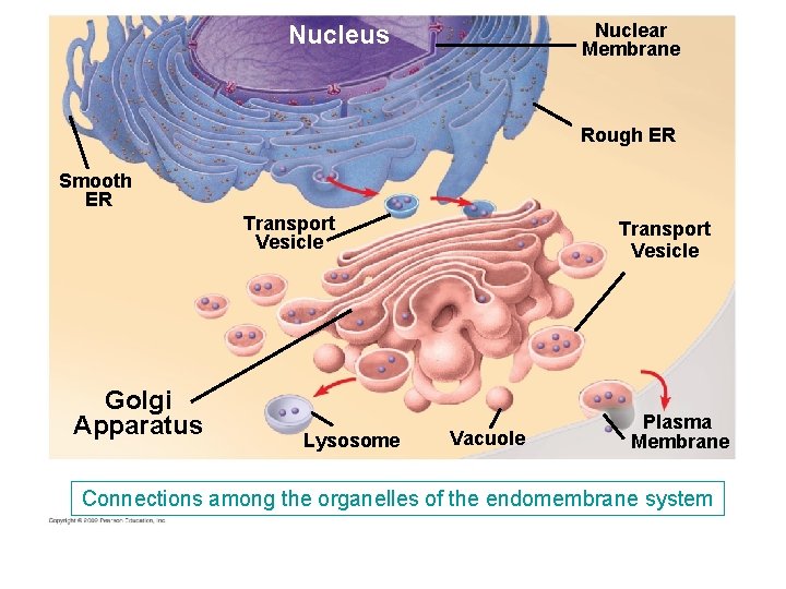
4. 8 Overview: Many cell organelles are connected through the endomembrane system § The membranes within an eukaryotic cell are physically connected and compose the endomembrane system – The endomembrane system includes the nuclear envelope, endoplasmic reticulum (ER), Golgi apparatus, lysosomes, vacuoles, and the plasma membrane Copyright © 2009 Pearson Education, Inc.
4. 8 Overview: Many cell organelles are connected through the endomembrane system ﻳﺔ § Some components of the endomembrane system are able to communicate with others with formation and transfer of small membrane segments called vesicles – One important result of communication is the synthesis, storage, and export of molecules Copyright © 2009 Pearson Education, Inc.
4. 9 The endoplasmic reticulum is a biosynthetic factory § There are two kinds of endoplasmic reticulum—smooth and rough § Smooth ER lacks attached ribosomes § Rough ER lines the outer surface of membranes – They differ in structure and function – However, they are connected Copyright © 2009 Pearson Education, Inc.
4. 9 The endoplasmic reticulum is a biosynthetic factory § Smooth ER is involved in a variety of diverse metabolic processes – For example, enzymes of the smooth ER are involved in the synthesis of lipids, oils, phospholipids, and steroids Copyright © 2009 Pearson Education, Inc.
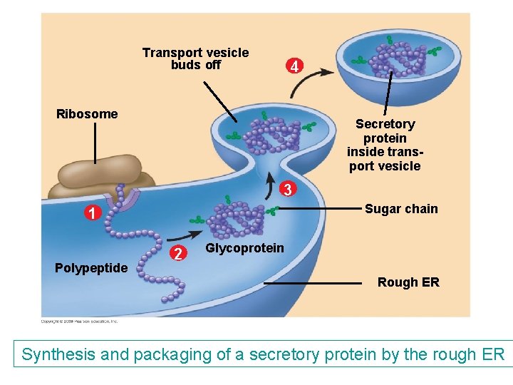
4. 9 The endoplasmic reticulum is a biosynthetic factory § Rough ER makes additional membrane for itself and proteins destined for secretion – Once proteins are synthesized, they are transported in vesicles to other parts of the endomembrane system Copyright © 2009 Pearson Education, Inc.
Transport vesicle buds off 4 Ribosome Secretory protein inside transport vesicle 3 Sugar chain 1 Polypeptide 2 Glycoprotein Rough ER Synthesis and packaging of a secretory protein by the rough ER
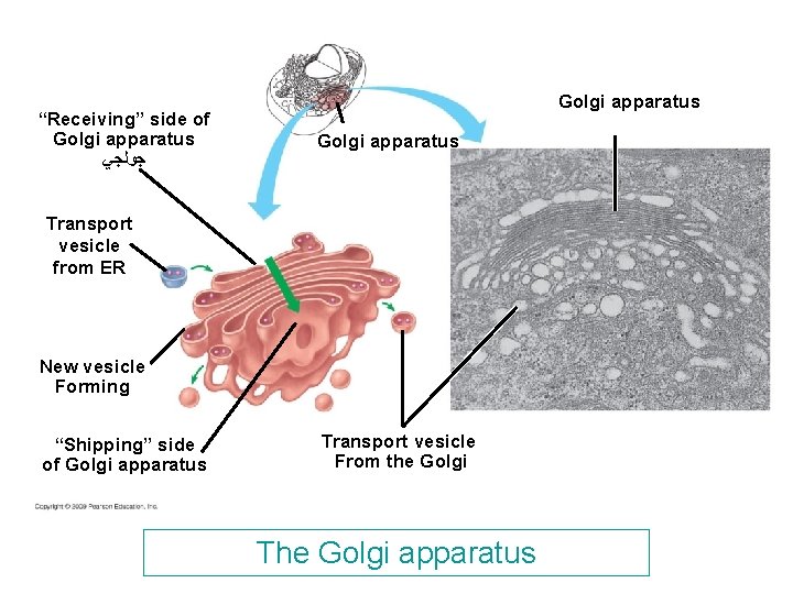
“Receiving” side of Golgi apparatus ﺟﻮﻟﺠﻲ Golgi apparatus Transport vesicle from ER New vesicle Forming “Shipping” side of Golgi apparatus Transport vesicle From the Golgi The Golgi apparatus
4. 10 The Golgi apparatus finishes, sorts, and ships cell products § The Golgi apparatus functions in conjunction with the ER by modifying products of the ER – Products travel in transport vesicles from the ER to the Golgi apparatus – One side of the Golgi apparatus functions as a receiving dock for the product and the other as a shipping dock – Products are modified as they go from one side of the Golgi apparatus to the other and travel in vesicles to other sites Copyright © 2009 Pearson Education, Inc.
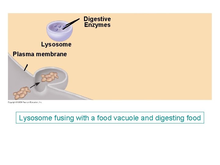
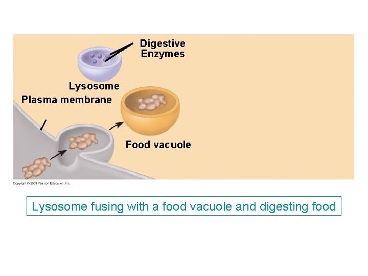
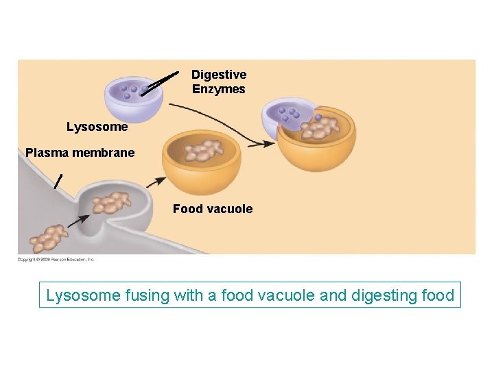
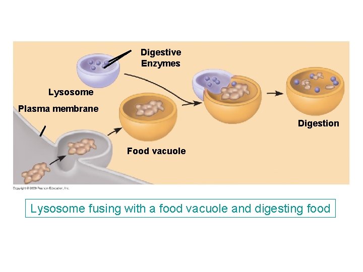
4. 11 Lysosomes are digestive compartments within a cell § A lysosome is a membranous sac containing digestive enzymes – The enzymes and membrane are produced by the ER and transferred to the Golgi apparatus for processing – The membrane serves to safely isolate these potent enzymes from the rest of the cell Copyright © 2009 Pearson Education, Inc.
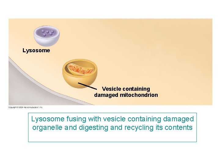
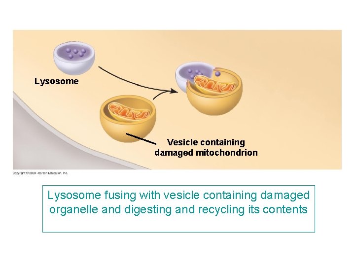
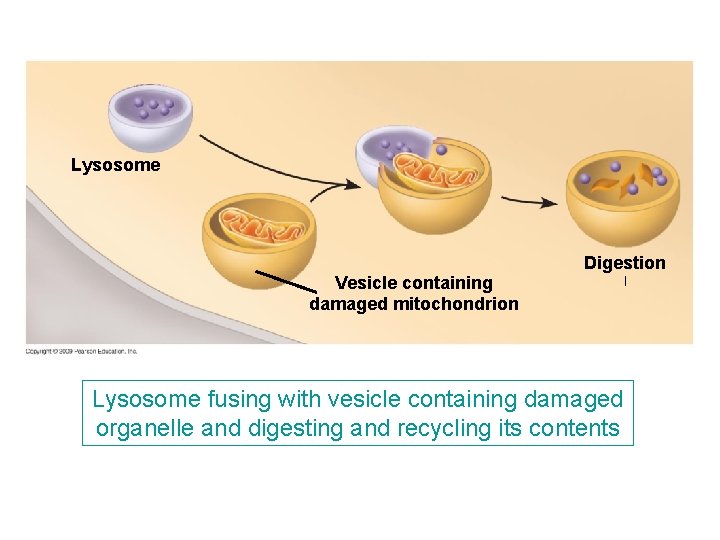
4. 11 Lysosomes are digestive compartments within a cell § One of the several functions of lysosomes is to remove or recycle damaged parts of a cell – The damaged organelle is first enclosed in a membrane vesicle – Then a lysosome fuses with the vesicle, dismantling its contents and breaking down the damaged organelle Animation: Lysosome Formation Copyright © 2009 Pearson Education, Inc.
Digestive Enzymes Lysosome Plasma membrane Lysosome fusing with a food vacuole and digesting food
Digestive Enzymes Lysosome Plasma membrane Food vacuole Lysosome fusing with a food vacuole and digesting food
Digestive Enzymes Lysosome Plasma membrane Food vacuole Lysosome fusing with a food vacuole and digesting food
Digestive Enzymes Lysosome Plasma membrane Digestion Food vacuole Lysosome fusing with a food vacuole and digesting food
Lysosome Vesicle containing damaged mitochondrion Lysosome fusing with vesicle containing damaged organelle and digesting and recycling its contents
Lysosome Vesicle containing damaged mitochondrion Lysosome fusing with vesicle containing damaged organelle and digesting and recycling its contents
Lysosome Vesicle containing damaged mitochondrion Digestion ﺍ Lysosome fusing with vesicle containing damaged organelle and digesting and recycling its contents
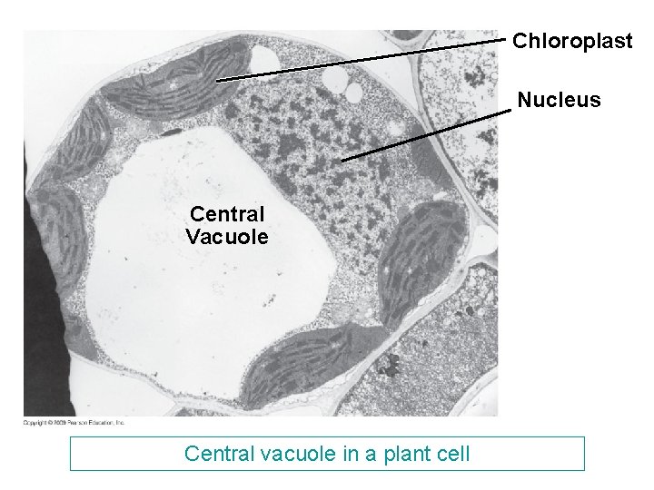
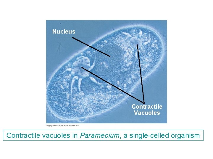
4. 12 Vacuoles function in the general maintenance of the cell § Vacuoles are membranous sacs that are found in a variety of cells and possess an assortment of functions – Examples are the central vacuole in plants with hydrolytic functions, pigment vacuoles in plants to provide color to flowers, and contractile vacuoles in some protists to expel water from the cell Video: Paramecium Vacuole Copyright © 2009 Pearson Education, Inc.
Chloroplast Nucleus Central Vacuole Central vacuole in a plant cell
Nucleus Contractile Vacuoles Contractile vacuoles in Paramecium, a single-celled organism
4. 13 A review of the structures involved in manufacturing and breakdown ( § The following figure summarizes the relationships among the major organelles of the endomembrane system Copyright © 2009 Pearson Education, Inc.
Nuclear Membrane Nucleus Rough ER Smooth ER Transport Vesicle Golgi Apparatus Lysosome Transport Vesicle Vacuole Plasma Membrane Connections among the organelles of the endomembrane system
ENERGY-CONVERTING ORGANELLES Copyright © 2009 Pearson Education, Inc.
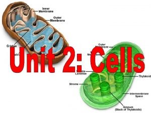
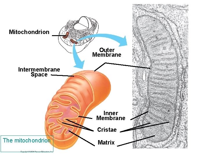
Mitochondrion Outer Membrane Intermembrane Space Inner Membrane Cristae The mitochondrion Matrix
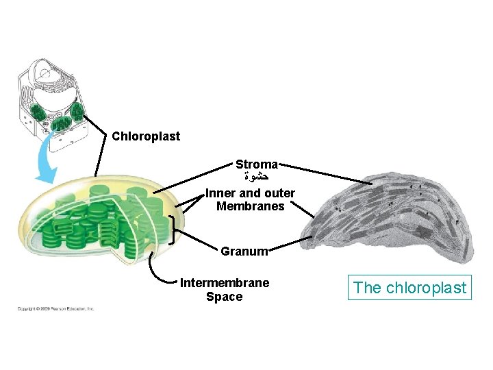
Chloroplast Stroma ﺣﺸﻮﺓ Inner and outer Membranes Granum Intermembrane Space The chloroplast
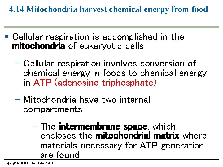
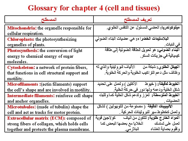
4. 14 Mitochondria harvest chemical energy from food § Cellular respiration is accomplished in the mitochondria of eukaryotic cells – Cellular respiration involves conversion of chemical energy in foods to chemical energy in ATP (adenosine triphosphate) – Mitochondria have two internal compartments – The intermembrane space, which encloses the mitochondrial matrix where materials necessary for ATP generation are found Copyright © 2009 Pearson Education, Inc.
4. 15 Chloroplasts convert solar energy to chemical energy § Chloroplasts are the photosynthesizing organelles of plants – Photosynthesis is the conversion of light energy to chemical energy of sugar molecules § Chloroplasts are partitioned into compartments – The important parts of chloroplasts are the stroma, thylakoids, and grana Copyright © 2009 Pearson Education, Inc.
INTERNAL AND EXTERNAL SUPPORT: THE CYTOSKELETON AND CELL SURFACES Copyright © 2009 Pearson Education, Inc.
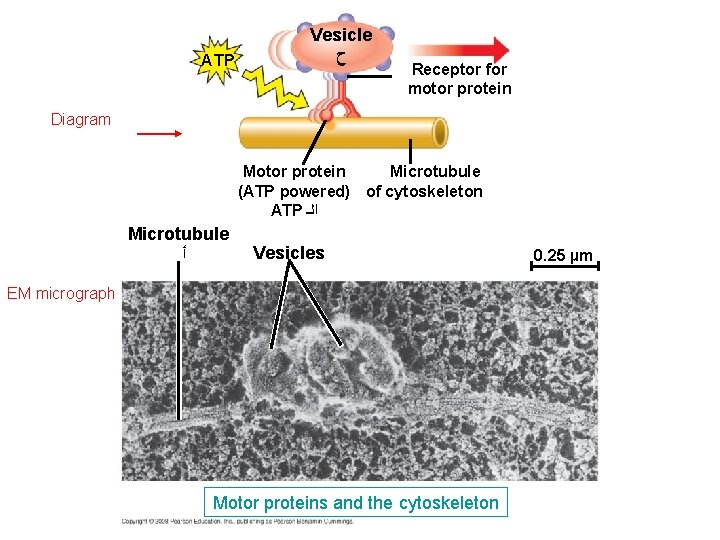
4. 17 The cell’s internal skeleton helps organize its structure and activities § Cells contain a network of protein fibers, called the cytoskeleton, that functions in cell structural support and motility – Scientists believe that motility and cellular regulation result when the cytoskeleton interacts with proteins called motor proteins Copyright © 2009 Pearson Education, Inc. Video: Cytoplasmic Streaming
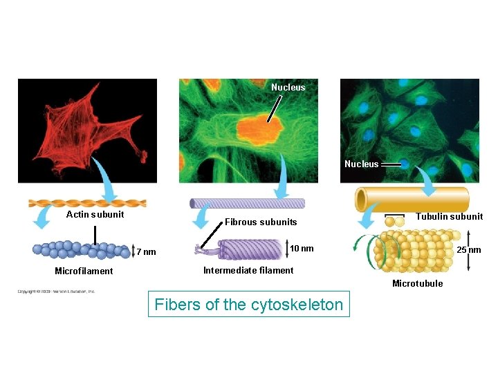
4. 17 The cell’s internal skeleton helps organize its structure and activities § The cytoskeleton is composed of three kinds of fibers – Microfilaments (actin filaments) support the cell’s shape and are involved in motility – Intermediate filaments reinforce cell shape and anchor organelles – Microtubules (made of tubulin) shape the cell and act as tracks for motor protein Copyright © 2009 Pearson Education, Inc.
ATP Vesicle ﺡ Receptor for motor protein Diagram Motor protein Microtubule (ATP powered) of cytoskeleton ATP ﺍﻟـ Microtubule ﺃ Vesicles EM micrograph Motor proteins and the cytoskeleton 0. 25 µm
Nucleus Actin subunit Fibrous subunits 7 nm Microfilament Tubulin subunit 10 nm 25 nm Intermediate filament Microtubule Fibers of the cytoskeleton
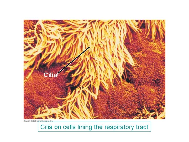
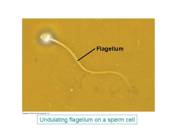
4. 18 Cilia and flagella move when microtubules bend § While some protists have flagella and cilia that are important in locomotion, some cells of multicellular organisms have them for different reasons – Cells that sweep mucus out of our lungs have cilia – Animal sperm are flagellated Copyright © 2009 Pearson Education, Inc.
Cilia ﺃ Cilia on cells lining the respiratory tract
Flagellum Undulating flagellum on a sperm cell
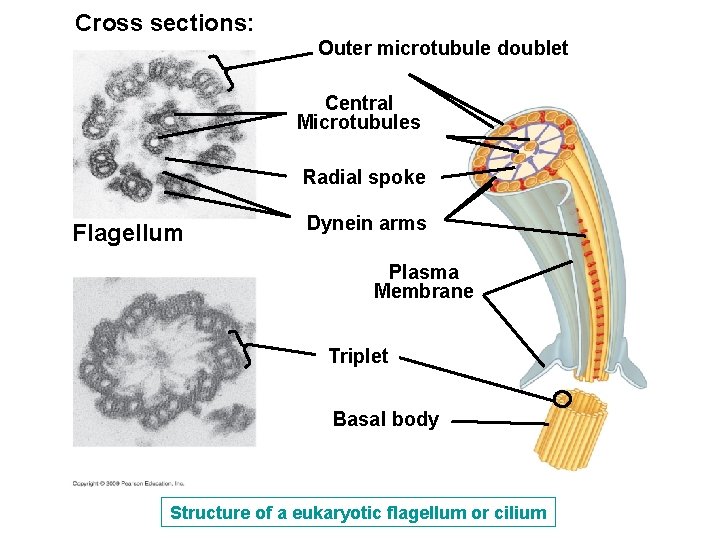
4. 18 Cilia and flagella move when microtubules bend § Although differences exist, flagella and cilia have a common structure and mechanism of movement § Both flagella and cilia are made of microtubules wrapped in an extension of the plasma membrane § A ring of nine microtubule doublets surrounds a central pair of microtubules Copyright © 2009 Pearson Education, Inc.
Cross sections: Outer microtubule doublet Central Microtubules Radial spoke Flagellum Dynein arms Plasma Membrane Triplet Basal body Structure of a eukaryotic flagellum or cilium
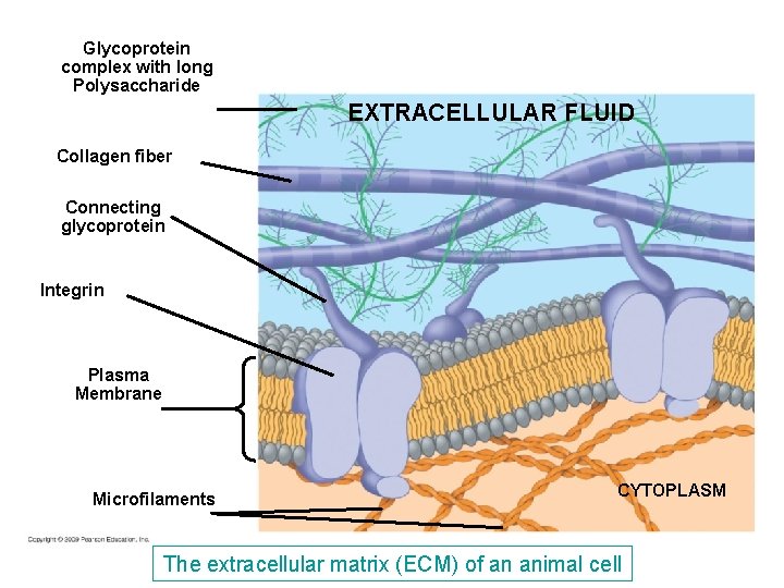
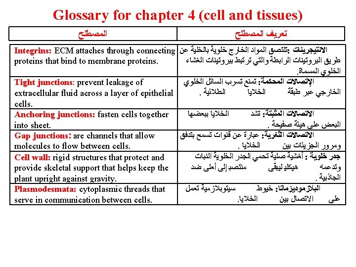
4. 20 The extracellular matrix of animal cells functions in support, movement, and regulation § Cells synthesize and secrete the extracellular matrix (ECM) that is essential to cell function – The ECM is composed of strong fibers of collagen, which holds cells together and protects the plasma membrane – ECM attaches through connecting proteins that bind to membrane proteins called integrins – Integrins span the plasma membrane and connect to microfilaments of the cytoskeleton Copyright © 2009 Pearson Education, Inc.
Glycoprotein complex with long Polysaccharide EXTRACELLULAR FLUID Collagen fiber Connecting glycoprotein Integrin Plasma Membrane Microfilaments CYTOPLASM The extracellular matrix (ECM) of an animal cell
4. 21 Three types of cell junctions are found in animal tissues § Adjacent cells communicate, interact, and adhere through specialized junctions between them – Tight junctions prevent leakage of extracellular fluid across a layer of epithelial cells – Anchoring junctions fasten cells together into sheets – Gap junctions are channels that allow molecules to flow between cells Copyright © 2009 Pearson Education, Inc.
Tight junctions Anchoring junction Gap junctions Plasma membranes of adjacent cells Extracellular matrix Three types of cell junctions in animal tissues
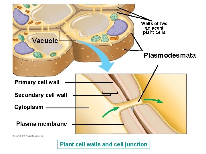
4. 22 Cell walls enclose and support plant cells § Plant, but not animal cells, have a rigid cell wall – It protects and provides skeletal support that helps keep the plant upright against gravity – Plant cell walls are composed primarily of cellulose § Plant cells have cell junctions called plasmodesmata that serve in communication between cells Copyright © 2009 Pearson Education, Inc.
Walls of two adjacent plant cells Vacuole Plasmodesmata Primary cell wall Secondary cell wall Cytoplasm Plasma membrane Plant cell walls and cell junction
FUNCTIONAL CATEGORIES OF CELL STRUCTURES Copyright © 2009 Pearson Education, Inc.
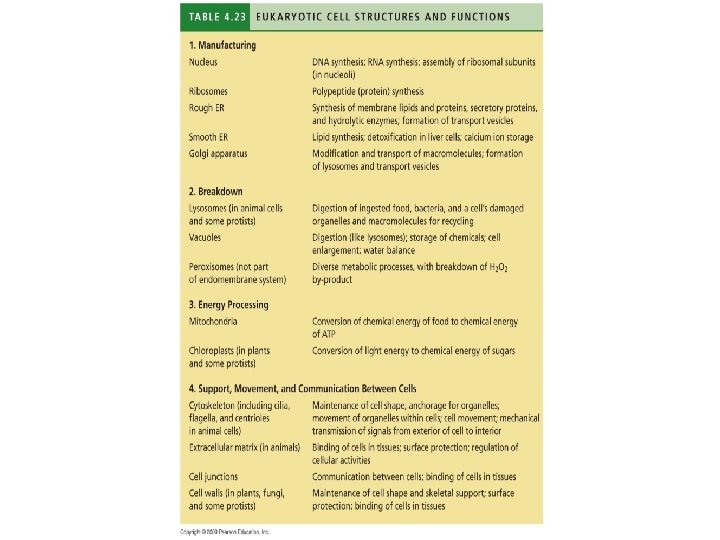
4. 23 Review: Eukaryotic cell structures can be grouped on the basis of four basic functions § It is possible to group cell organelles into four categories based on general functions of organelles – In each category structure is correlated with function Copyright © 2009 Pearson Education, Inc.
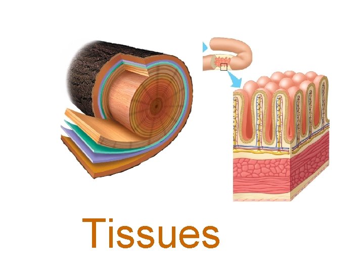
Tissues
ANIMAL TISSUES Copyright © 2009 Pearson Education, Inc.
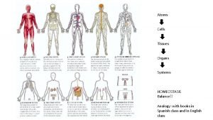
20. 1 Structure fits function at all levels of organization in the animal body § Anatomy—structure § Physiology—function § Animals consist of a hierarchy of levels of organization Copyright © 2009 Pearson Education, Inc.
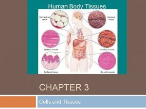
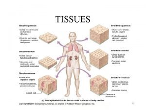
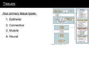
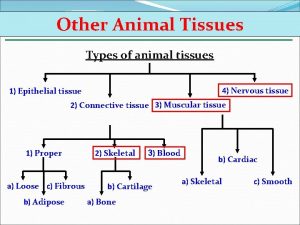
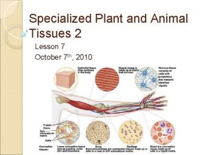
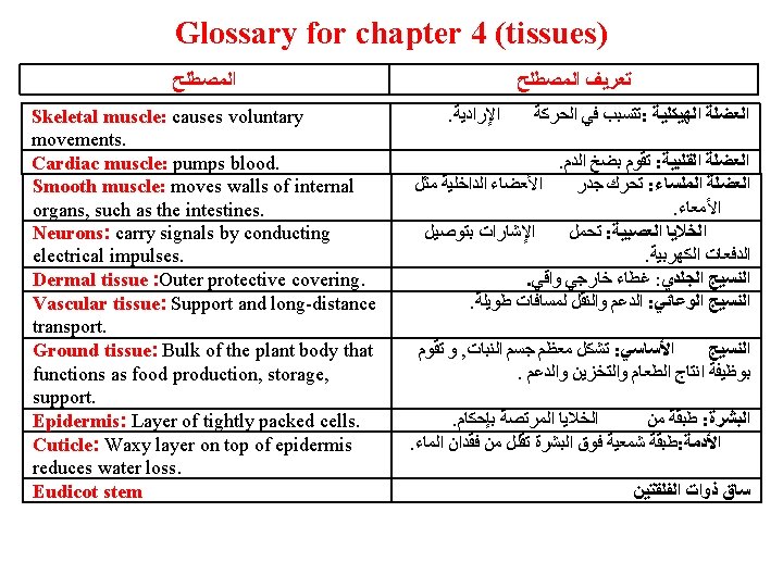
20. 3 Tissues are groups of cells with a common structure and function § Animals have four main categories of tissues – Epithelial tissue – Connective tissue – Muscle tissue – Nervous tissue Copyright © 2009 Pearson Education, Inc.
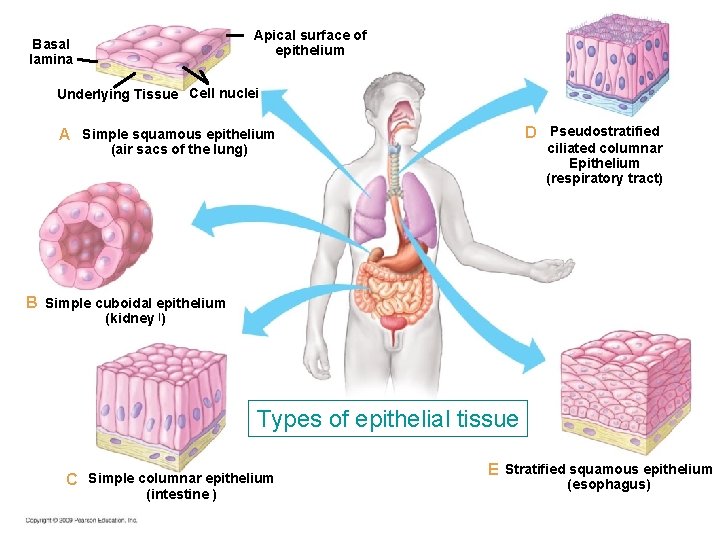
20. 4 Epithelial tissue covers the body and lines its organs and cavities § Epithelial cells come in three shapes – Squamous—like a fried egg – Cuboidal—as tall as they are wide – Columnar—taller than they are wide Copyright © 2009 Pearson Education, Inc.
Apical surface of epithelium Basal lamina Underlying Tissue Cell nuclei D Pseudostratified A Simple squamous epithelium ciliated columnar Epithelium (respiratory tract) (air sacs of the lung) B Simple cuboidal epithelium (kidney )ﺍ Types of epithelial tissue C Simple columnar epithelium (intestine ) E Stratified squamous epithelium (esophagus)
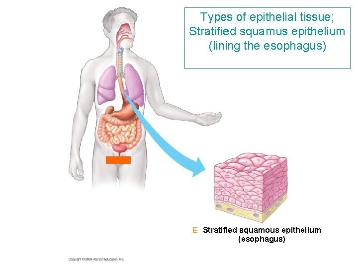
20. 4 Epithelial tissue covers the body and lines its organs and cavities § Stratified epithelial cells are stacked on top of each other Copyright © 2009 Pearson Education, Inc.
Types of epithelial tissue; Stratified squamus epithelium (lining the esophagus) E Stratified squamous epithelium (esophagus)
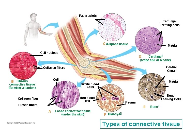
20. 5 Connective tissue binds and supports other tissues § Connective tissue can be grouped into six major types Copyright © 2009 Pearson Education, Inc.
Fat droplets Cartilage. Forming cells C Adipose tissue Cell nucleus Matrix Cartilage ﺍ (at the end of a bone) D Central Canal Collagen fibers Cell B Fibrous Matrix White blood Cells connective tissue (forming a tendon) Red blood cell Collagen fiber Bone. Forming Cells Plasma Elastic fibers E A Loose connective tissue (under the skin) Bone ﺍ F Blood ﺍﻟﺪﻡ Types of connective tissue
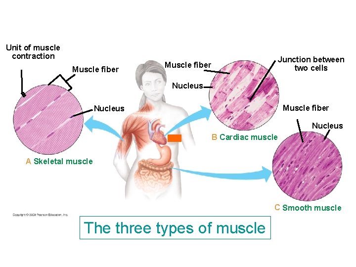
20. 6 Muscle tissue functions in movement § Skeletal muscle causes voluntary movements § Cardiac muscle pumps blood § Smooth muscle moves walls of internal organs, such as the intestines Copyright © 2009 Pearson Education, Inc.
Unit of muscle contraction Muscle fiber Junction between two cells Muscle fiber Nucleus B Cardiac muscle A Skeletal muscle C Smooth muscle The three types of muscle
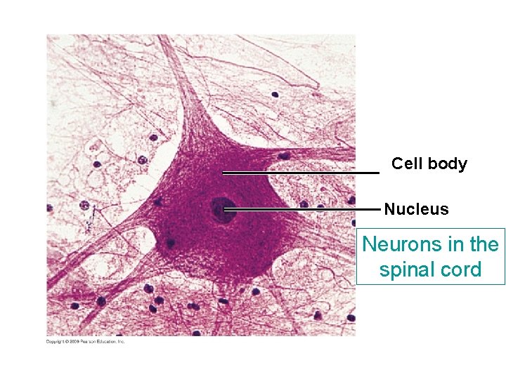
20. 7 Nervous tissue forms a communication network § Neurons carry signals by conducting electrical impulses § Supporting cells insulate axons and nourish neurons Copyright © 2009 Pearson Education, Inc.
Cell body Nucleus Neurons in the spinal cord
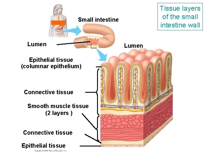
20. 8 Organs are made up of tissues § Each tissue performs specific functions § The heart has epithelial, connective, and nervous tissues – Epithelia line the heart chambers – Connective tissues make the heart elastic – Neurons regulate contractions Copyright © 2009 Pearson Education, Inc.
Tissue layers of the small intestine wall Small intestine Lumen Epithelial tissue (columnar epithelium) Connective tissue Smooth muscle tissue (2 layers ) Connective tissue Epithelial tissue Lumen
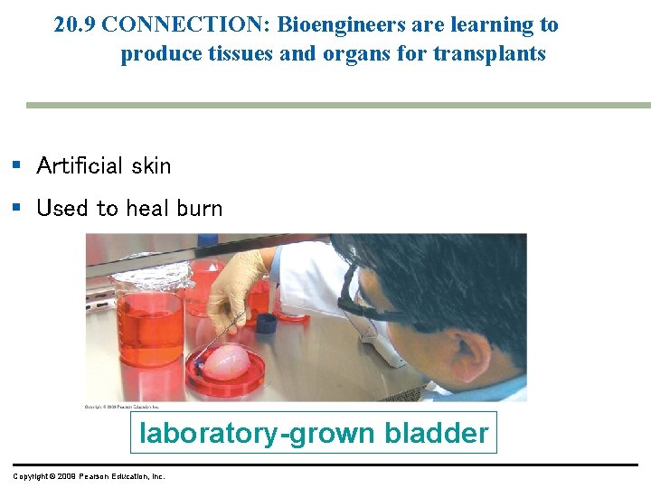
20. 9 CONNECTION: Bioengineers are learning to produce tissues and organs for transplants § Artificial skin § Used to heal burn laboratory-grown bladder Copyright © 2009 Pearson Education, Inc.
PLANT TISSUES Copyright © 2009 Pearson Education, Inc.
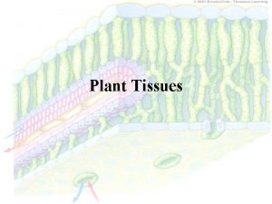
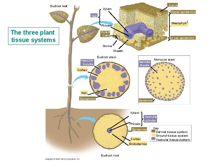
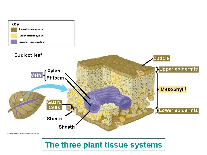
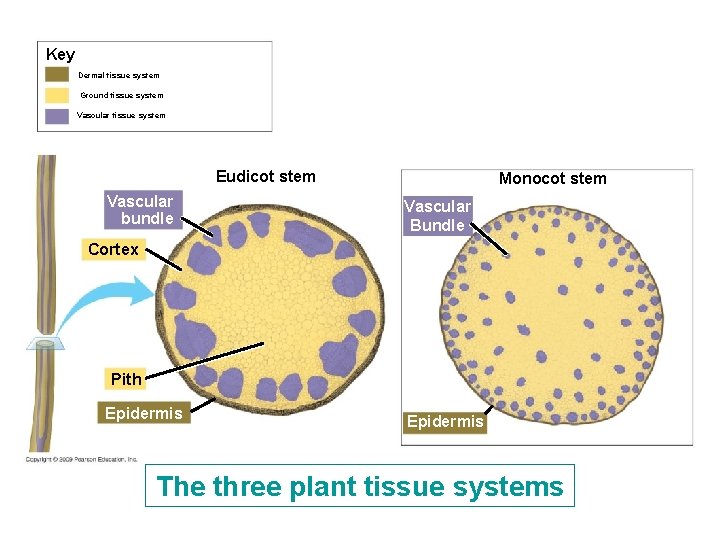
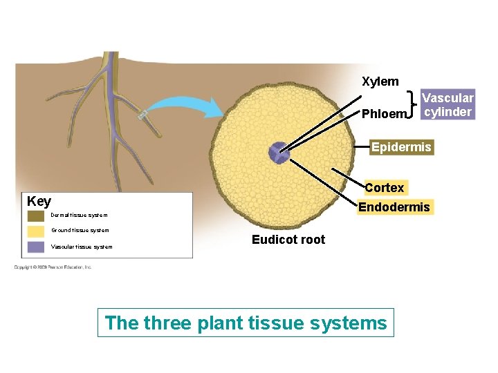
31. 5 Three tissue systems make up the plant body § Dermal tissue – Outer protective covering § Vascular tissue – Support and long-distance transport § Ground tissue – Bulk of the plant body – Food production, storage, support Copyright © 2009 Pearson Education, Inc.
31. 5 Three tissue systems make up the plant body § Dermal tissue – Layer of tightly packed cells called the epidermis – First line of defense against damage and infection – Waxy layer called cuticle on top of epidermis, reduces water loss Copyright © 2009 Pearson Education, Inc.
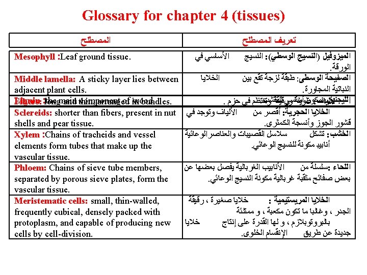
31. 5 Three tissue systems make up the plant body § Vascular tissue – Composed of xylem and phloem – Arranged in bundles § Ground tissue – Lies between dermal and vascular tissue – Eudicot stem ground tissue is divided into pith and cortex – Leaf ground tissue is called mesophyll Copyright © 2009 Pearson Education, Inc.
Eudicot leaf Cuticle Xylem Upper epidermis Vein ﺍ Mesophyll Phloem The three plant tissue systems Guard Cells Lower epidermis Stoma Sheath Eudicot stem Vascular bundle Vascular Bundle Monocot stem Cortex Pith Epidermis Xylem Vascular cylinder Phloem Epidermis Cortex Endodermis Eudicot root Key Dermal tissue system Ground tissue system Vascular tissue system
Key Dermal tissue system Ground tissue system Vascular tissue system Eudicot leaf Vein Cuticle Xylem Phloem Upper epidermis Mesophyll ﺍ Guard Cells Lower epidermis Stoma Sheath The three plant tissue systems
Key Dermal tissue system Ground tissue system Vascular tissue system Eudicot stem Vascular bundle Monocot stem Vascular Bundle Cortex Pith Epidermis The three plant tissue systems
Xylem Phloem Vascular cylinder Epidermis Cortex Key Endodermis Dermal tissue system Ground tissue system Vascular tissue system Eudicot root The three plant tissue systems
31. 5 Three tissue systems make up the plant body § Plants cells have three structures that distinguish them from animals cells – Chloroplasts used in photosynthesis – A large, fluid-filled vacuole – A cell wall composed of cellulose Copyright © 2009 Pearson Education, Inc.
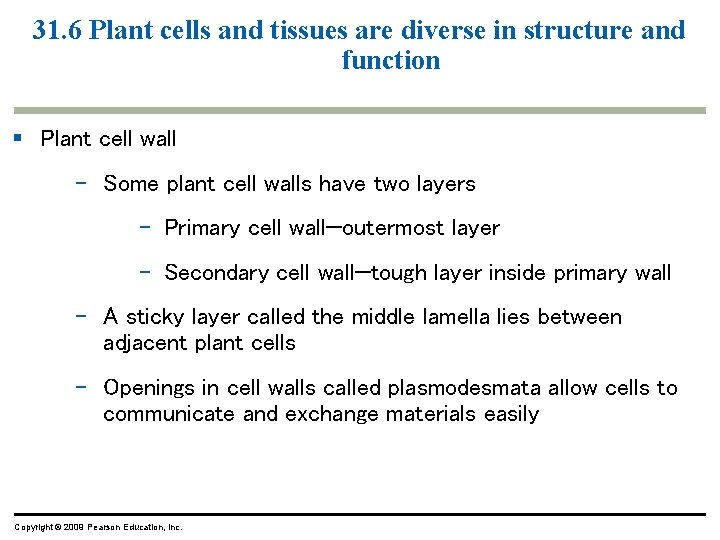
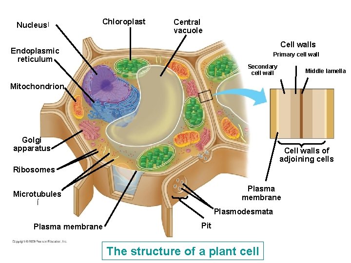
31. 6 Plant cells and tissues are diverse in structure and function § Plant cell wall – Some plant cell walls have two layers – Primary cell wall—outermost layer – Secondary cell wall—tough layer inside primary wall – A sticky layer called the middle lamella lies between adjacent plant cells – Openings in cell walls called plasmodesmata allow cells to communicate and exchange materials easily Copyright © 2009 Pearson Education, Inc.
Nucleus ﺍ Chloroplast Central vacuole Cell walls Endoplasmic reticulum Primary cell wall Secondary cell wall Middle lamella Mitochondrion Golgi apparatus Cell walls of adjoining cells Ribosomes Plasma membrane Microtubules ﺃ Plasma membrane Plasmodesmata Pit The structure of a plant cell
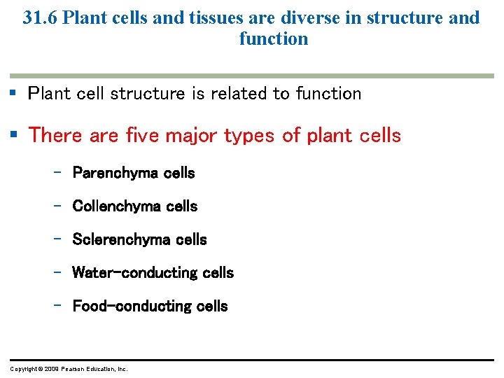
31. 6 Plant cells and tissues are diverse in structure and function § Plant cell structure is related to function § There are five major types of plant cells – Parenchyma cells – Collenchyma cells – Sclerenchyma cells – Water-conducting cells – Food-conducting cells Copyright © 2009 Pearson Education, Inc.
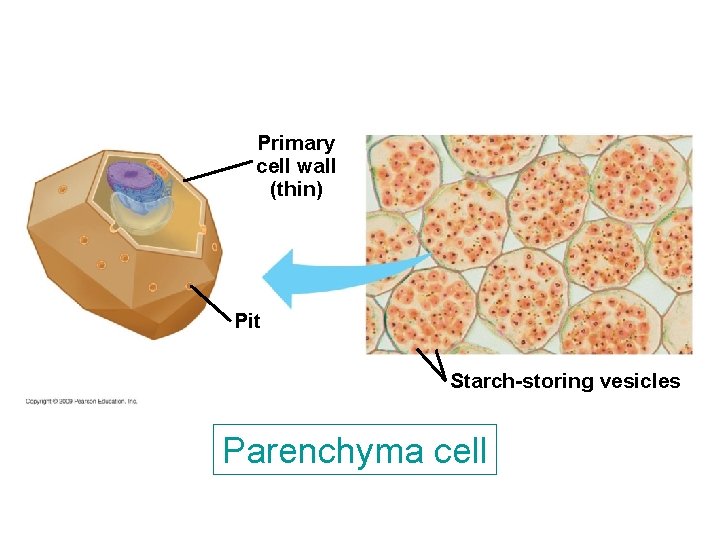
31. 6 Plant cells and tissues are diverse in structure and function § Parenchyma cells – Most abundant cell type – Thin primary cell wall – Lack secondary cell wall – Alive at maturity – Function in photosynthesis, food and water storage Copyright © 2009 Pearson Education, Inc.
Primary cell wall (thin) Pit Starch-storing vesicles Parenchyma cell
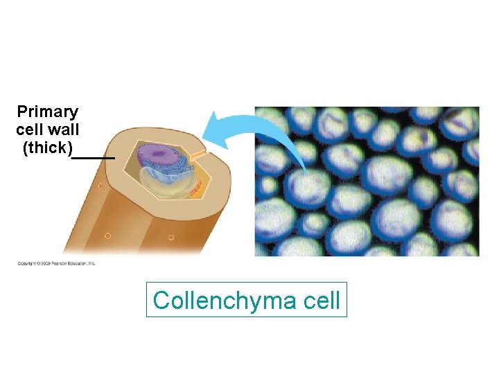
31. 6 Plant cells and tissues are diverse in structure and function § Collenchyma cells – Unevenly thickened primary cell wall – Lack secondary cell wall – Alive at maturity – Provide flexible support Copyright © 2009 Pearson Education, Inc.
Primary cell wall (thick) Collenchyma cell
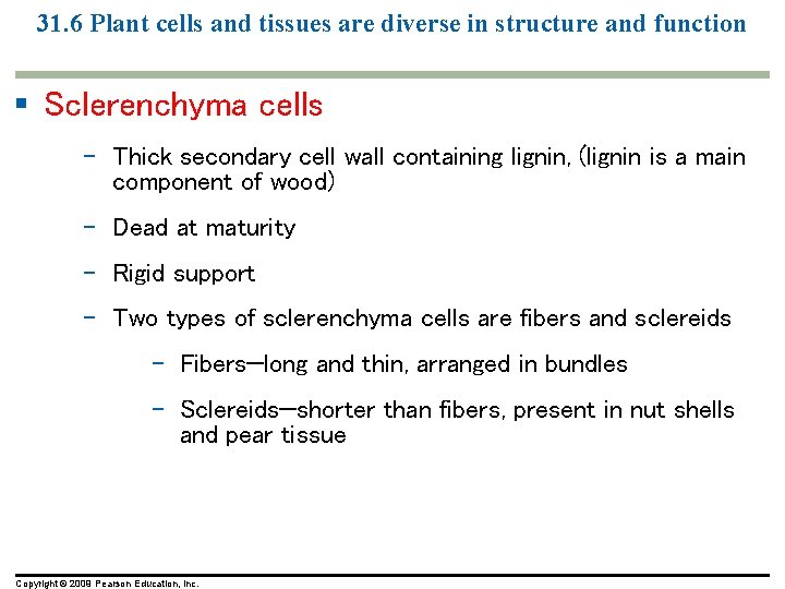
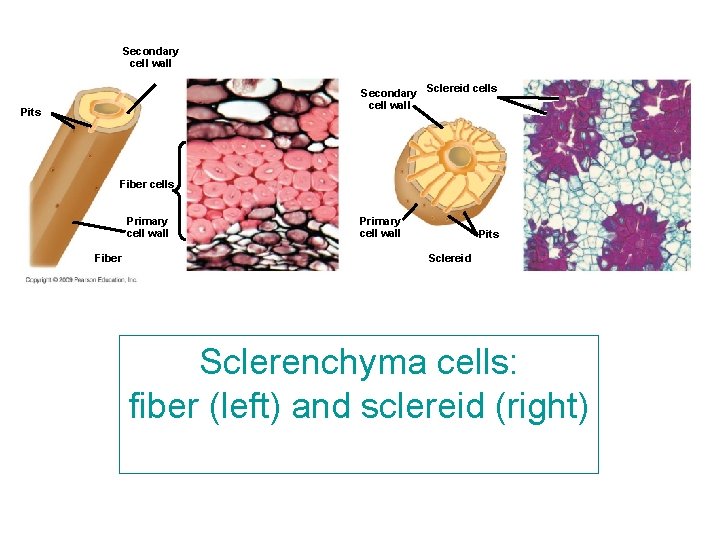
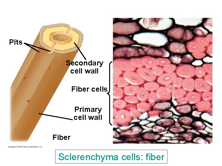
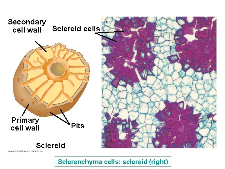
31. 6 Plant cells and tissues are diverse in structure and function § Sclerenchyma cells – Thick secondary cell wall containing lignin, (lignin is a main component of wood) – Dead at maturity – Rigid support – Two types of sclerenchyma cells are fibers and sclereids – Fibers—long and thin, arranged in bundles – Sclereids—shorter than fibers, present in nut shells and pear tissue Copyright © 2009 Pearson Education, Inc.
Secondary cell wall Secondary Sclereid cells cell wall Pits Fiber cells Primary cell wall Fiber Primary cell wall Pits Sclereid Sclerenchyma cells: fiber (left) and sclereid (right)
Pits Secondary cell wall Fiber cells Primary cell wall Fiber Sclerenchyma cells: fiber
Secondary cell wall Sclereid cells Primary cell wall Pits Sclereid Sclerenchyma cells: sclereid (right)
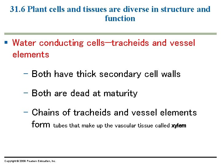
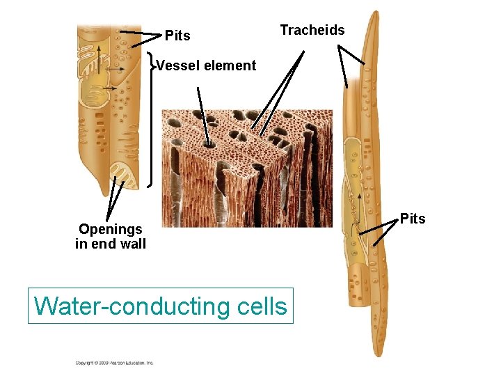
31. 6 Plant cells and tissues are diverse in structure and function § Water conducting cells—tracheids and vessel elements – Both have thick secondary cell walls – Both are dead at maturity – Chains of tracheids and vessel elements form tubes that make up the vascular tissue called xylem Copyright © 2009 Pearson Education, Inc.
Pits Tracheids Vessel element Openings in end wall Water-conducting cells Pits
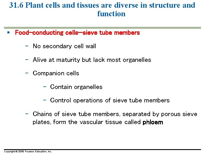
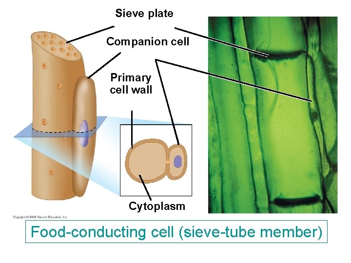
31. 6 Plant cells and tissues are diverse in structure and function § Food-conducting cells—sieve tube members – No secondary cell wall – Alive at maturity but lack most organelles – Companion cells – Contain organelles – Control operations of sieve tube members – Chains of sieve tube members, separated by porous sieve plates, form the vascular tissue called phloem Copyright © 2009 Pearson Education, Inc.
Sieve plate Companion cell Primary cell wall Cytoplasm Food-conducting cell (sieve-tube member)
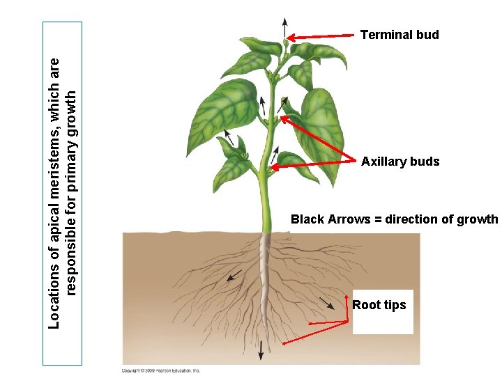
31. 6 Plant cells and tissues are diverse in structure and function Meristematic tissues § These are located at the tips of roots and stems, between the water - and food-conducting tissues of stems, and at various other places in plant bodies
Locations of apical meristems, which are responsible for primary growth Terminal bud Axillary buds Black Arrows = direction of growth Root tips
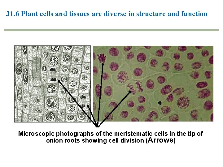
31. 6 Plant cells and tissues are diverse in structure and function § Meristematic cells are small, thin-walled, frequently cubical, densely packed with protoplasm, and capable of producing new cells by cell-division. § permanent tissues do not become changed into other kinds of tissues as do Meristematic tissues. (source of differentiation : they give rise to all other kinds of tissues)
31. 6 Plant cells and tissues are diverse in structure and function Microscopic photographs of the meristematic cells in the tip of onion roots showing cell division (Arrows)