
CHAPTER 4 A Tour of the Cell THE


- Slides: 49
CHAPTER 4 A Tour of the Cell
THE MICROSCOPIC WORLD OF CELLS • Cells are the building blocks of all life • Cells must be tiny for materials to move in and out of them fast enough to meet the cell’s metabolic needs Copyright © 2004 Pearson Education, Inc. publishing as Benjamin Cummings
• Organisms are either: – Single-celled (unicellular), such as most bacteria and protists – Multi-celled (multi-cellular), such as plants, animals, and most fungi Copyright © 2004 Pearson Education, Inc. publishing as Benjamin Cummings
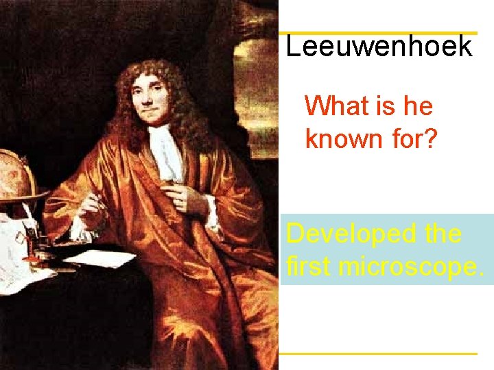
Leeuwenhoek What is he known for? Developed the first microscope. Copyright © 2004 Pearson Education, Inc. publishing as Benjamin Cummings
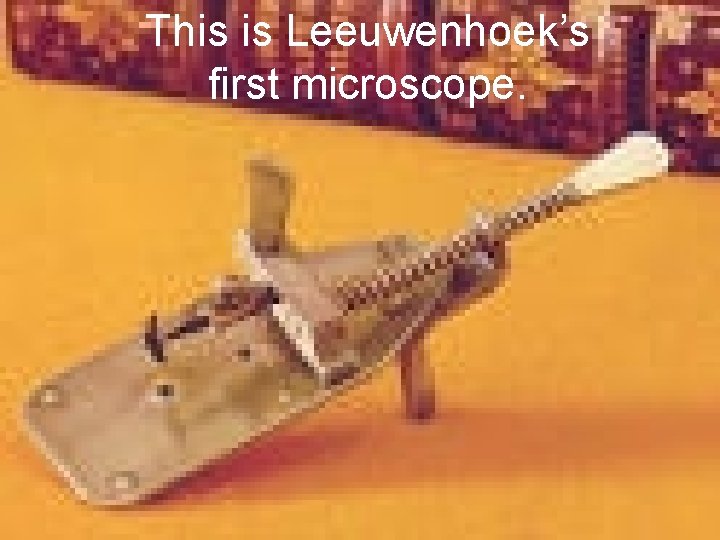
This is Leeuwenhoek’s first microscope. Copyright © 2004 Pearson Education, Inc. publishing as Benjamin Cummings
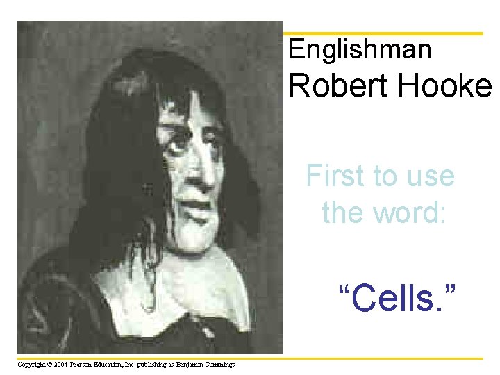
Englishman Robert Hooke First to use the word: “Cells. ” Copyright © 2004 Pearson Education, Inc. publishing as Benjamin Cummings
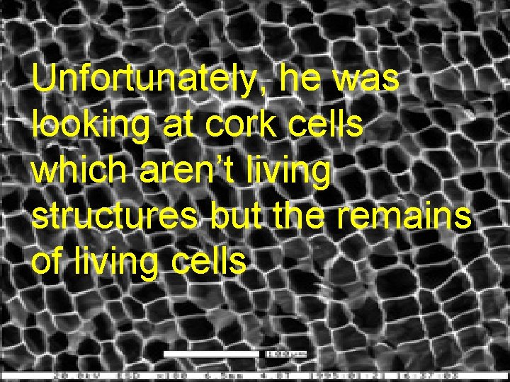
Unfortunately, he was looking at cork cells which aren’t living structures but the remains of living cells Copyright © 2004 Pearson Education, Inc. publishing as Benjamin Cummings
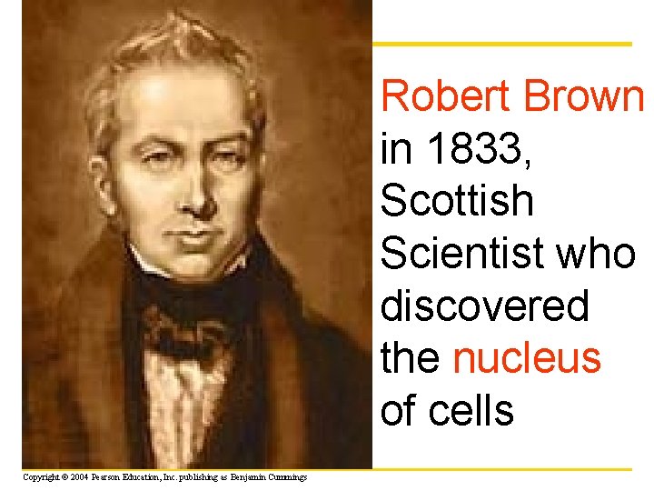
Robert Brown in 1833, Scottish Scientist who discovered the nucleus of cells Copyright © 2004 Pearson Education, Inc. publishing as Benjamin Cummings
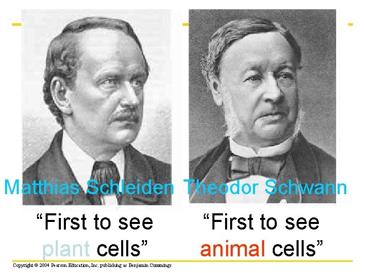
Matthias Schleiden Theodor Schwann “First to see plant cells” Copyright © 2004 Pearson Education, Inc. publishing as Benjamin Cummings “First to see animal cells”
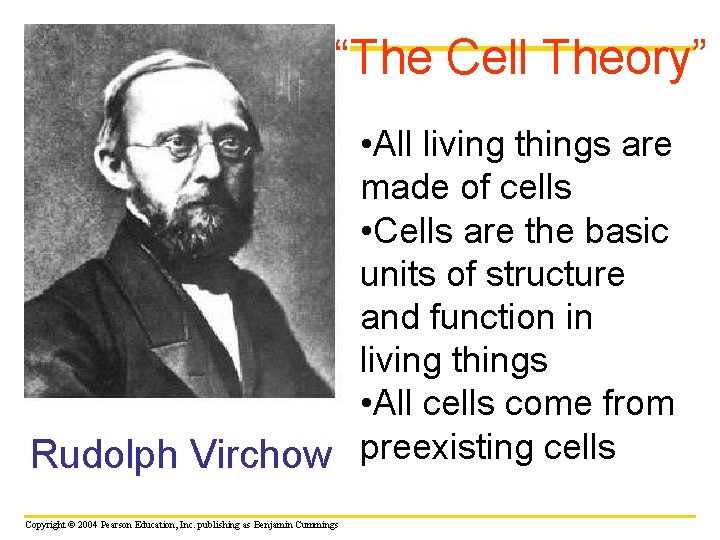
“The Cell Theory” • All living things are made of cells • Cells are the basic units of structure and function in living things • All cells come from Rudolph Virchow preexisting cells Copyright © 2004 Pearson Education, Inc. publishing as Benjamin Cummings
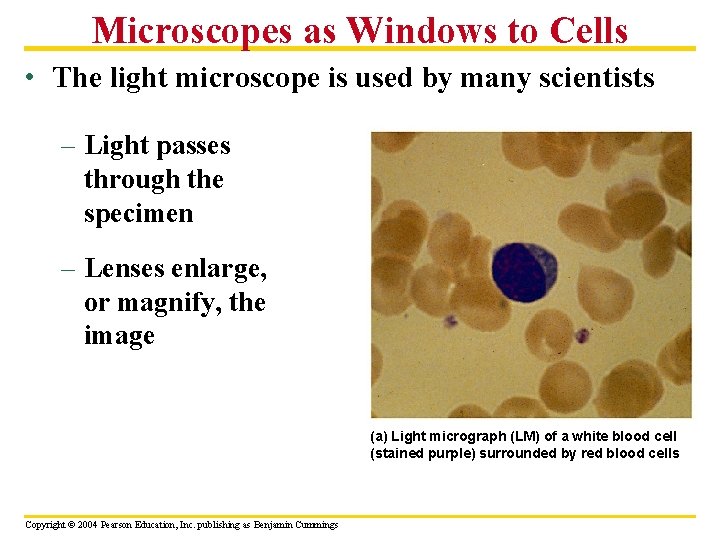
Microscopes as Windows to Cells • The light microscope is used by many scientists – Light passes through the specimen – Lenses enlarge, or magnify, the image (a) Light micrograph (LM) of a white blood cell (stained purple) surrounded by red blood cells Copyright © 2004 Pearson Education, Inc. publishing as Benjamin Cummings
• The electron microscope (EM) uses a beam of electrons – It has a higher resolving power than the light microscope Copyright © 2004 Pearson Education, Inc. publishing as Benjamin Cummings
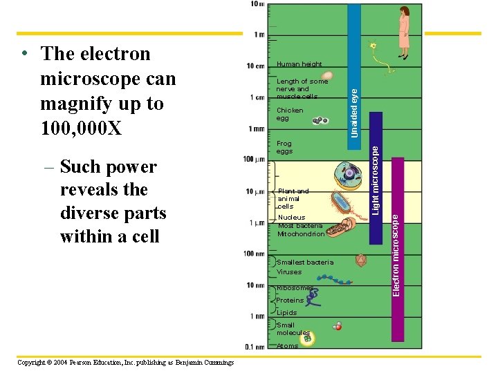
Chicken egg Frog eggs Plant and animal cells Nucleus Most bacteria Mitochondrion Smallest bacteria Viruses Ribosomes Proteins Lipids Small molecules Atoms Copyright © 2004 Pearson Education, Inc. publishing as Benjamin Cummings Electron microscope Length of some nerve and muscle cells Light microscope – Such power reveals the diverse parts within a cell Human height Unaided eye • The electron microscope can magnify up to 100, 000 X
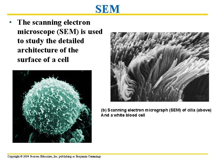
SEM • The scanning electron microscope (SEM) is used to study the detailed architecture of the surface of a cell (b) Scanning electron micrograph (SEM) of cilia (above) And a white blood cell Copyright © 2004 Pearson Education, Inc. publishing as Benjamin Cummings
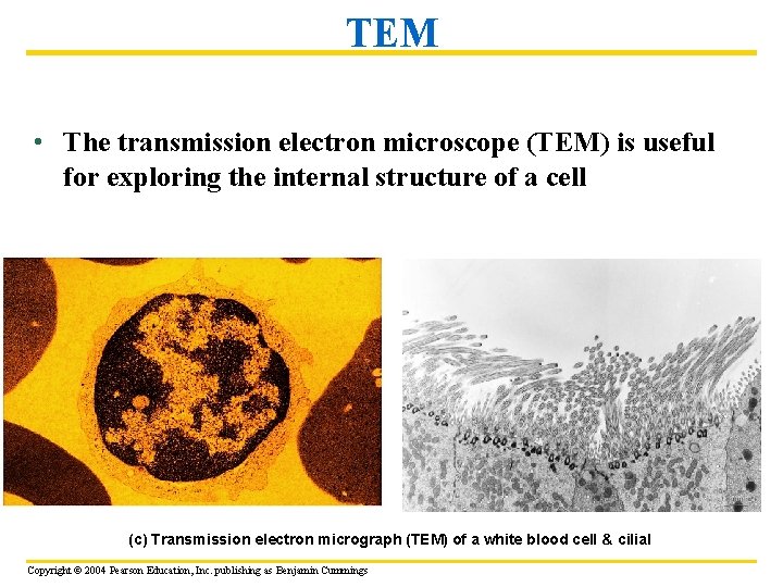
TEM • The transmission electron microscope (TEM) is useful for exploring the internal structure of a cell (c) Transmission electron micrograph (TEM) of a white blood cell & cilial Copyright © 2004 Pearson Education, Inc. publishing as Benjamin Cummings
The Two Major Categories of Cells • The countless cells on earth fall into two categories – Prokaryotic cells – Eukaryotic cells Copyright © 2004 Pearson Education, Inc. publishing as Benjamin Cummings
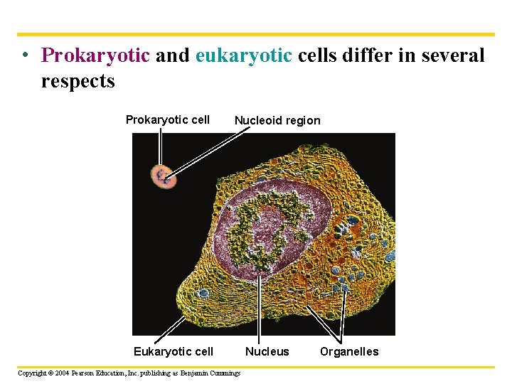
• Prokaryotic and eukaryotic cells differ in several respects Prokaryotic cell Nucleoid region Eukaryotic cell Copyright © 2004 Pearson Education, Inc. publishing as Benjamin Cummings Nucleus Organelles
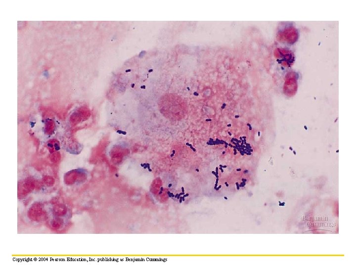
• Prokaryotic cells – Are smaller than eukaryotic cells – Lack internal structures surrounded by membranes – Lack a nucleus Copyright © 2004 Pearson Education, Inc. publishing as Benjamin Cummings
Copyright © 2004 Pearson Education, Inc. publishing as Benjamin Cummings
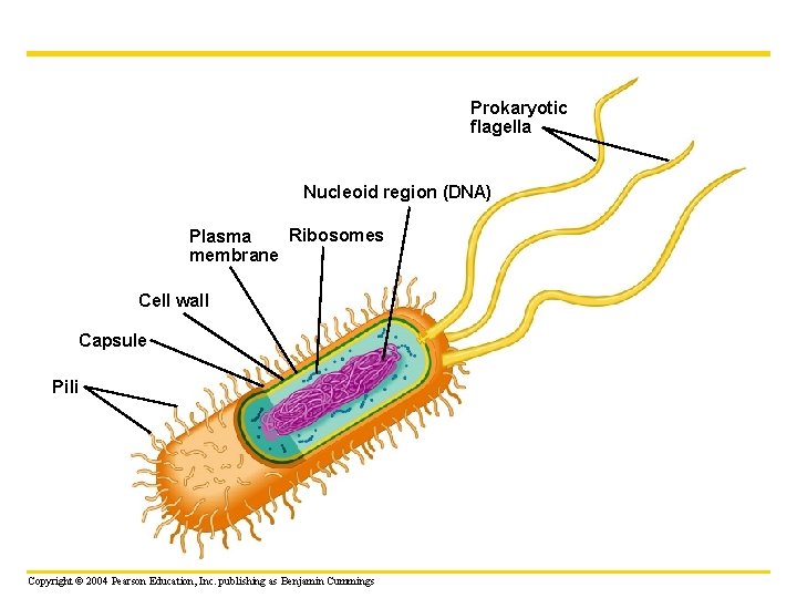
Prokaryotic flagella Nucleoid region (DNA) Ribosomes Plasma membrane Cell wall Capsule Pili Copyright © 2004 Pearson Education, Inc. publishing as Benjamin Cummings
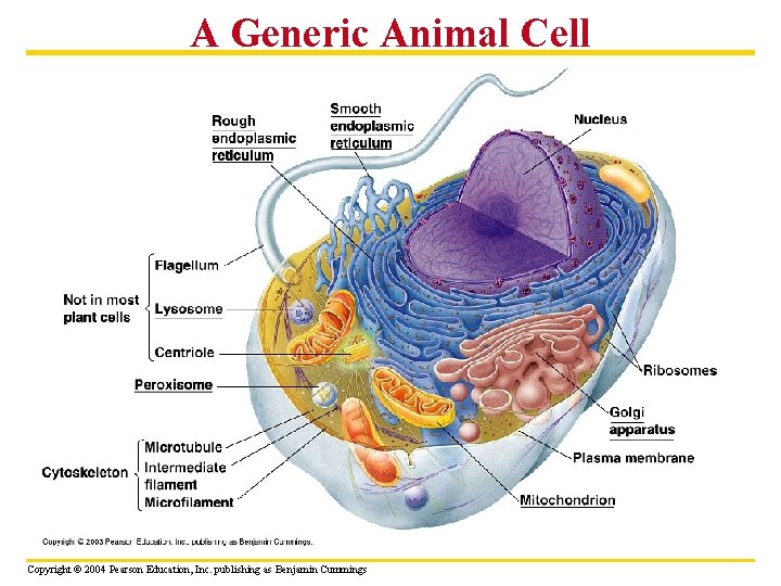
A Generic Animal Cell Copyright © 2004 Pearson Education, Inc. publishing as Benjamin Cummings
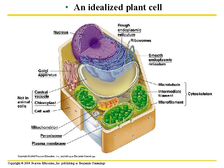
• An idealized plant cell Copyright © 2004 Pearson Education, Inc. publishing as Benjamin Cummings
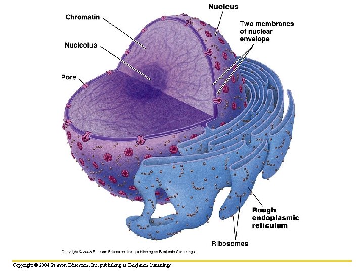
Structure and Function of the Nucleus • The nucleus is bordered by a double membrane called the nuclear envelope – It contains chromatin -a DNA-protein structure – It contains a nucleolus - which produces ribosomal parts Copyright © 2004 Pearson Education, Inc. publishing as Benjamin Cummings
Copyright © 2004 Pearson Education, Inc. publishing as Benjamin Cummings
Ribosomes • Ribosomes build all the cell’s proteins – Are not membrane bound Copyright © 2004 Pearson Education, Inc. publishing as Benjamin Cummings
How DNA Controls the Cell • DNA controls the cell by transferring its coded information into RNA – The information in the RNA is used to make proteins DNA 1 Synthesis of m. RNA in the nucleus Nucleus Cytoplasm 2 Movement of m. RNA into cytoplasm via nuclear pore 3 Synthesis of protein in the cytoplasm Copyright © 2004 Pearson Education, Inc. publishing as Benjamin Cummings m. RNA Ribosome Protein
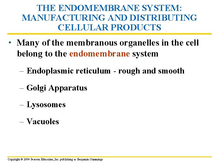
THE ENDOMEMBRANE SYSTEM: MANUFACTURING AND DISTRIBUTING CELLULAR PRODUCTS • Many of the membranous organelles in the cell belong to the endomembrane system – Endoplasmic reticulum - rough and smooth – Golgi Apparatus – Lysosomes – Vacuoles Copyright © 2004 Pearson Education, Inc. publishing as Benjamin Cummings
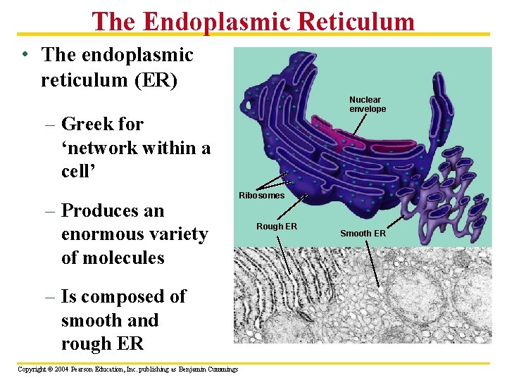
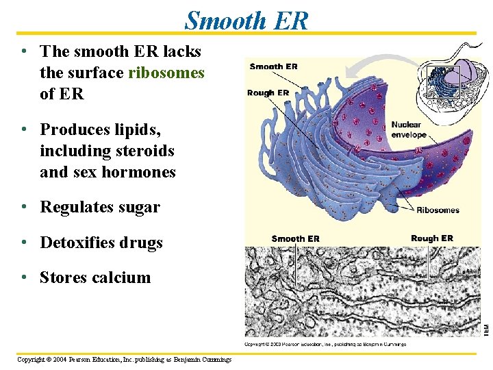
The Endoplasmic Reticulum • The endoplasmic reticulum (ER) Nuclear envelope – Greek for ‘network within a cell’ – Produces an enormous variety of molecules – Is composed of smooth and rough ER Copyright © 2004 Pearson Education, Inc. publishing as Benjamin Cummings Ribosomes Rough ER Smooth ER
Rough ER • The “roughness” of the rough ER is due to ribosomes that stud the outside of the ER membrane Copyright © 2004 Pearson Education, Inc. publishing as Benjamin Cummings
• The functions of the rough ER include – Producing proteins – Producing new membrane Copyright © 2004 Pearson Education, Inc. publishing as Benjamin Cummings
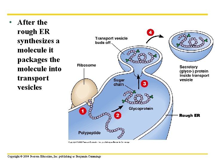
• After the rough ER synthesizes a molecule it packages the molecule into transport vesicles 1 Copyright © 2004 Pearson Education, Inc. publishing as Benjamin Cummings
Smooth ER • The smooth ER lacks the surface ribosomes of ER • Produces lipids, including steroids and sex hormones • Regulates sugar • Detoxifies drugs • Stores calcium Copyright © 2004 Pearson Education, Inc. publishing as Benjamin Cummings
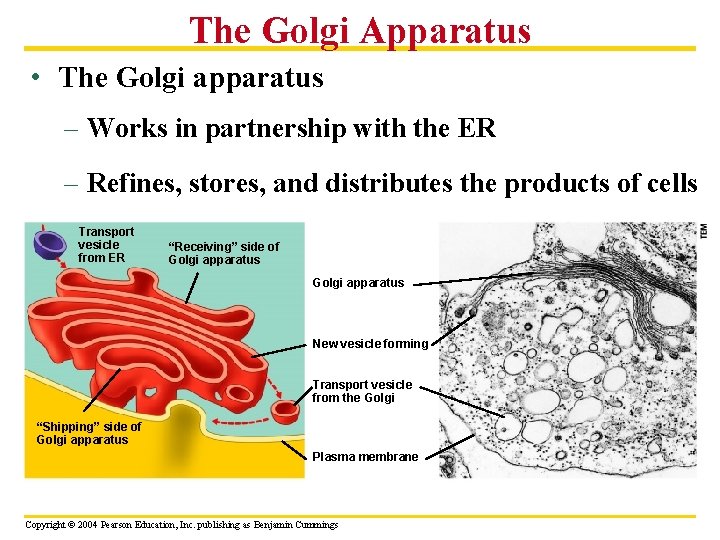
The Golgi Apparatus • The Golgi apparatus – Works in partnership with the ER – Refines, stores, and distributes the products of cells Transport vesicle from ER “Receiving” side of Golgi apparatus New vesicle forming Transport vesicle from the Golgi “Shipping” side of Golgi apparatus Plasma membrane Copyright © 2004 Pearson Education, Inc. publishing as Benjamin Cummings
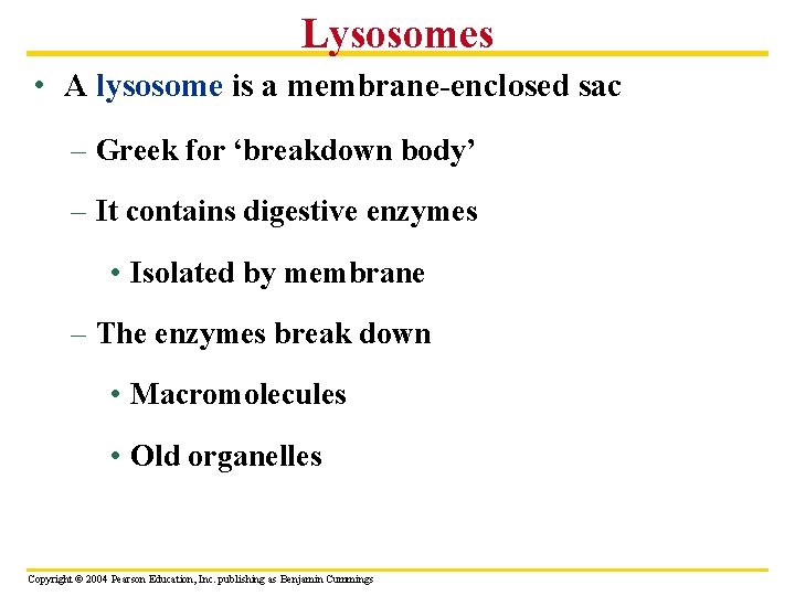
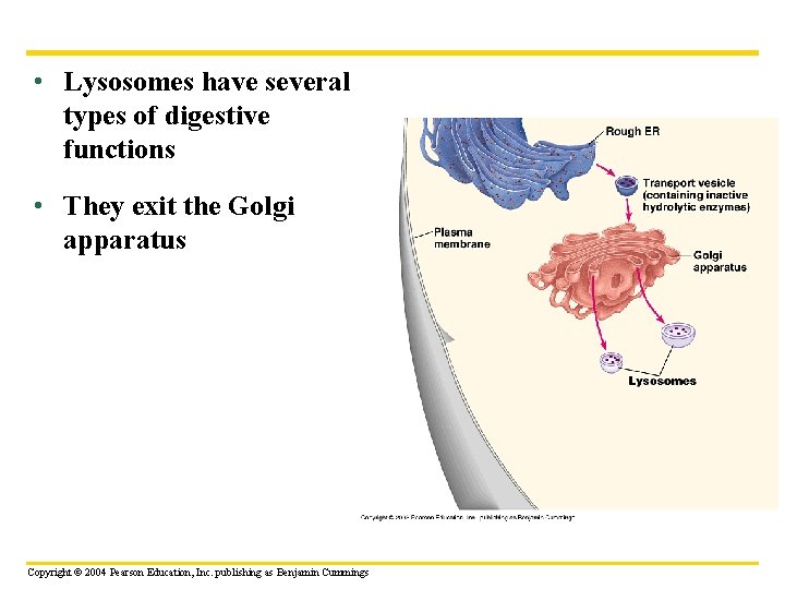
Lysosomes • A lysosome is a membrane-enclosed sac – Greek for ‘breakdown body’ – It contains digestive enzymes • Isolated by membrane – The enzymes break down • Macromolecules • Old organelles Copyright © 2004 Pearson Education, Inc. publishing as Benjamin Cummings
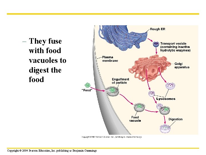
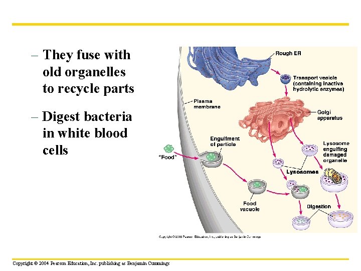
• Lysosomes have several types of digestive functions • They exit the Golgi apparatus Copyright © 2004 Pearson Education, Inc. publishing as Benjamin Cummings
– They fuse with food vacuoles to digest the food Copyright © 2004 Pearson Education, Inc. publishing as Benjamin Cummings
– They fuse with old organelles to recycle parts – Digest bacteria in white blood cells Copyright © 2004 Pearson Education, Inc. publishing as Benjamin Cummings
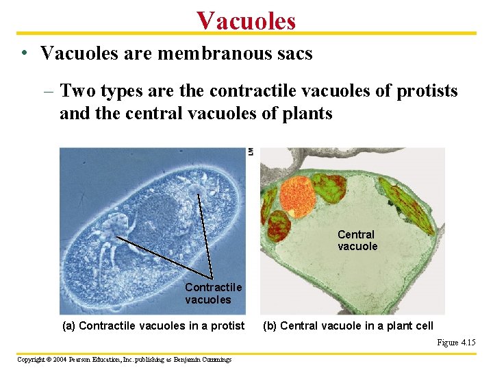
Vacuoles • Vacuoles are membranous sacs – Two types are the contractile vacuoles of protists and the central vacuoles of plants Central vacuole Contractile vacuoles (a) Contractile vacuoles in a protist (b) Central vacuole in a plant cell Figure 4. 15 Copyright © 2004 Pearson Education, Inc. publishing as Benjamin Cummings
CHLOROPLASTS AND MITOCHONDRIA: ENERGY CONVERSION • Cells require a constant energy supply to do all the work of life Copyright © 2004 Pearson Education, Inc. publishing as Benjamin Cummings
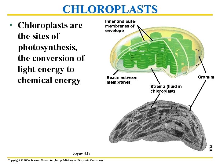
CHLOROPLASTS • Chloroplasts are the sites of photosynthesis, the conversion of light energy to chemical energy Figure 4. 17 Copyright © 2004 Pearson Education, Inc. publishing as Benjamin Cummings Inner and outer membranes of envelope Space between membranes Granum Stroma (fluid in chloroplast)
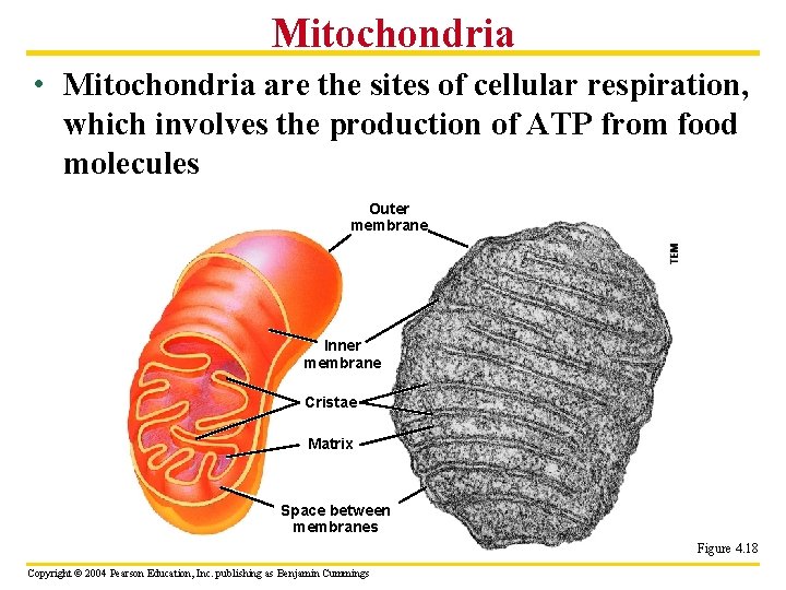
Mitochondria • Mitochondria are the sites of cellular respiration, which involves the production of ATP from food molecules Outer membrane Inner membrane Cristae Matrix Space between membranes Figure 4. 18 Copyright © 2004 Pearson Education, Inc. publishing as Benjamin Cummings
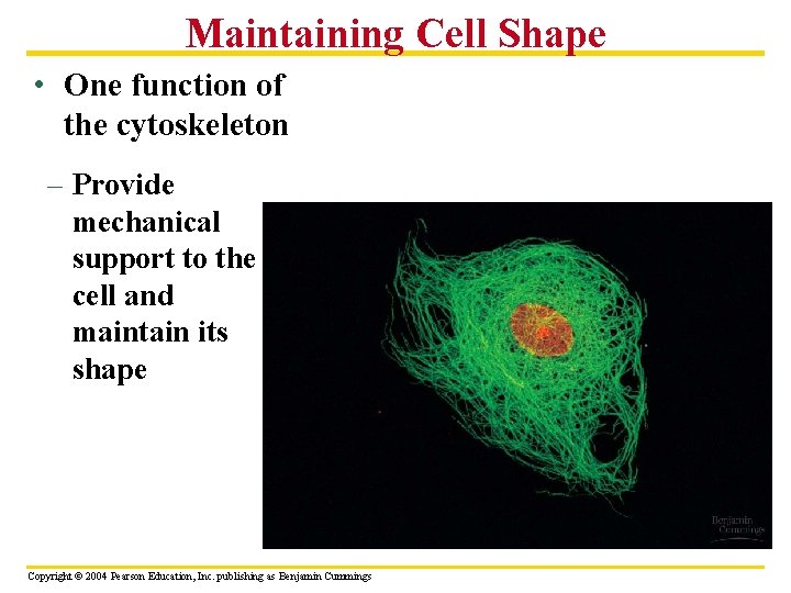
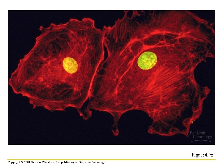
THE CYTOSKELETON: CELL SHAPE AND MOVEMENT • The cytoskeleton is an infrastructure of the cell consisting of a network of fibers – Microfilaments - small threads – Intermediate filaments - ropelike – Microtubules - small tubes Copyright © 2004 Pearson Education, Inc. publishing as Benjamin Cummings
Maintaining Cell Shape • One function of the cytoskeleton – Provide mechanical support to the cell and maintain its shape Copyright © 2004 Pearson Education, Inc. publishing as Benjamin Cummings
Figure 4. 9 x Copyright © 2004 Pearson Education, Inc. publishing as Benjamin Cummings
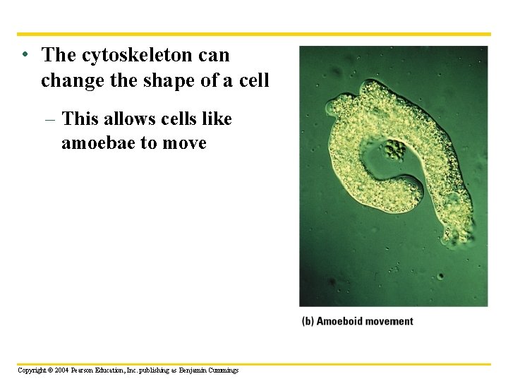
• The cytoskeleton can change the shape of a cell – This allows cells like amoebae to move Copyright © 2004 Pearson Education, Inc. publishing as Benjamin Cummings
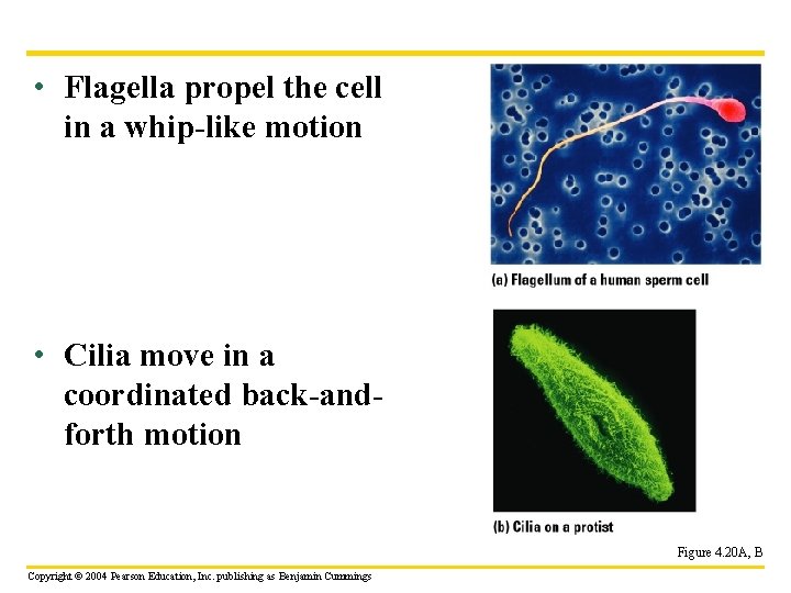
Cilia and Flagella • Cilia and flagella are motile appendages Copyright © 2004 Pearson Education, Inc. publishing as Benjamin Cummings
• Flagella propel the cell in a whip-like motion • Cilia move in a coordinated back-andforth motion Figure 4. 20 A, B Copyright © 2004 Pearson Education, Inc. publishing as Benjamin Cummings
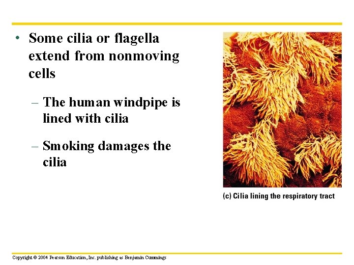
• Some cilia or flagella extend from nonmoving cells – The human windpipe is lined with cilia – Smoking damages the cilia Copyright © 2004 Pearson Education, Inc. publishing as Benjamin Cummings
CELL SURFACES: PROTECTION, SUPPORT, AND CELL-CELL INTERACTIONS • Most cells secrete materials that are external to the plasma membrane • This extra cellular matrix – Regulates – Protects – Supports Copyright © 2004 Pearson Education, Inc. publishing as Benjamin Cummings