
A Tour of the Cell Chapter 3 Answer







- Slides: 48
A Tour of the Cell Chapter 3
Answer the Following: • What functions must a cell • What cellular structures perform in order to perform each of these maintain life? properties.
A Typical Cell • 3 main structural components – Plasma or Cell Membrane – surrounds the cell – Cytoplasm – gel like substance, holds organelles – Nucleus – surrounded by a membrane
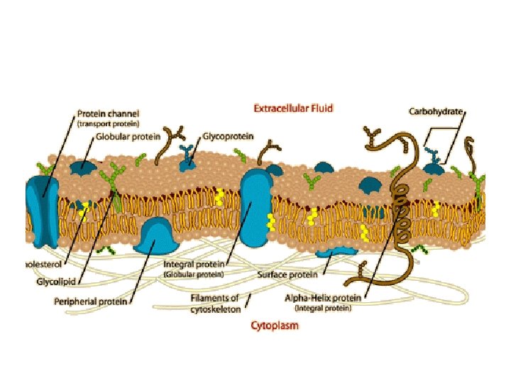
Cell Membrane • Fluid Mosaic Model – – Membrane molecules can float around the membrane • Double layer of phospholipid molecules – Hydrophilic phosphate heads – Hydrophobic fatty acid tails • In aqueous solutions these phospholipids form a sheet with fatty acid tails away from water and phosphate heads toward the water
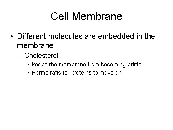
Cell Membrane • Different molecules are embedded in the membrane – Cholesterol – • keeps the membrane from becoming brittle • Forms rafts for proteins to move on
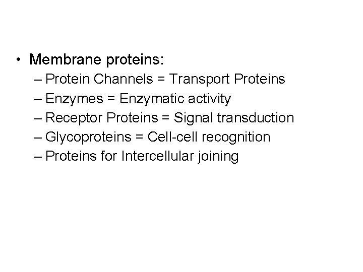
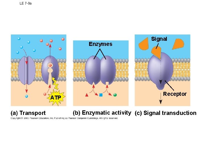
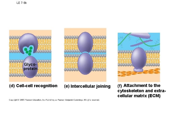
• Membrane proteins: – Protein Channels = Transport Proteins – Enzymes = Enzymatic activity – Receptor Proteins = Signal transduction – Glycoproteins = Cell-cell recognition – Proteins for Intercellular joining
LE 7 -9 a Enzymes Receptor ATP Transport Signal Enzymatic activity Signal transduction
LE 7 -9 b Glycoprotein Cell-cell recognition Intercellular joining Attachment to the cytoskeleton and extracellular matrix (ECM)
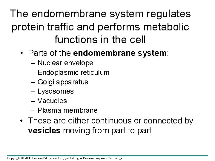
The endomembrane system regulates protein traffic and performs metabolic functions in the cell • Parts of the endomembrane system: – – – Nuclear envelope Endoplasmic reticulum Golgi apparatus Lysosomes Vacuoles Plasma membrane • These are either continuous or connected by vesicles moving from part to part Copyright © 2008 Pearson Education, Inc. , publishing as Pearson Benjamin Cummings
Endomembrane Animation • http: //users. uma. maine. edu/SBaker/nucleu s_endo. html
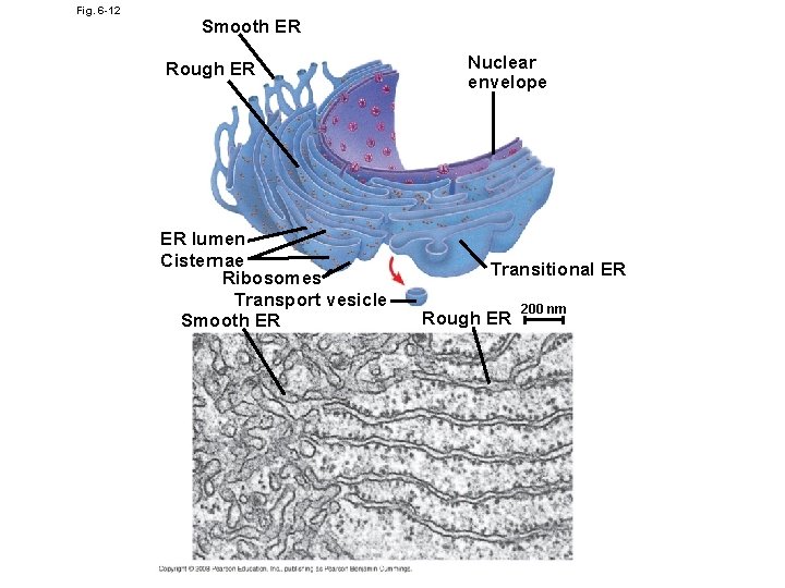
Endoplasmic Reticulum • 2 Types – Rough ER – with ribosomes – Smooth ER – without ribosomes • Structure – – Broad, flattened sacs – Extend outward from the nucleus
Fig. 6 -12 Smooth ER Rough ER ER lumen Cisternae Ribosomes Transport vesicle Smooth ER Nuclear envelope Transitional ER Rough ER 200 nm
Functions of ER • The smooth ER (no Ribosomes) – Makes lipids for making membranes throughout the cell – Removes Calcium ions from the cytoplasm and stores them – Detoxifies poison • The rough ER (Has Ribosomes) – Has bound ribosomes – Ribosomes produce polypeptide strands that enter the ER and are folded or joined with other proteins – Proteins may be embedded in the ER membrane or move to the Golgi Apparatus in transport vesicles
Ribosomes • Made of RNA • 2 sub units, not membrane bound • May be free or attached to the endoplasmic reticulum • Site of protein synthesis – Translation of m. RNA into polypeptide chain
The Golgi Apparatus • The Golgi apparatus – Receives many of the transport vesicles produced in the rough ER – Consists of flattened membranous sacs • Functions of the Golgi apparatus include – To sort and modify of the products of the rough ER • Packages these into vesicles to be transported – Produces lysosomes
Golgi Animation • http: //bcs. whfreeman. com/thelifewire/conte nt/chp 04/0402002. html
• Lysosomes carry out intracellular digestion by 1. Phagocytosis 2. Break down proteins that are not needed The monomers can be reused • Peroxisomes contain peroxidase to break down hydrogen peroxide 1 µm Nucleus Lysosome contains active hydrolytic enzymes Food vacuole fuses with lysosome Hydrolytic enzymes digest food particles Digestive enzymes Lysosome Plasma membrane Digestion Food vacuole (a) Phagocytosis: lysosome digesting food
Lysosome Animation • http: //highered. mcgrawhill. com/sites/0072507470/student_view 0/ chapter 3/animation__lysosomes. html
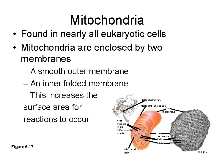
Mitochondria • Found in nearly all eukaryotic cells • Mitochondria are enclosed by two membranes – A smooth outer membrane – An inner folded membrane – This increases the surface area for reactions to occur Free ribosomes in the mitochondrial matrix Figure 6. 17 Mitochondrion Intermembrane space Outer membrane Inner membrane Cristae Matrix Mitochondrial DNA 100 µm
Mitochondria change energy from one form to another • Mitochondria – Are the sites of cellular respiration – Breaks down large molecules to produce ATP – Much of this process takes place in the inner membrane
Eukaryotes: The Nucleus • DNA is located in the nucleus and does not leave • Transcription of DNA into m. RNA occurs • Pre-m. RNA is edited before leaving the nucleus • Nuclear envelope – has nuclear pores – Is double sided • Nucleolus – ribosomes are produced
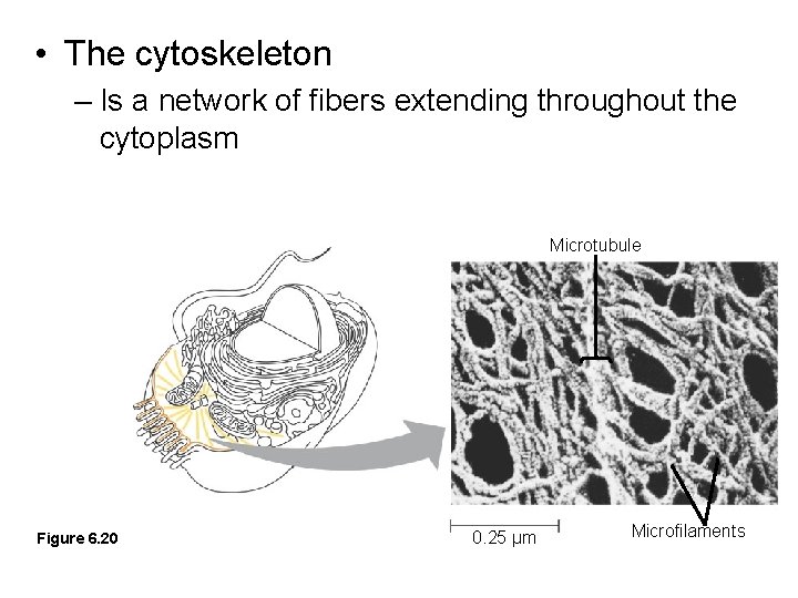
• The cytoskeleton – Is a network of fibers extending throughout the cytoplasm Microtubule Figure 6. 20 0. 25 µm Microfilaments
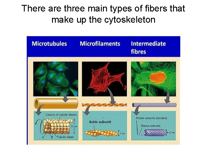
There are three main types of fibers that make up the cytoskeleton
Microtubules • Microtubules – Shape the cell – Guide movement of organelles – Help separate the chromosome copies in dividing cells
Microfilaments (Actin Filaments) • Microfilaments – Are built from molecules of the protein actin
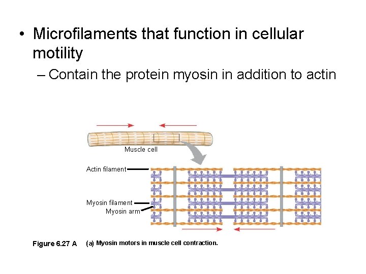
• Microfilaments that function in cellular motility – Contain the protein myosin in addition to actin Muscle cell Actin filament Myosin arm Figure 6. 27 A (a) Myosin motors in muscle cell contraction.
Centrosomes and Centrioles • The centrosome – Is considered to be a “microtubule-organizing center” – Plants and yeast cells lack centrosomes
Molecular Motors • http: //www. youtube. com/watch? feature=pl ayer_detailpage&v=k. Oe. Jw. Q 0 OXc 4
Cell Extensions • Microvilli – Found on cells where absorption is important – Functions • Increase surface area • Allow for quick absorption
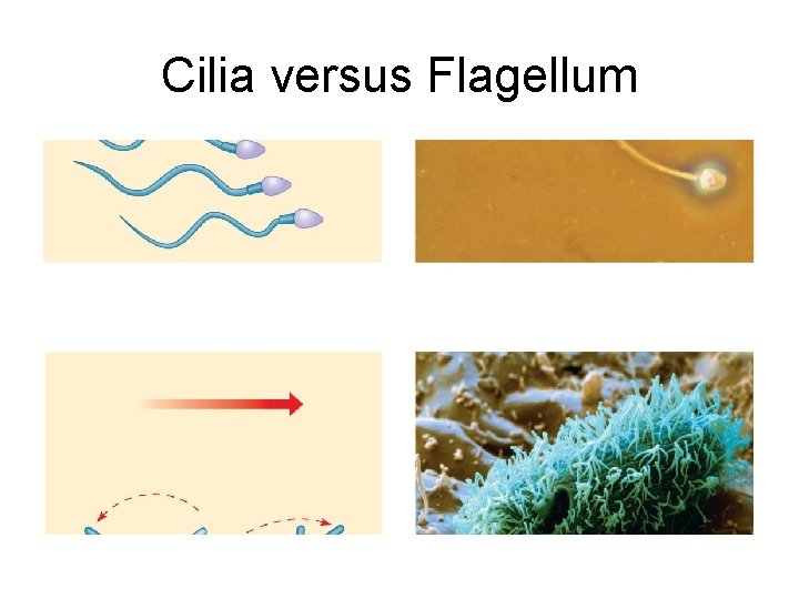
Cell Extensions • Cilia and flagella – Contain specialized arrangements of microtubules – Are locomotor appendages of some cells
Cilia versus Flagellum
Cell Connections • In animals, there are three types of intercellular junctions – Desmosomes – • like spot welds, hold cells together like velcro • Example – Skin Cells
Cell Connections – Gap junctions – • When membranes of adjacent cells stick together – Form tunnels that join cytoplasm – Fuse the two membranes into one – Tight Junctions – • Membrane proteins from different cells fuse • Like soft drink cans held by plastic
Cell Animation • http: //aimediaserver 4. com/studiodaily/vide oplayer/? src=ai 4/harvard. swf&widt h=640&height=520
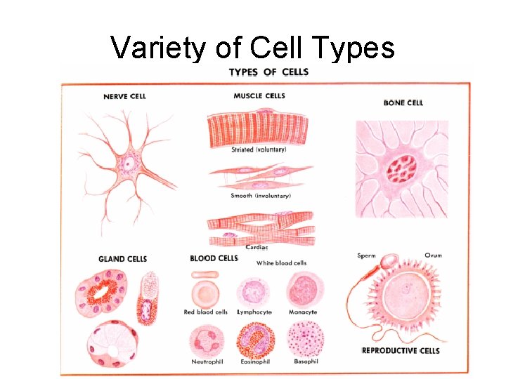
Variety of Cell Types
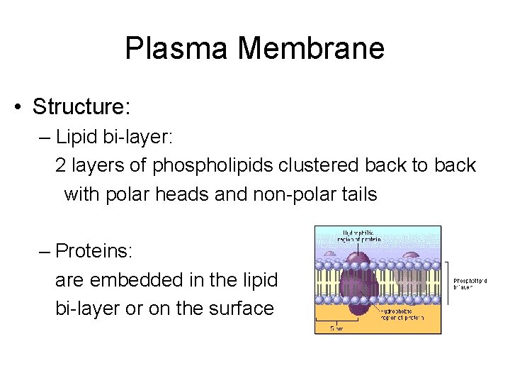
Plasma Membrane • Structure: – Lipid bi-layer: 2 layers of phospholipids clustered back to back with polar heads and non-polar tails – Proteins: are embedded in the lipid bi-layer or on the surface
Cell Membrane • Function: – Regulates what enters and leaves the cell. – Cell membranes are selectively permeable. – Also provides protection and support • 2 ways in which molecules can be transported into a cell: – Passive transport – Active transport
Passive Transport = Diffusion • Osmosis – diffusion of water through a selectively permeable membrane • Facilitated diffusion - the use of membrane proteins to aid diffusion of a substance through a membrane
• http: //highered. mcgrawhill. com/sites/0072495855/student_view 0/ chapter 2/animation__how_diffusion_works. html
• http: //highered. mcgrawhill. com/sites/0072495855/student_view 0/ chapter 2/animation__how_osmosis_works. html
• http: //highered. mcgrawhill. com/sites/0072495855/student_view 0/ chapter 2/animation__how_facilitated_diffu sion_works. html
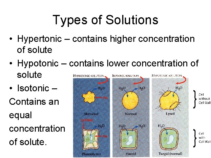
Types of Solutions • Hypertonic – contains higher concentration of solute • Hypotonic – contains lower concentration of solute • Isotonic – Contains an equal concentration of solute.
• http: //www. youtube. com/watch? v=7 -QJUUX 0 i. Y
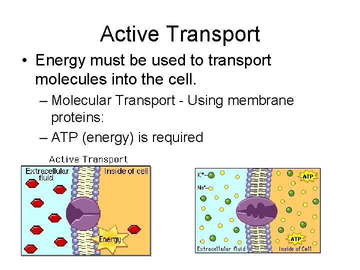
Active Transport • Energy must be used to transport molecules into the cell. – Molecular Transport - Using membrane proteins: – ATP (energy) is required
• http: //highered. mcgrawhill. com/sites/0072495855/student_view 0/ chapter 2/animation__how_the_sodium_po tassium_pump_works. html
Review of Diffusion • http: //www. youtube. com/watch? v=2 UPq. L m-u. Dn. I
Active Transport Other types of active transport: • Endocytosis –Taking particles into the cell – Phagocytosis – endocytosis of solids – Pinocytosis – endocytosis of liquids • Exocytosis – Secreting particles from the cell