AUTOIMMUNE DISEASES Martina Vachov Department of Immunology and









- Slides: 63
AUTOIMMUNE DISEASES Martina Vachová Department of Immunology and Allergology Faculty of Medicine and Faculty Hospital in Pilsen
27. 11. 2014 MUDr. Martina Vachová 54. Non-organ-specific autoantibodies and their diagnostic significance. 55. Autoantibodies in organ specific autoimmune diseases. Organ-localized autoimmune diseases with non-organ specific autoantibodies. 56. Autoimmune diseases - pathophysiology, division, laboratory diagnostics, treatment. 57. Examples of systemic autoimmune diseases. 58. Examples of organ-specific autoimmune diseases. 59. Immunosupressive treatment - examples of drugs, indications, risks.
AUTOIMMUNE DISEASES § chronic and usually irreversible § incidence: 5%-7% of population, higher frequencies in women, increases with age
Autoimmune diseases § Result from a failure of self-tolerance § Immunological tolerance is specific unresponsiveness to an antigen § All individuals are tolerant of their own (self) antigens
Autoimmunity § is defined as an immune response against self antigens § The principal factors in the development of autoimmunity are the inheritance of susceptibility genes and environmental triggers, such as infections § Infections may activate self-reactive lymphocytes, thereby trigger the development of autoimmune diseases § Most autoimmune diseases are polygenic and are asssociated with multiple gene loci, the most important of which are the MHC genes
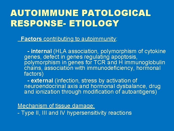
AUTOIMMUNE PATOLOGICAL RESPONSE- ETIOLOGY Factors contributing to autoimmunity: - internal (HLA association, polymorphism of cytokine genes, defect in genes regulating apoptosis, polymorphism in genes for TCR and H immunoglobulin chains, association with immunodeficiency, hormonal factors) - external (infection, stress by activation of neuroendocrinal axis and hormonal dysbalance, drug and ionization through modification of autoantigens) Mechanism of tissue damage: - Type II, III and IV hypersensitivity reactions
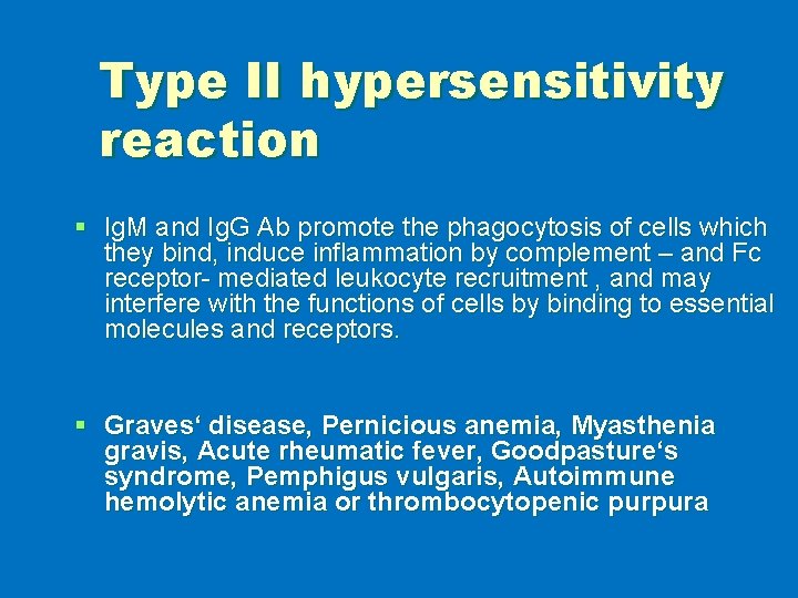
Type II hypersensitivity reaction § Ig. M and Ig. G Ab promote the phagocytosis of cells which they bind, induce inflammation by complement – and Fc receptor- mediated leukocyte recruitment , and may interfere with the functions of cells by binding to essential molecules and receptors. § Graves‘ disease, Pernicious anemia, Myasthenia gravis, Acute rheumatic fever, Goodpasture‘s syndrome, Pemphigus vulgaris, Autoimmune hemolytic anemia or thrombocytopenic purpura
Type III hypersensitivity reaction § Ab may bind to circulating antigens to form immune complexes, which deposit in vessels and cause tissue injury § Injury is mainly due to leukocyte recruitment and inflammation § Systemic lupus erythematosus, Polyarteritis nodosa, Poststreptococcal glomerulonephritis
Type IV hypersensitivity reaction § T cell- mediated diseases are caused by Th 1 -mediated delayed-type hypersensitivity reactions or Th 17 - mediated inflammatory reactions, or by killing of host cells by CD 8+ CTLs (cytotoxic lymphocytes). § Diabetes mellitus (insulin-dependent), Rheumatoid arthritis, Multiple sclerosis, Inflammatory bowel disease
Autoimmune diseases ¬ Systemic ¬ Organ-specific ¬ Organ-localised
CLINICAL CATEGORIES § systemic - affect many organs and tissue - organ non-specific autoantibodies § organ specific - affect one organ - organ specific autoantibodies or autoreactive T lymphocytes § organ localised - affect predominantly one organ accompanied by affection of other organs (inflammatory bowel diseases, coeliac disease, AI hepatitis, pulmonary fibrosis) - organ non-specific autoantibodies
EXAMPLES OF SYSTEMIC AUTOIMMUNE DISEASES § examples § autoantibodies
SYSTEMIC AUTOIMMUNE DISEASES § § § § Systemic lupus erythematosus Rheumathoid arthritis Sjögren‘s syndrome Dermatopolymyositis Systemic sclerosis Mixed connective tissue disease Vasculitis
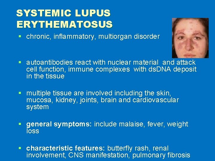
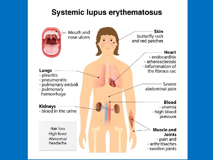
SYSTEMIC LUPUS ERYTHEMATOSUS § chronic, inflammatory, multiorgan disorder § autoantibodies react with nuclear material and attack cell function, immune complexes with ds. DNA deposit in the tissue § multiple tissue are involved including the skin, mucosa, kidney, joints, brain and cardiovascular system § general symptoms: include malaise, fever, weight loss § characteristic features: butterfly rash, renal involvement, CNS manifestation, pulmonary fibrosis
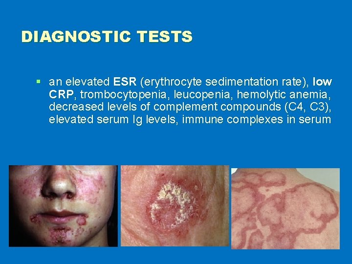
DIAGNOSTIC TESTS § an elevated ESR (erythrocyte sedimentation rate), low CRP, trombocytopenia, leucopenia, hemolytic anemia, decreased levels of complement compounds (C 4, C 3), elevated serum Ig levels, immune complexes in serum
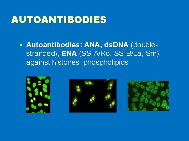
AUTOANTIBODIES § Autoantibodies: ANA, ds. DNA (doublestranded), ENA (SS-A/Ro, SS-B/La, Sm), against histones, phospholipids
§ https: //www. youtube. com/watch? v=Wgf. F cp. AD 9 XQ
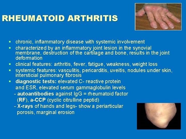
RHEUMATOID ARTHRITIS § chronic, inflammatory disease with systemic involvement § characterized by an inflammatory joint lesion in the synovial membrane, destruction of the cartilage and bone, results in the joint deformation § clinical features: arthritis, fever, fatigue, weakness, weight loss § systemic features: vasculitis, pericarditis, uveitis, nodules under skin, intersticial pulmonary fibrosis § diagnostic tests: elevated C- reactive protein and ESR, elevated serum gammaglobulin levels - autoantibodies against Ig. G = rheumatoid factor (RF), a-CCP (cyclic citrulline peptid) - X-rays of hands and legs- show a periarticular porosis, marginal erosion
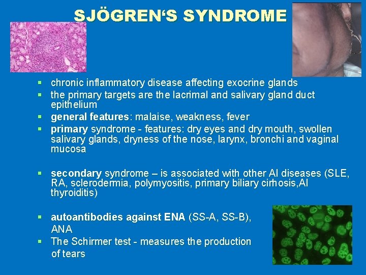
SJÖGREN‘S SYNDROME § chronic inflammatory disease affecting exocrine glands § the primary targets are the lacrimal and salivary gland duct epithelium § general features: malaise, weakness, fever § primary syndrome - features: dry eyes and dry mouth, swollen salivary glands, dryness of the nose, larynx, bronchi and vaginal mucosa § secondary syndrome – is associated with other AI diseases (SLE, RA, sclerodermia, polymyositis, primary biliary cirhosis, AI thyroiditis) § autoantibodies against ENA (SS-A, SS-B), ANA § The Schirmer test - measures the production of tears
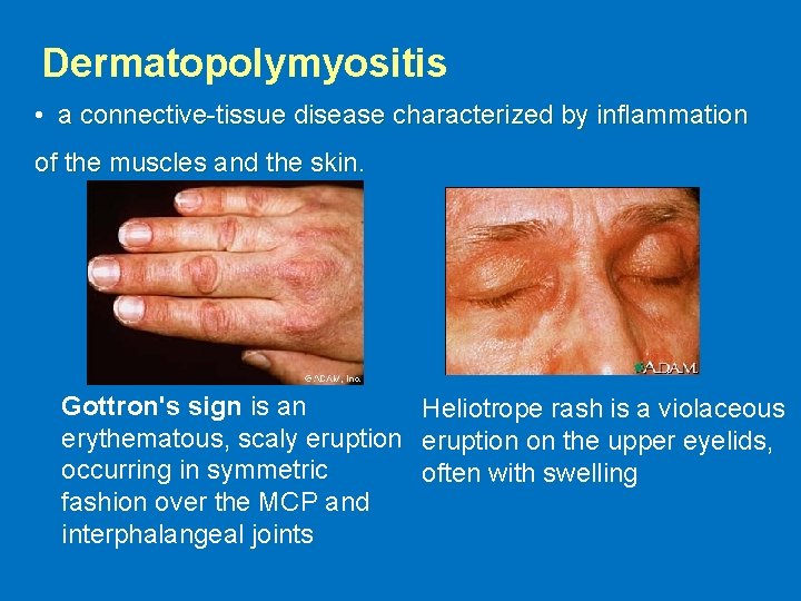
Dermatopolymyositis • a connective-tissue disease characterized by inflammation of the muscles and the skin. Gottron's sign is an Heliotrope rash is a violaceous erythematous, scaly eruption on the upper eyelids, occurring in symmetric often with swelling fashion over the MCP and interphalangeal joints
Dermatopolymyositis § elevated creatine phosphokinase (CPK) § muscle biopsy (a mixed B- and T-cell perivascular inflammatory infiltrate, perifascicular muscle fiber atrophy) § EMG (electromyogram) § autoantibodies - ENA (Jo-1)
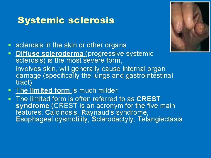
Systemic sclerosis § sclerosis in the skin or other organs § Diffuse scleroderma (progressive systemic sclerosis) is the most severe form, involves skin, will generally cause internal organ damage (specifically the lungs and gastrointestinal tract) § The limited form is much milder § The limited form is often referred to as CREST syndrome (CREST is an acronym for the five main features: Calcinosis, Raynaud's syndrome, Esophageal dysmotility, Sclerodactyly, Telangiectasia
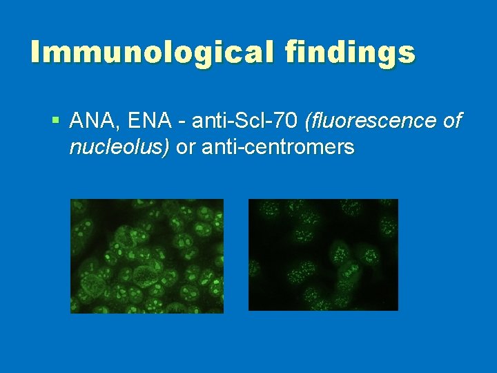
Immunological findings § ANA, ENA - anti-Scl-70 (fluorescence of nucleolus) or anti-centromers
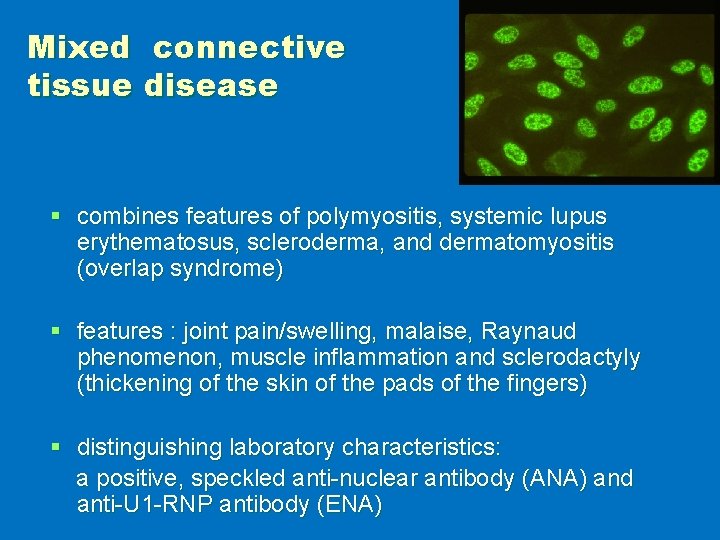
Mixed connective tissue disease § combines features of polymyositis, systemic lupus erythematosus, scleroderma, and dermatomyositis (overlap syndrome) § features : joint pain/swelling, malaise, Raynaud phenomenon, muscle inflammation and sclerodactyly (thickening of the skin of the pads of the fingers) § distinguishing laboratory characteristics: a positive, speckled anti-nuclear antibody (ANA) and anti-U 1 -RNP antibody (ENA)
Vasculitis § characterized by inflammatory destruction of vessels leading to thrombosis and aneurysms § affect mostly lung, kidneys, skin § diagnostic tests: elevated ESR, CRP, leucocytosis, biopsy of affected organ (necrosis, granulomas), angiography, ANCA (antineutrophil cytoplasmic antibodies)
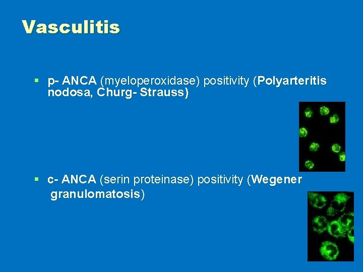
Vasculitis § p- ANCA (myeloperoxidase) positivity (Polyarteritis nodosa, Churg- Strauss) § c- ANCA (serin proteinase) positivity (Wegener granulomatosis)
Classification § Large vessel vasculitis (Takayasu arteritis, Giant cell (temporal) arteritis) § Medium vessel vasculitis (Polyarteritis nodosa, Wegener's granulomatosis, Kawasaki disease) § Small vessel vasculitis (Churg-Strauss arteritis, Microscopic polyarteritis, Henoch-Schönlein purpura) § Symptoms: fatigue, weakness, fever, arthralgias, abdominal pain, hypertension, renal insufficiency, and neurologic dysfunction
§ https: //www. youtube. com/watch? v=A 4 b_u. UNv 7 w
EXAMPLES OF ORGAN LOCALISED AUTOIMMUNE DISEASES § diseases § autoantibodies
ORGAN LOCALIZED AUTOIMMUNE DISEASES § § § Ulcerative colitis Crohn‘s disease Autoimmune hepatitis Primary biliary cirhosis Pulmonary fibrosis
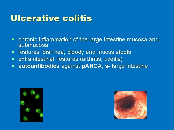
Ulcerative colitis § chronic inflammation of the large intestine mucosa and submucosa § features: diarrhea, bloody and mucus stools § extraintestinal features (arthritis, uveitis) § autoantibodies against p. ANCA, a- large intestine
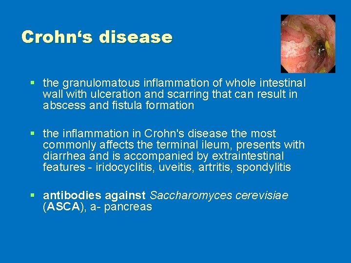
Crohn‘s disease § the granulomatous inflammation of whole intestinal wall with ulceration and scarring that can result in abscess and fistula formation § the inflammation in Crohn's disease the most commonly affects the terminal ileum, presents with diarrhea and is accompanied by extraintestinal features - iridocyclitis, uveitis, artritis, spondylitis § antibodies against Saccharomyces cerevisiae (ASCA), a- pancreas
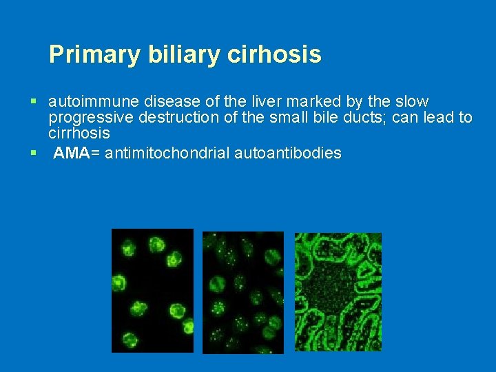
Primary biliary cirhosis § autoimmune disease of the liver marked by the slow progressive destruction of the small bile ducts; can lead to cirrhosis § AMA= antimitochondrial autoantibodies
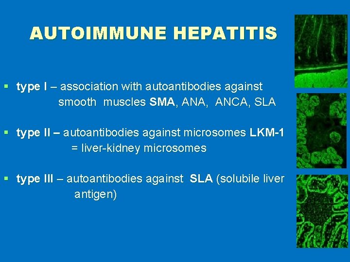
AUTOIMMUNE HEPATITIS § type I – association with autoantibodies against smooth muscles SMA, ANA, ANCA, SLA § type II – autoantibodies against microsomes LKM-1 = liver-kidney microsomes § type III – autoantibodies against SLA (solubile liver antigen)
ORGAN SPECIFIC AUTOIMMUNE DISEASES § § § Autoimmune endocrinopathy Autoimmune neurological diseases Autoimmune cytopenia
AUTOIMMUNE ENDOCRINOPATHY § § § Hashimoto‘s thyroiditis Graves-Basedow disease Diabetes mellitus I. type Addison‘s disease Autoimmune polyglandular syndrome Pernicious anemia
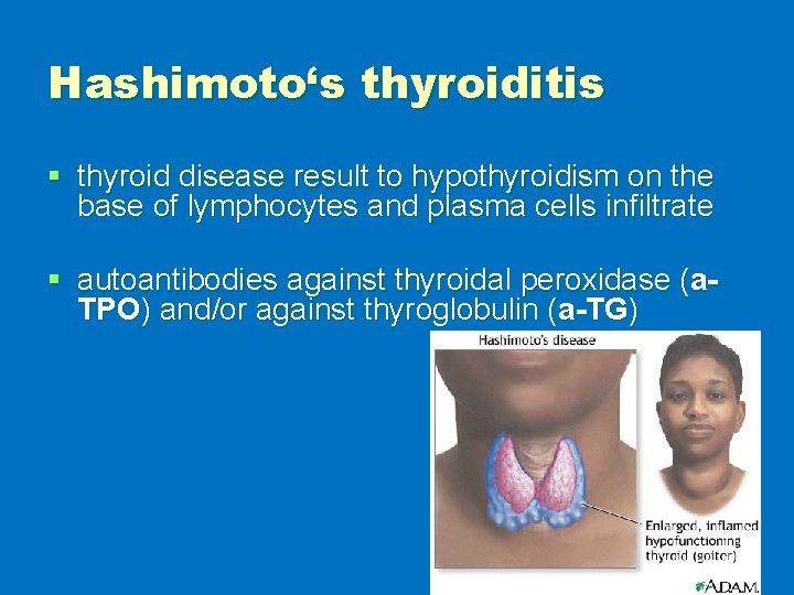
Hashimoto‘s thyroiditis § thyroid disease result to hypothyroidism on the base of lymphocytes and plasma cells infiltrate § autoantibodies against thyroidal peroxidase (a. TPO) and/or against thyroglobulin (a-TG)
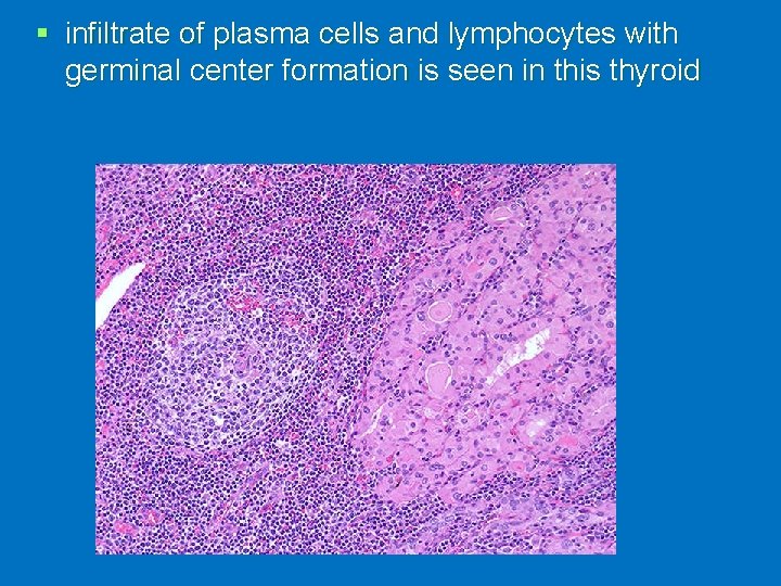
§ infiltrate of plasma cells and lymphocytes with germinal center formation is seen in this thyroid
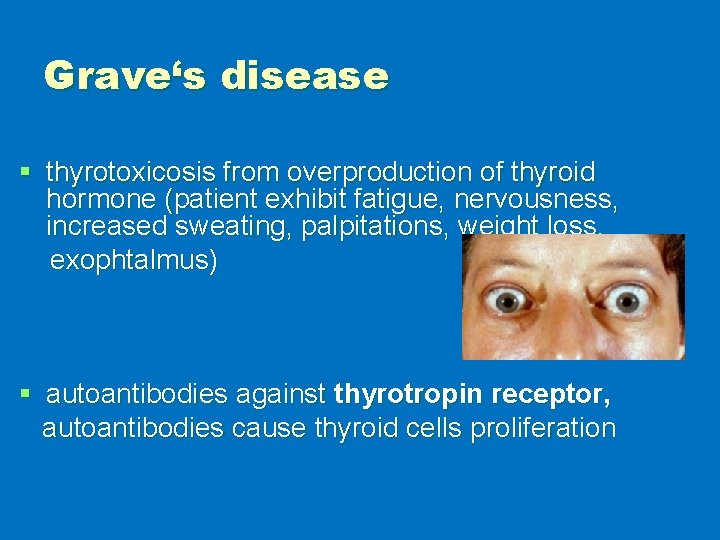
Grave‘s disease § thyrotoxicosis from overproduction of thyroid hormone (patient exhibit fatigue, nervousness, increased sweating, palpitations, weight loss, exophtalmus) § autoantibodies against thyrotropin receptor, autoantibodies cause thyroid cells proliferation
Diabetes mellitus (insulindependent) § characterized by an inability to process sugars in the diet, due to a decrease in or total absence of insulin production § results from immunologic destruction of the insuline- producing β-cells of the islets of Langerhans in the pancreas § autoantibodies against GAD- glutamic acid decarboxylase = primary antigen), autoantibodies anti- islet cell, anti- insulin § islets are infiltrated with B and T cells
Polyglandular autoimmune syndrome § combination of several different AI endocrinopathies § autoantibodies appear in according with the connected disorders
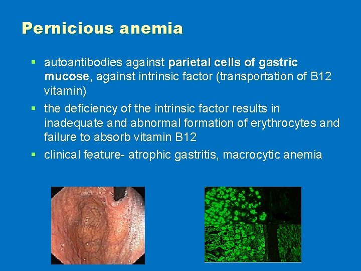
Pernicious anemia § autoantibodies against parietal cells of gastric mucose, against intrinsic factor (transportation of B 12 vitamin) § the deficiency of the intrinsic factor results in inadequate and abnormal formation of erythrocytes and failure to absorb vitamin B 12 § clinical feature- atrophic gastritis, macrocytic anemia
AUTOIMMUNE NEUROPATHY § Guillain-Barré syndrome (acute idiopathic polyneuritis) § Myasthenia gravis § Multiple sclerosis
Guillain-Barré syndrome § inflammation demyelinates peripheral nerves that causes progressive muscle weakness and paralysis § the cause is the loss of myelin § occurs often 1 -3 weeks after infection (Campylobacter jej. ) § features: progressive weakness and paresthesia of the lower and later upper extremitas and respiratory muscles, weakness can leads to paralysis and respiratory failure § immunologic findings: autoantibodies against ganglioside membrane
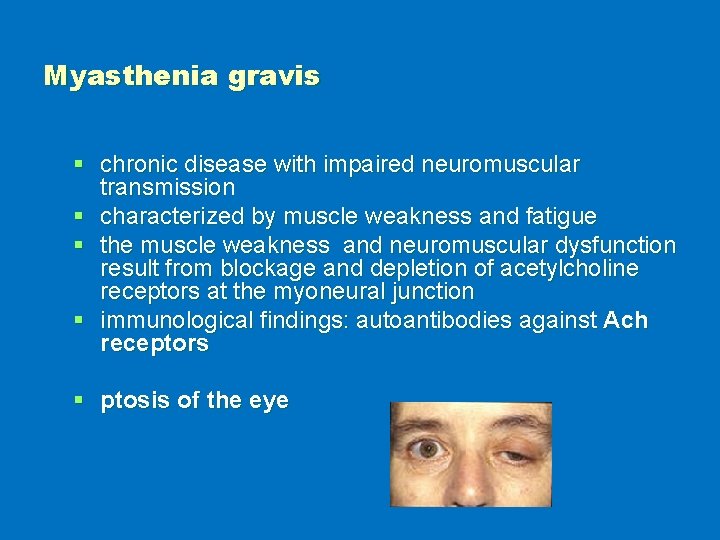
Myasthenia gravis § chronic disease with impaired neuromuscular transmission § characterized by muscle weakness and fatigue § the muscle weakness and neuromuscular dysfunction result from blockage and depletion of acetylcholine receptors at the myoneural junction § immunological findings: autoantibodies against Ach receptors § ptosis of the eye
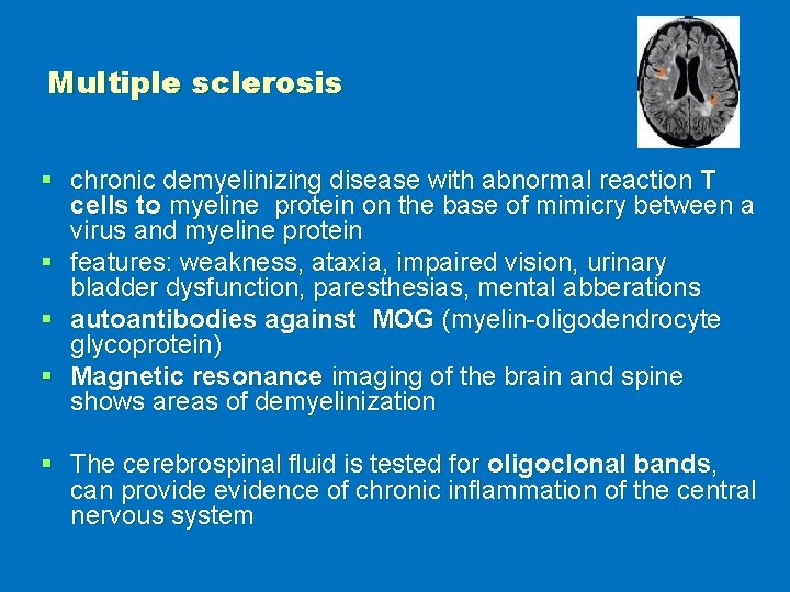
Multiple sclerosis § chronic demyelinizing disease with abnormal reaction T cells to myeline protein on the base of mimicry between a virus and myeline protein § features: weakness, ataxia, impaired vision, urinary bladder dysfunction, paresthesias, mental abberations § autoantibodies against MOG (myelin-oligodendrocyte glycoprotein) § Magnetic resonance imaging of the brain and spine shows areas of demyelinization § The cerebrospinal fluid is tested for oligoclonal bands, can provide evidence of chronic inflammation of the central nervous system
AUTOIMMUNE CYTOPENIA § AI hemolytic disease- autoantibodies against membrane erythrocyte antigens § AI trombocytopenia - autoantibodies against trombocyte antigens (GPIIb/IIIa) § AI neutropenia - autoantibodies against membrane neutrofil antigens
IMMUNOSUPPRESSION § non-specific treatment § examples of drugs § indication § risks
Immunosuppressants § Drugs that inhibit or prevent activity of the immune system § They are used to: § Prevent the rejection of transplanted organs and tissues (bone marrow, heart, kidney, liver) § Treat autoimmune diseases or diseases that are most likely of autoimmune origin (rheumatoid arthritis, multiple sclerosis, myasthenia gravis, systemic lupus erythematosus, Crohn's disease, pemphigus, ulcerative colitis). § Treat some other non-autoimmune inflammatory diseases (allergic asthma, atopic eczema).
Immunosuppressants § Glucocorticoids § Drugs affecting the proliferation of both T cells and B cells § Drugs blocking the activation of lymphocytes § Monoclonal antibodies
Glucocorticoids § suppress the cell-mediated immunity- decrease cytokine production reduces the T cell proliferation. § suppress the humoral immunity, causing B cells to express smaller amounts of IL-2 and IL-2 receptors- this diminishes both B cell clone expansion and antibody synthesis.
Glucocorticoids § Glucocorticoids also stimulate the lipocortin-1 escaping to the extracellular space, where it binds to the leucocyte membrane receptors and inhibits : epithelial adhesion, migration, chemotaxis, phagocytosis, respiratory burst, and the release of various inflammatory mediators from neutrophils, macrophages, and mastocytes.
Drugs affecting the proliferation of both T cells and B cells § Cyclophosphamide -very efficient in therapy of systemic lupus erythematosus, autoimmune hemolytic anemias high doses cause pancytopenia and hemorrhagic cystitis § Methotrexate is a folic acid antagonist, acts during DNA and RNA synthesis, and thus it is cytotoxic during the S-phase of the cell cycle; used in the treatment of autoimmune diseases (RA, Crohn's disease) and in transplantations.
Drugs affecting the proliferation of both T cells and B cells § Azathioprine is a purine synthesis inhibitor, inhibiting the proliferation of cells, especially leucocytes; SLE, RA, sclerosis multiplex, transplantation § Mycophenolate mofetil – affects the enzyme that controls the purine synthesis Used in transplantation of solid organ
Drugs blocking the activation of lymphocytes § Tacrolimus Used to prevent rejection reactions, atopic eczema § Cyclosporin A (inhibits calcineurin, which is responsible for activating the transcription of interleukin-2; inhibits cytokines production and interleukin release) § Used to prevent rejection reactions § Side effects: nephrotoxicity, neurotoxicity, hypertension, dyslipidemia, hyperglycemia
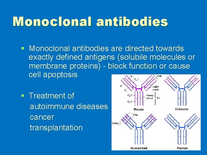
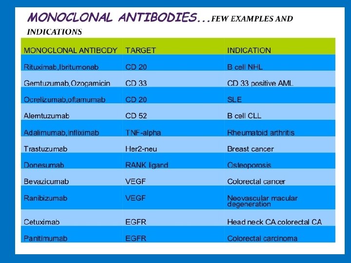
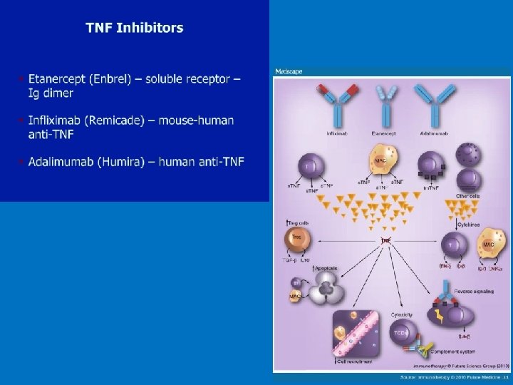
Monoclonal antibodies § Monoclonal antibodies are directed towards exactly defined antigens (solubile molecules or membrane proteins) - block function or cause cell apoptosis § Treatment of autoimmune diseases cancer transplantation
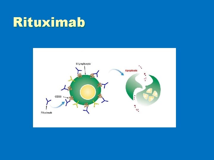
Rituximab
§ https: //www. youtube. com/watch? v=y. Z 6 w Wu. AQn. ME
Thank you for your attention!