Approach to Chest Pain Adel Altamimi MD EM


 Acute Aortic Dissection (AAD)")


. • PMHx •")


: • ECG within 10 minutes of arriving to ED. •")


: • They are regulatory proteins that control the")
: • The ultrasound can assess for pericardial effusion as")

, nausea/vomiting, diaphoresis, and radiation to")


 or left bundle branch")


. • Fibrinolytics.")


- Slides: 63
Approach to Chest Pain Adel Altamimi, MD EM Consultant KSU-MC
• Chest pain is one of the highest risk chief complaints you will see in the Emergency Department (ED). • Approximately 6 million patients visit the emergency department (ED) each year with complaints of chest pain, constituting 9% of all patients seen in ED in USA.
Definition of Acute Chest Pain: • It is the recent onset of pain, pressure, or tightness in the anterior thorax between the xiphoid, supra sternal notch, and both midaxillary lines.
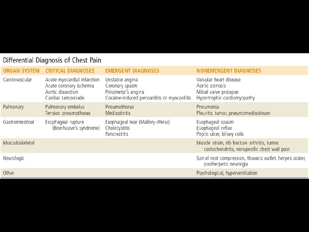
Chest pain challenges: • It can caused by several life-threatening as well as non– life-threatening diseases and has a broad differential diagnosis. • The diagnosis of chest pain in ED can be difficult because neither the quality nor intensity of the pain is specific to any organ or diagnosis. • The presentations can vary significantly even amongst the most common life-threatening diseases.
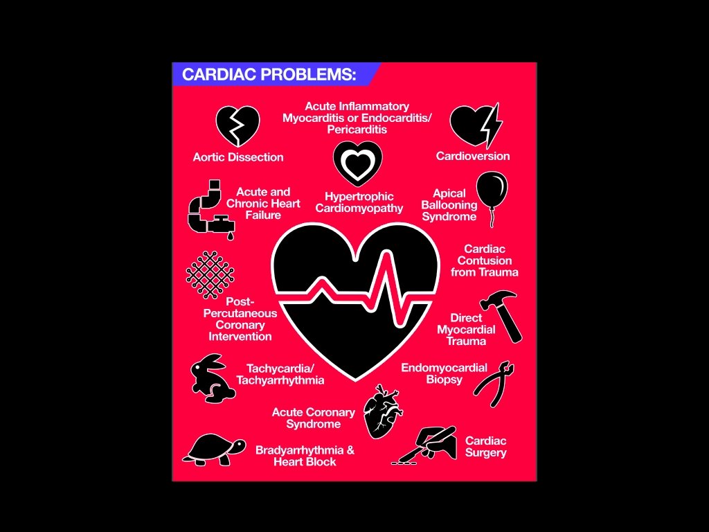
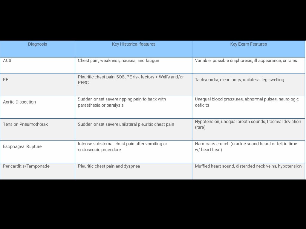
Life Threatening Causes of Chest Pain Acute Coronary Syndrome (ACS) Acute Aortic Dissection (AAD) Pulmonary Embolism (PE) Cardiac Tamponade Esophageal Rupture Tension Pneumothorax
Pathophysiology: • Afferent fibers from the heart, lungs, great vessels, and esophagus enter the same thoracic dorsal ganglia. • Through these visceral fibers, each organ produces the same indistinct quality and location of pain. • Because dorsal segments overlap three segments above and below a level, disease of a thoracic origin can produce pain anywhere from the jaw to the epigastrium.
• Chest pain evaluation is challenging but very manageable. • Most patients who present with chest pain are actually not that sick and many can be safely discharged from the ED.
Initial Evaluation: • Determine “Sick vs “Not Sick”. • Assesses primary survey: ABC’s and vital signs (Stable vs unstable) • Obtain focused history and exam. • Obtain ECG.
History: Character of the pain: • Squeezing. • Crushing • Pressure like Think of ACS Sharp or stabbing more in pulmonary
• relation to exercise. • onset. • relation to food. • severity. • location. • radiation
• Duration. • aggravation factors. • associated symptoms ( diaphoresis). • PMHx • Risk Factors.
Classical cardiac chest pain: • Retrosternal left anterior chest crushing, squeezing, tightness, or pressure. • Aggravated by exertion and relieved by rest. • Radiation. • Diaphoresis.
Examination: • General appearance. • V/S. • Cardiac exam. • Pulmonary exam. • Abdominal exam. • Extremities exam. • Neurologic exam.
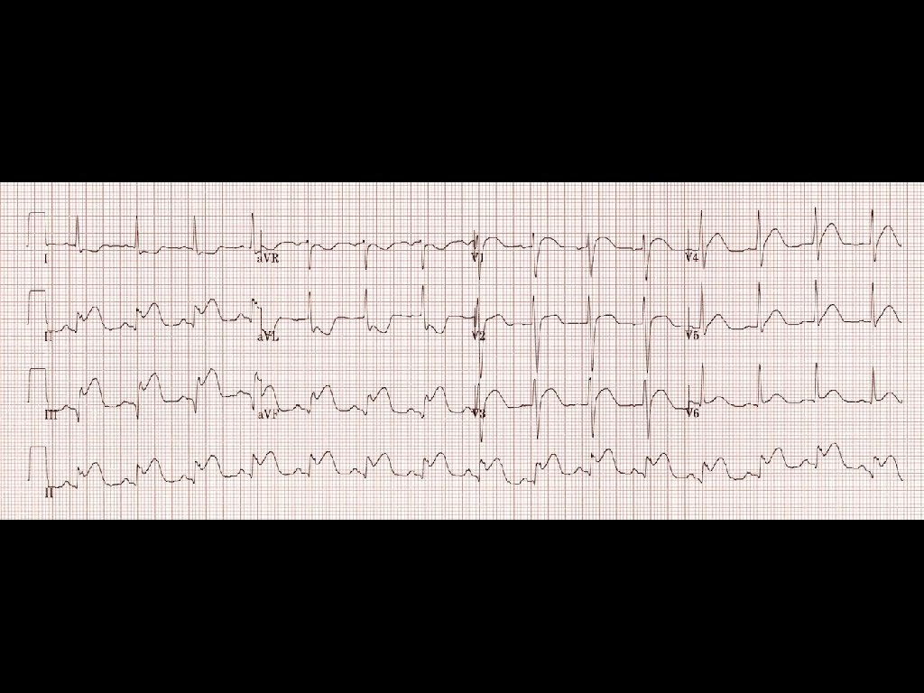
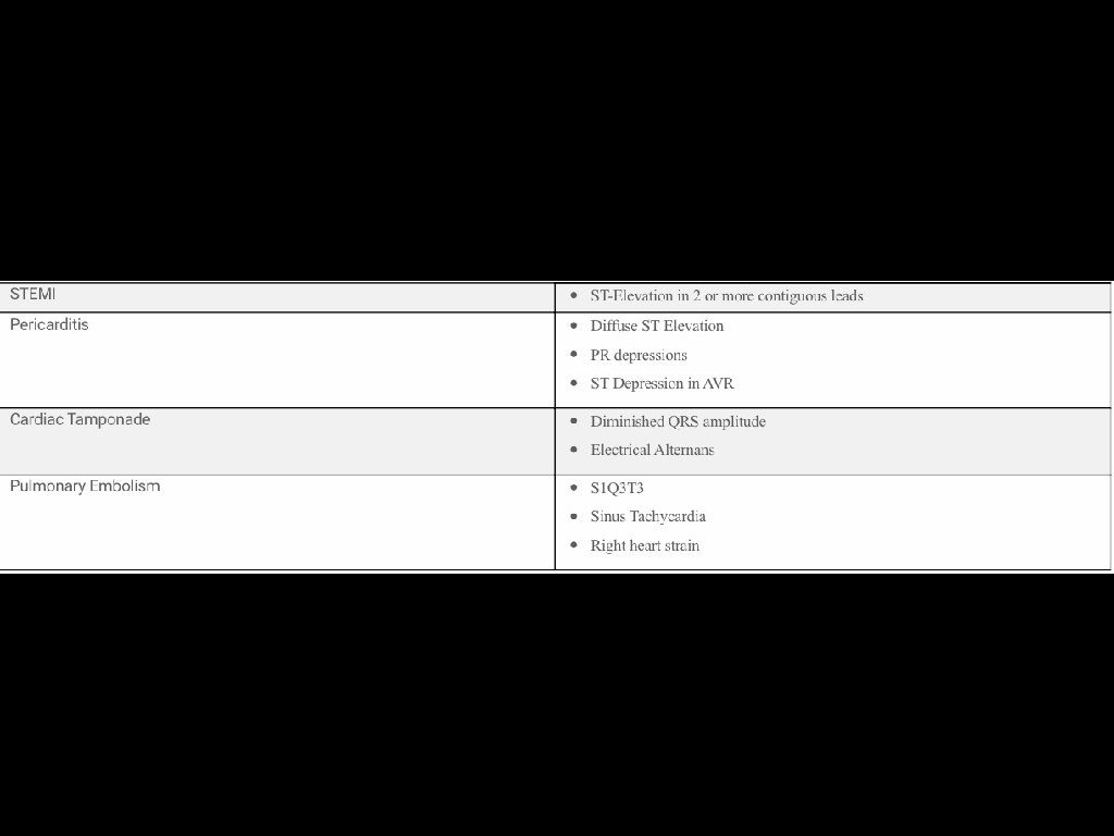
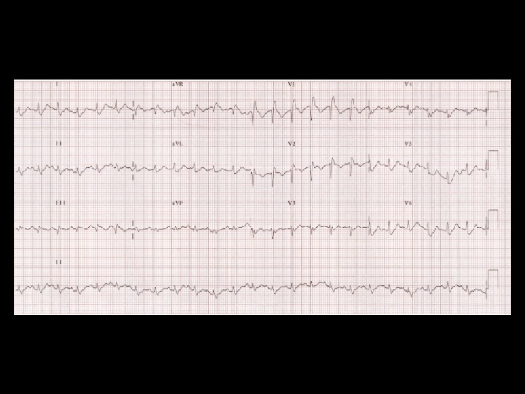
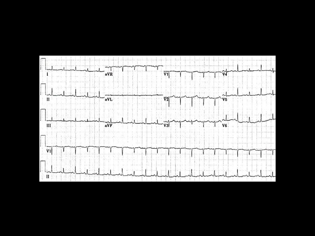
Diagnostic Testing: Electrocardiogram (ECG): • ECG within 10 minutes of arriving to ED. • It can gives the clinician a lot of information. • A rapid screen is done to look for STEMI/ ischemia and arrthymia. • Serial ECGs.
• ST-segment elevation also occurs in patients with pericarditis, myocarditis, early repolarization, left ventricular hypertrophy, and ventricular aneurysms. • A normal ECG lacks the sensitivity to exclude ACS.
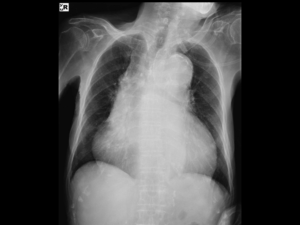
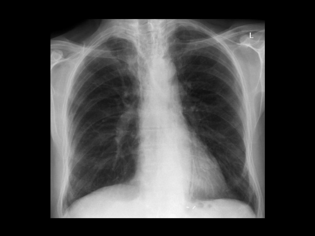
Imaging: • CXR is commonly performed in the evaluation of ED patients with chest pain. • Most patients with ACS have a normal CXR. • Useful to exclude other conditions such as pneumonia and pneumothorax.
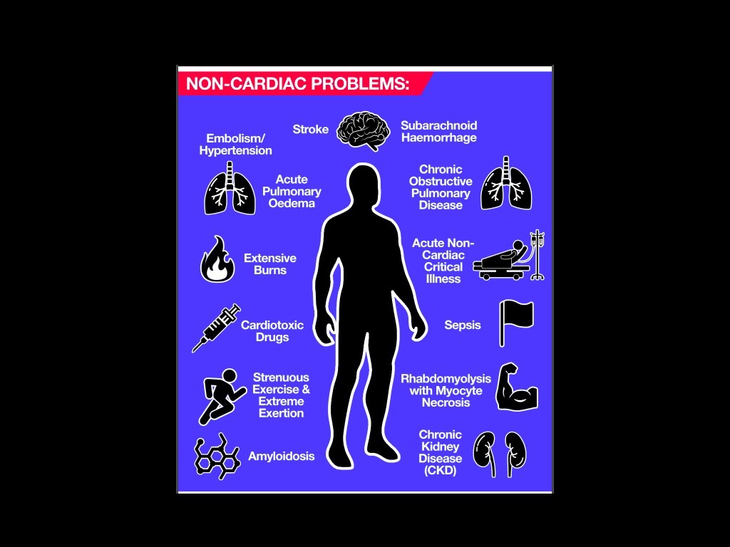
Laboratory Studies: Cardiac troponins (c. Tn): • They are regulatory proteins that control the calciummediated interaction of actin and myosin, producing myocardial contraction. • Myocardial injury, including but not limited to ischemia, causes release of these proteins into blood. • There are numerous non- ischemic causes of c. Tn elevations.
Point of Care Ultrasound (POCUS): • The ultrasound can assess for pericardial effusion as well as tamponade. • It can assess the cardiac contractility to assess the left ventricular function. • It can determined the t presence of right ventricular strain.
ACS • Incidence: ~12% of ED patients with Chest pain have ACS. • ACS is comprised of ST-elevation myocardial infarction (STEMI), non-ST-elevation myocardial infarction (NSTEMI), and unstable angina (UA).
History: • Classically associated with shortness of breath (SOB), nausea/vomiting, diaphoresis, and radiation to neck/arm/shoulder. • Pain worsens upon exertion. • Response to nitroglycerin has no predictive value for ACS.
Non-classic presentations of ACS occur more frequently in women, black race, diabetics, the elderly, and patients with psychiatric disease or altered mental status
Chest pain that significantly decreases the likelihood of ACS include: • Pleuritic in nature. • Positional pain. • Pain described as sharp. • Pain that is fully reproducible with palpation. • Non-exertional
Diagnosis: • In the absence of left ventricular hypertrophy (LVH) or left bundle branch block (LBBB), ST elevation (STE) is defined as follows: ≥ 1 mm STE in at least 2 contiguous leads except for V 2 -V 3. If V 2 -V 3, STE must be ≥ 2 mm in men ≥ 40 y old, ≥ 2. 5 mm in men <40 y old, or ≥ 1. 5 mm in women. • In AMI, initial ECGs are often non-specific (40%-70%) or normal (<10%).
• Troponin is the test of choice for diagnosing MI. Along with the history and ECG, troponin plays an essential role in diagnosing MI. • Elevated troponin is proportional to increased mortality in ACS.
Time course of Troponin: • Rise 4 -8 hours post onset of symptoms in MI • Peaks at 18 -24 hours. • Levels stay elevated for 10 days.
Therapeutic Interventions: • Address ABC. • Start an IV, oxygen to maintain Sp. O 2 >90% and start cardiac monitoring. • Aspirin 162 -325 mg chewed. If the patient is unable to tolerate aspirin, clopidogrel 75 mg PO is an alternative. • Nitroglycerin 0. 4 mg SL q 5 min × 3 as needed for CP.
Contraindication to Nitroglycerin: • Hypotension • Inferior STEMI • History of use phosphodiesterase inhibitors.
STEMI Patients: • Percutaneous intervention (PCI). • Fibrinolytics.
PE • Incidence is 0. 5% of ED patients with Chest pain. • Presentation varies greatly, ranging from asymptomatic to shock.
Massive PE: • Accounts for only 5% of acute PE. • Mortality rate 25 -65% • Hypotension: SBP <90 mm Hg or decrease >40 mm Hg for 15 min. • Signs of shock secondary to PE.
Submassive PE: PE without hypotension but with either of the following: • Myocardial necrosis defined by elevation of troponin or • Right ventricular (RV) dysfunction defined by RV dilation on imaging (echocardiographic or CT) and elevation of B-type natriuretic peptide (BNP) or ECG criteria.
Presentation: • SOB is the most common symptoms followed by pleuritic chest pain. • Look for risk factors. • Tachypnea is the most common sign. • Risk stratification by clinical rules , e. g Wells Criteria.
Diagnosis: • ECG and CXR: Often non-specific. • Low to moderate risk of having PE → D-dimer. • High risk of having PE → send directly to imaging. • CT pulmonary angiogram is the test of choice.
Therapy: • Anticoagulation is the main step of treatment. • For massive PE, thrombolytics can be used if there is no contraindication.
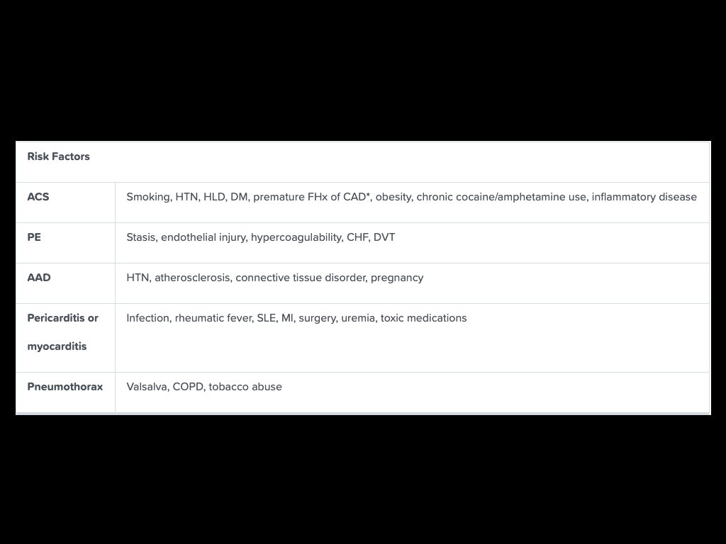
AAD • Incidence is approximately 5 cases/million population/y. • Risk factors: HTN, atherosclerosis and connective tissue disease.
History: • Classic presentation is severe tearing or ripping pain radiating to the back with severe distress and diaphoresis. • May cause signs of infarction/ischemia anywhere along the aorta (arms, legs, bowels, kidneys, brain, spinal cord). • Approximately a third of patients have neurologic complaints/findings.
Exam: • Pulse deficit, caused by a hematoma or intimal flap, may unilaterally obstruct flow. This is observed in fewer than one-third of patients. • Diastolic murmur may be due to aortic regurgitation. • Abnormal neurologic exam may be due to stroke from cervical artery dissection or infarct of the spinal cord.
Diagnosis: • ECG mostly non specific or may show a STEMI as a result of dissection of coronary ostia. • CXR may shows widened mediastinum (61%) or abnormal aortic contour (50%). • Normal x-ray does not rule out the diagnosis. • CTA is the test of choice.
Therapy: • Pain control. • BP control • Surgical consultation
Esophageal Rupture • Boerhaave’s syndrome. • Severe CP, usually following forceful vomiting. • Often associated with subcutaneous crepitus. • Think of this diagnosis in alcoholics or with history of recent procedures.
Diagnosis: • Ill appearing, septic and febrile. • CXR may show left side pleural effusion and mediastinal air. • CT Chest will help in defining injury.
Therapy: • IV antibiotics. • Emergent surgical consultation
Pericardial Tamponade • Can present acutely from trauma or aortic injury, or slowly from cancer and infection. • Consider this diagnosis if the patient is in shock.
Diagnosis: • Tachycardia. • Pericardial rub. • Muffled heart sounds. • Jugular venous distention. • POCUS is the test of choice and it usually show diastolic collapse of the cardiac chambers.
• ECG can show low voltage, tachycardia or electrical alternans. • POCUS is the test of choice and it usually show diastolic collapse of the cardiac chambers.
Therapy: • The treatment is pericardiocentesis.
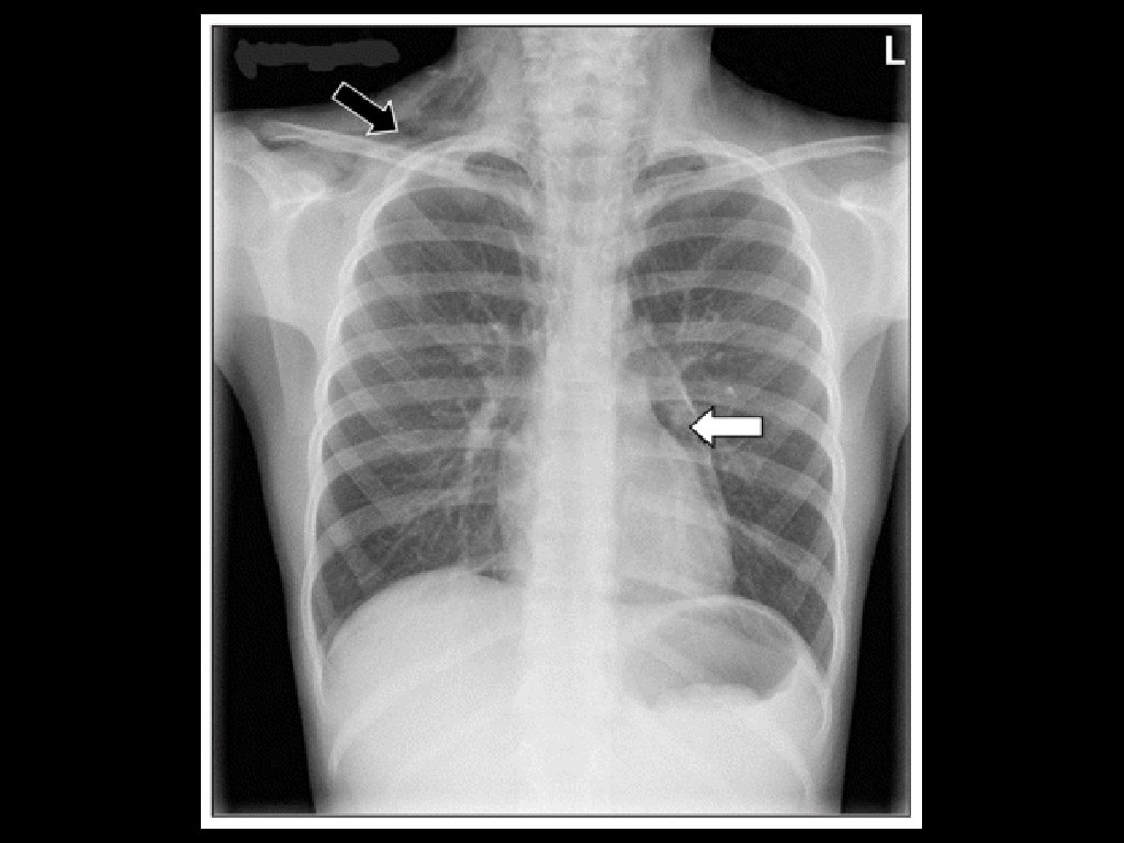
Tension Pneumothorax • Can occur for many reasons including iatrogenic (e. g. from central lines). • Shock and respiratory symptoms due to pneumothorax compressing the mediastinum, resulting in decreased venous return to the heart. • Often secondary to thoracic trauma.
• It can also caused by a simple spontaneous pneumothorax progressing to tension pneumothorax. • Diagnosis is clinical, Don’t wait for the X-ray to make the diagnosis. • Treat with needle decompression while preparing for ED Thoracostomy.