Adel Hasanin Ahmed MRCP UK Generation and Conduction

- Slides: 25
Adel Hasanin Ahmed, MRCP (UK)
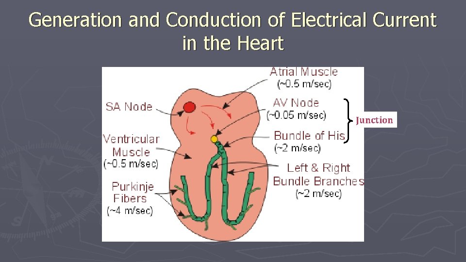
Generation and Conduction of Electrical Current in the Heart Junction
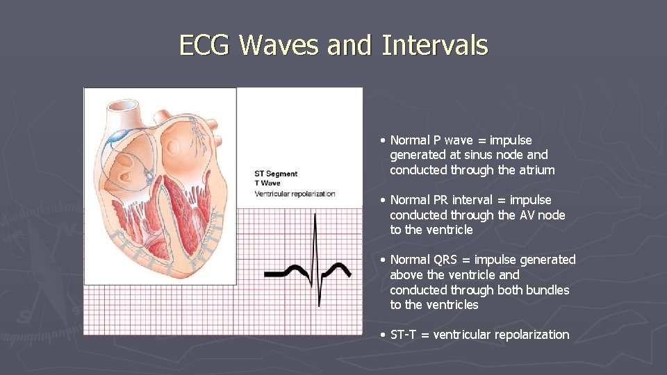
ECG Waves and Intervals • Normal P wave = impulse generated at sinus node and conducted through the atrium • Normal PR interval = impulse conducted through the AV node to the ventricle • Normal QRS = impulse generated above the ventricle and conducted through both bundles to the ventricles • ST-T = ventricular repolarization
Why 12 Leads? If you wanted the best description of the elephant, whom would you ask?
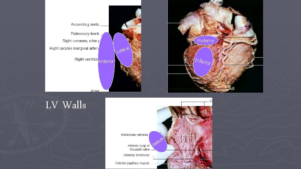
Posterior ral Late Infer Anterior LV Walls S m tu p e
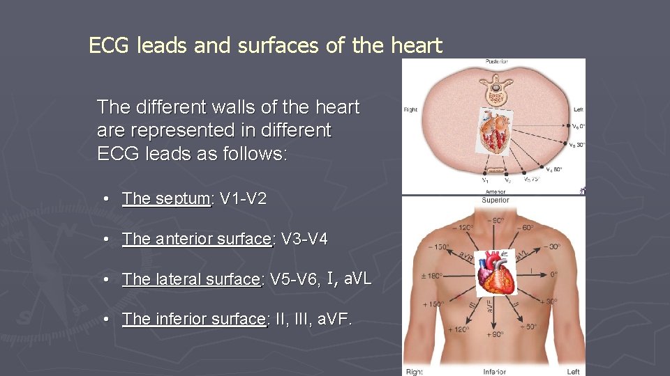
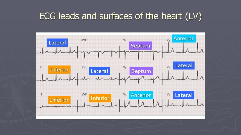
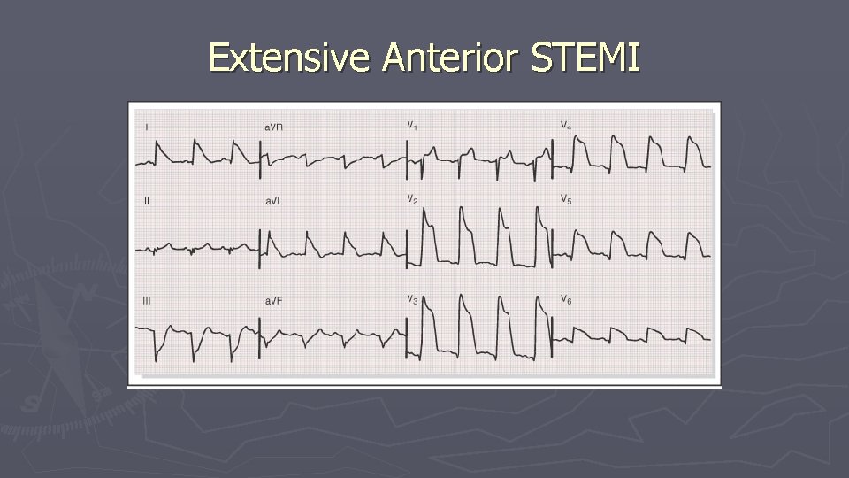
ECG leads and surfaces of the heart The different walls of the heart are represented in different ECG leads as follows: • The septum: V 1 -V 2 • The anterior surface: V 3 -V 4 • The lateral surface: V 5 -V 6, I, a. VL • The inferior surface: II, III, a. VF.
ECG leads and surfaces of the heart (LV) Anterior Lateral Septum Inferior Lateral Inferior Septum Anterior Lateral
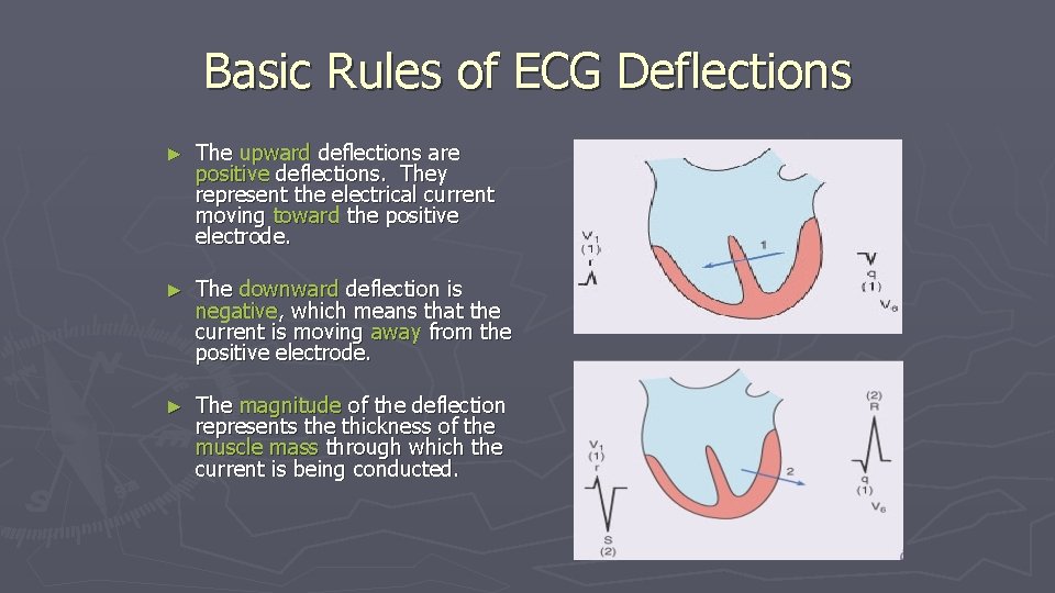
Basic Rules of ECG Deflections ► The upward deflections are positive deflections. They represent the electrical current moving toward the positive electrode. ► The downward deflection is negative, which means that the current is moving away from the positive electrode. ► The magnitude of the deflection represents the thickness of the muscle mass through which the current is being conducted.
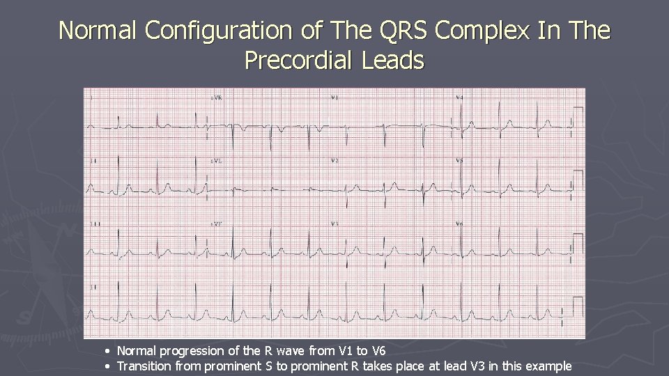
Normal Configuration of The QRS Complex In The Precordial Leads • Normal progression of the R wave from V 1 to V 6 • Transition from prominent S to prominent R takes place at lead V 3 in this example
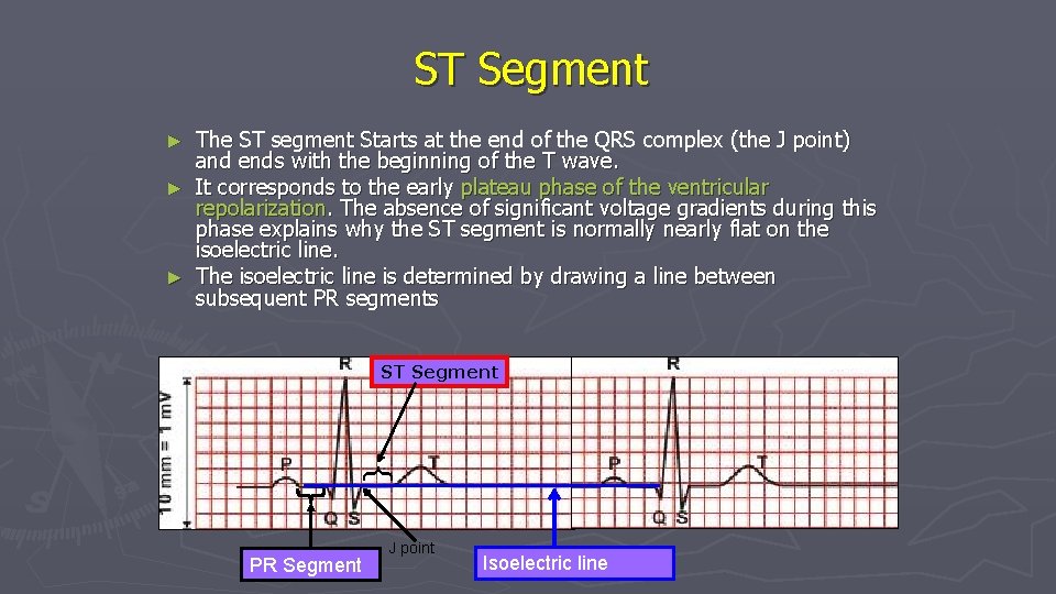
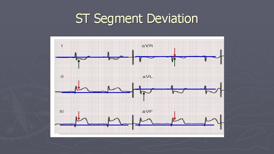
ST Segment The ST segment Starts at the end of the QRS complex (the J point) and ends with the beginning of the T wave. ► It corresponds to the early plateau phase of the ventricular repolarization. The absence of significant voltage gradients during this phase explains why the ST segment is normally nearly flat on the isoelectric line. ► The isoelectric line is determined by drawing a line between subsequent PR segments ► ST Segment { PR Segment J point Isoelectric line
ST Segment Deviation
Extensive Anterior STEMI
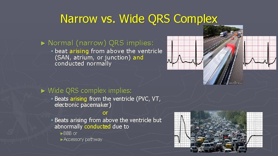
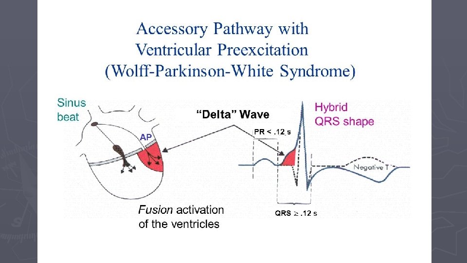
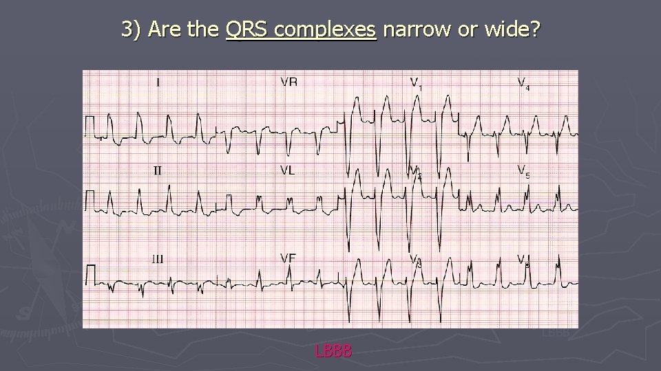
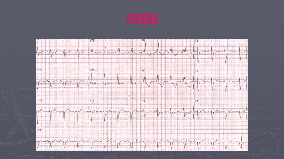
Narrow vs. Wide QRS Complex ► Normal (narrow) QRS implies: § beat arising from above the ventricle (SAN, atrium, or junction) and conducted normally ► Wide QRS complex implies: § Beats arising from the ventricle (PVC, VT, electronic pacemaker) or § Beats arising from above the ventricle but abnormally conducted due to ►BBB or ►Accessory pathway
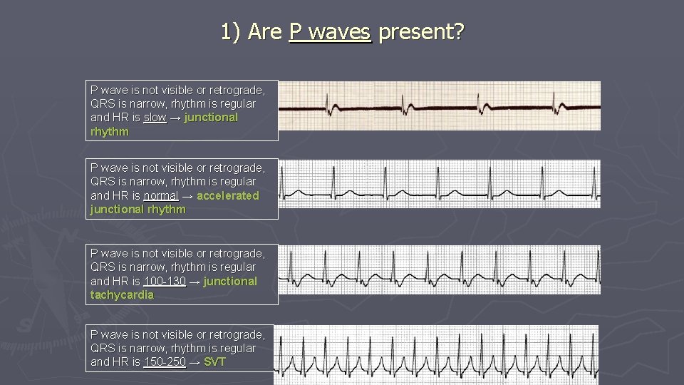
1) Are P waves present? P wave is not visible or retrograde, QRS is narrow, rhythm is regular and HR is slow → junctional rhythm P wave is not visible or retrograde, QRS is narrow, rhythm is regular and HR is normal → accelerated junctional rhythm P wave is not visible or retrograde, QRS is narrow, rhythm is regular and HR is 100 -130 → junctional tachycardia P wave is not visible or retrograde, QRS is narrow, rhythm is regular and HR is 150 -250 → SVT
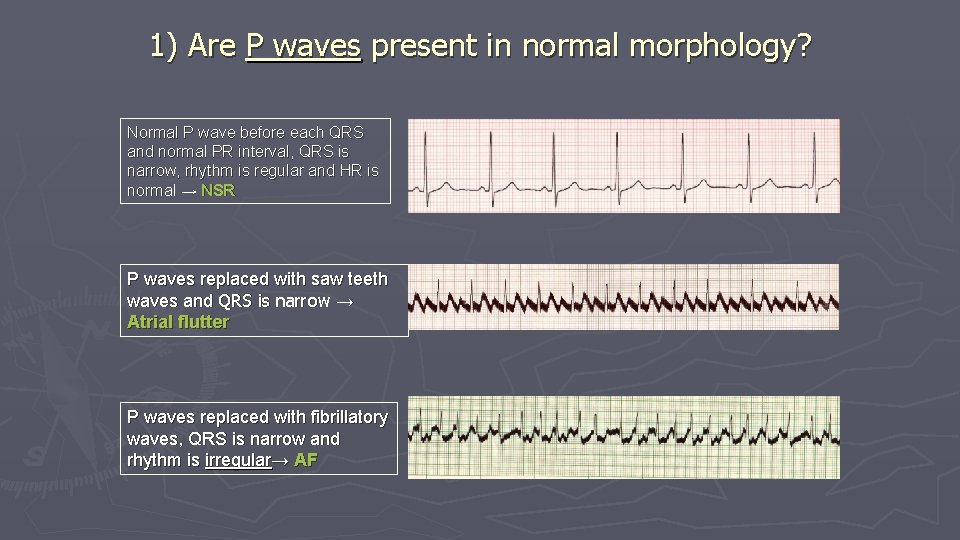
1) Are P waves present in normal morphology? Normal P wave before each QRS and normal PR interval, QRS is narrow, rhythm is regular and HR is normal → NSR P waves replaced with saw teeth waves and QRS is narrow → Atrial flutter P waves replaced with fibrillatory waves, QRS is narrow and rhythm is irregular→ AF
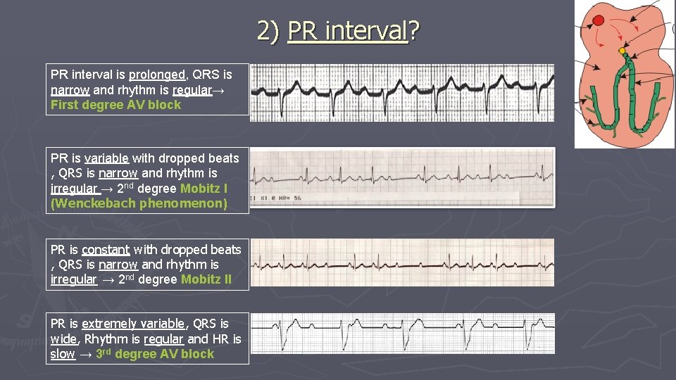
2) PR interval? PR interval is prolonged, QRS is narrow and rhythm is regular→ First degree AV block PR is variable with dropped beats , QRS is narrow and rhythm is irregular → 2 nd degree Mobitz I (Wenckebach phenomenon) PR is constant with dropped beats , QRS is narrow and rhythm is irregular → 2 nd degree Mobitz II PR is extremely variable, QRS is wide, Rhythm is regular and HR is slow → 3 rd degree AV block
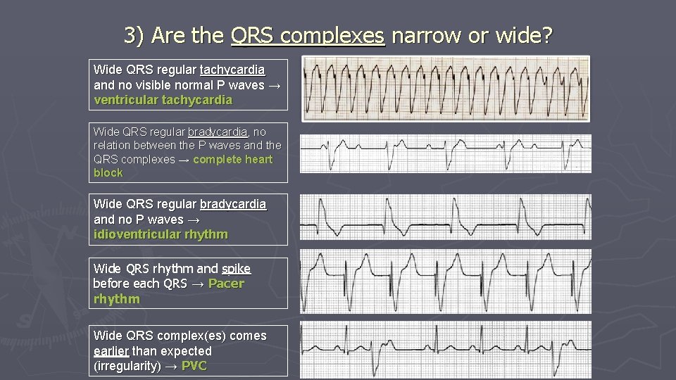
3) Are the QRS complexes narrow or wide? Wide QRS regular tachycardia and no visible normal P waves → ventricular tachycardia Wide QRS regular bradycardia, no relation between the P waves and the QRS complexes → complete heart block Wide QRS regular bradycardia and no P waves → idioventricular rhythm Wide QRS rhythm and spike before each QRS → Pacer rhythm Wide QRS complex(es) comes earlier than expected (irregularity) → PVC
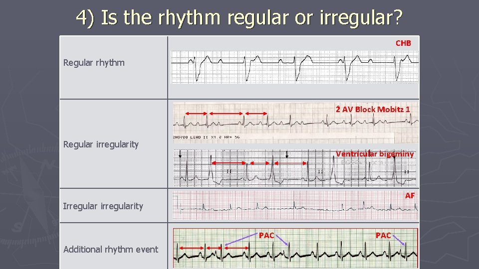
4) Is the rhythm regular or irregular? CHB Regular rhythm 2 AV Block Mobitz 1 Regular irregularity Ventricular bigeminy AF Irregular irregularity PAC Additional rhythm event PAC
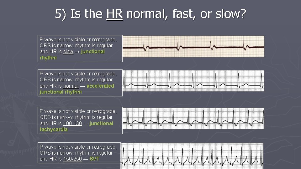
5) Is the HR normal, fast, or slow? P wave is not visible or retrograde, QRS is narrow, rhythm is regular and HR is slow → junctional rhythm P wave is not visible or retrograde, QRS is narrow, rhythm is regular and HR is normal → accelerated junctional rhythm P wave is not visible or retrograde, QRS is narrow, rhythm is regular and HR is 100 -130 → junctional tachycardia P wave is not visible or retrograde, QRS is narrow, rhythm is regular and HR is 150 -250 → SVT
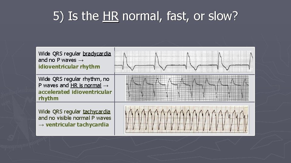
5) Is the HR normal, fast, or slow? Wide QRS regular bradycardia and no P waves → idioventricular rhythm Wide QRS regular rhythm, no P waves and HR is normal → accelerated idioventricular rhythm Wide QRS regular tachycardia and no visible normal P waves → ventricular tachycardia
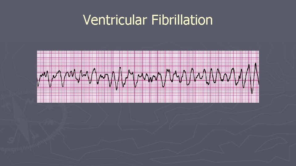
Ventricular Fibrillation
3) Are the QRS complexes narrow or wide? Sinus Tachycardia AF SVT VT Pacemaker Rhythm LBBB
RBBB