ACG Clinical Guideline Epidemiology Risk Factors Patterns of


 and EMBASE (1980 to present) with")


")


 q Blood tests")


 can be used")


 because it is")


 should be performed")


![q Serology value § Hgb <12 mg/dl, § hyponatremia (Na <136 m. Eq/l[mmol/l]), §](https://slidetodoc.com/presentation_image_h2/414b12e0db7abde12c1571823e824034/image-76.jpg "q Serology value § Hgb <12 mg/dl, § hyponatremia (Na <136 m. Eq/l[mmol/l]), §")


- Slides: 89
ACG Clinical Guideline: Epidemiology, Risk Factors, Patterns of Presentation, Diagnosis, and Management of Colon Ischemia (CI) published online 23 December 2014
Dr gavidel � Journal club�
INTRODUCTION �A search of MEDLINE (1946 to present) and EMBASE (1980 to present) with language restriction to English was conducted using the search terms ischemic colitis, ischaemic colitis, colon ischemia, colonic ischemia, colon ischaemia, colonic ischaemia, colon gangrene, colonic gangrene, colon infarction, colonic infarction, rectal ischemia, rectal ischaemia, ischemic proctitis, ischaemic proctitis, cecal ischemia, cecal ischaemia, ischemic colon stricture, ischaemic colon stricture, ischemic colonic stricture, ischaemic colonic stricture, ischemic megacolon, ischaemic megacolon, colon cast, and colonic cast.
DEFINITION � CI is the condition that results when blood flow to the colon is reduced to a level insufficient to maintain cellular metabolic function. � We now know that blood flow need not stop but only diminish significantly to cause ischemic damage. � The degree to which colonic blood flow must diminish before ischemia results varies with the acuteness of the event, the degree of preexisting vascular collateralization, and the length of time the low flow state persists. � CI may manifest with reversible or irreversible damage.
� Reversible damage includes colopathy, i. e. , subepithelial hemorrhage or edema, and colitis; � Colitis reflects an evolutionary stage in which the overlying mucosa ulcerates as the subepithelial edema and blood are resorbed. � In reversible disease, such resorption occurs rather promptly, usually within 3 days. � Ulcerations may persist for several months before resolving, although during this time, the patient usually is asymptomatic. � Irreversible manifestations of CI include gangrene, fulminant colitis, stricture formation, and, rarely, chronic ischemic colitis. � Recurrent sepsis due to bacterial translocation is another rare manifestation of irreversibly damaged bowel.
EPIDEMIOLOGY Children with CI are only rarely reported , but CI occurs in adults of all ages and increases with age, especially after age 49 years. � CI is more common in women than in men, and 57– 76% of patients in large series have been female. � Mortality rates in large series range from 4 to 12%, but inclusion criteria, case ascertainment methods, and rates of comorbidity and surgery in these studies differed. � Recurrent CI increases over time; for example, estimated cumulative recurrence rates at 1, 2– 3, 4, and 5– 6 years were 3%, 5%, 6%, and 10%, respectively, in one study and 3. 3% at 2 years and 7. 5% at 5 years in another study. � Particular predisposing illnesses have been reported with recurrent disease, such as hypercoagulable states. �
PATHOPHYSIOLOGY � CI can result from alterations in the systemic circulation or from anatomic or functional changes in the mesenteric vasculature; � Type I disease: In most cases, no specific cause for ischemia is identified, and such episodes are attributed to localized nonocclusive ischemia, likely a result of small-vessel disease. � Type. II disease the etiology is identified and most commonly follows an episode of systemic hypotension, decreased cardiac output, or aortic surgery. � In practice, patients with Type II disease can have therapy targeted toward the underlying cause, whereas Type I CI is treated in a broader and supportive manner.
� Abnormalities seen on angiography rarely correlate with clinical manifestations of CI, and age-related abnormalities in the splanchnic vessels are not uncommon, including narrowing of small vessels, and tortuosity of the long colic arteries; �Fibromuscular dysplasia of the superior rectal artery has been associated with CI. � The colon is particularly susceptible to ischemia, perhaps owing to its relatively low blood flow, its unique decrease in blood flow during periods of functional activity, and its sensitivity to autonomic stimulation. � What triggers the episode of CI, however, usually is not identified.
RISK FACTORS � Coronary artery disease and atrial fibrillation were approximately twice as common in patients with isolated right-colon ischemia (IRCI) compared with other anatomic patterns of CI that are generally less severe. � In patients with CI, a French group found a “proven” potential cardiac source of embolism in 35% of patients, primarily those with sustained or paroxysmal atrial fibrillation. � CI occurred within 3 days of acute myocardial infarction in 0. 13% of patients, and complications and mortality were higher in patients with both diseases than in those with either CI or myocardial infarction alone.
� Among patients with severe hematochezia, patients with CI more often had moderate or severe lung disease than did patients with other colonic causes of hemorrhage and chronic obstructive pulmonary disease independently predicted mortality. � Hypertension and diabetes mellitus independently predicted CI among patients with acute lower abdominal pain. � Endothelial dysfunction could contribute to the effects of hypertension and diabetes and has been offered to explain the increased risk of CI associated with rheumatic autoimmune diseases , including rheumatoid arthritis, although association of hypercoagulable states with these diseases is another potential risk factor.
� Excessive sympathetic activity in IBS could impair vasodilation in the mesenteric vessels, the most reactive vascular bed in the body. � Constipation could mediate increased risk through increased intracolonic pressure and reduced blood flow as a result of fecal impaction, and constipation preceding the symptoms of CI could be a clinical clue for differential diagnosis. � Diarrhea was a risk factor in one study but, as with constipation , CI could have been a diagnosis applied to patients with IBS.
� Among patients with severe hematochezia, the mean serum creatinine was twice as high in patients with CI as it was in those with other colonic causes and hemodialysis independently predicted CI in patients with acute lower abdominal pain � Thrombophilia is another potential risk factor for CI. � There are numerous case reports of various coagulopathies in patients with CI, including deficiencies of protein C, protein S, antithrombin III, and factor V Leiden mutation. � In particular, the “catastrophic”variant of the antiphospholipid syndrome causes multiple vascular occlusions, especially in small vessels, but typically causes more widespread intestinal ischemia than CI alone.
� genetic polymorphisms associated with thrombophilia and vascular hyperactivity were found in more young patients with CI than in controls. � At this time, routine testing for a coagulation disorder in most patients with CI does not appear justified, although it seems reasonable in young patients with CI and in patients with recurrent disease. � Sickle cell crisis with microvascular occlusion can also cause CI. � Sickle cell disease is only rarely mentioned as an etiology for CI, but the rate of this disease causing CI could be higher in populations with a greater proportion of patients who are of African ancestry.
� CI has also rarely followed colonoscopy. � Postcolonoscopy CI could result from reduced colonic blood flow as a consequence of luminal distention and increased intraluminal pressure, both of which are more pronounced with insufflation by air than by carbon dioxide
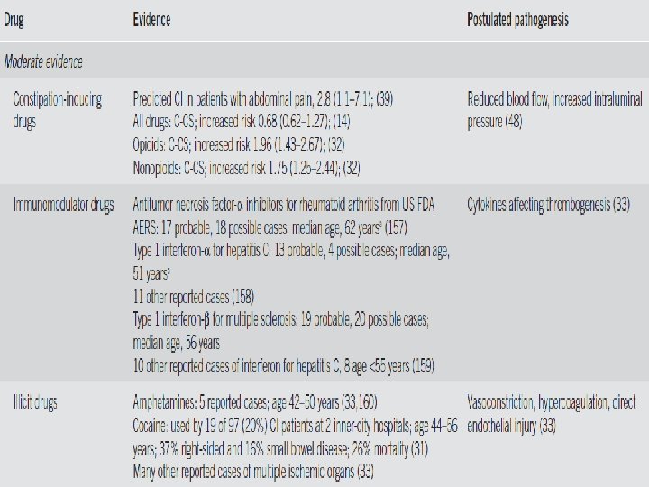
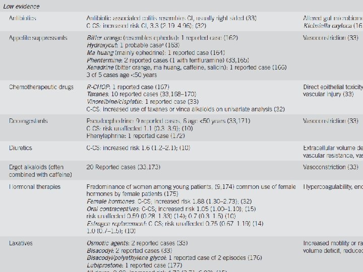
� Surgical history; Surgical procedures in which the IMA has been sacrificed, such as abdominal aortic aneurysm repair and other abdominal operations, should increase consideration of CI in patients with typical clinical features. � Drug use. When drug-induced CI was reviewed in 2007, there was evidence to propose various major classes of pharmacologic agents as predisposing to Cl. � constipation-inducing drugs, immunomodulators, and illicit drugs are best supported as etiologic agents, but there is some support for a role of many disparate drug classes.
� Despite extensive observations on the above clinical factors associated with CI, there are no specific identifiable risk factors for CI in most patients, and some patients have multiple risk factors. � The heterogeneous risk factors for CI support its multifactorial pathogenesis and indicate the importance of careful assessment of the medical, surgical, and drug use history in every patient with CI. �Further research into the cause of CI and its risk factors is needed.
Summary statements 1. CVD and DM should increase consideration of CI in patients with typical clinical features. 2. A history of IBS and constipation should be sought in patients suspected to have CI. 3. Selective cardiology consultation is justified in patients with CI, particularly if a cardiac source of embolism is suspected. 4. CKD and COPD are associated with increased mortality from CI. 5. Evaluation for thrombophilia should be considered in young patients with CI and in all patients with recurrent CI. 6. Surgical procedures in which the IMA has been sacrificed. 7. In patients suspected of having CI, a history of medication and drug use should be sought, especially constipation inducing medications, immunomodulators, and illicit drugs.
CLINICAL PRESENTATION � Summary of evidence � CI generally manifests with sudden cramping, mild, left lower abdominal pain; an urgent desire to defecate; and passage within 24 h of bright red or maroon blood per rectum or bloody diarrhea. � Abdominal pain, urgent need to defecate, and bloody diarrhea are the major features, and all three symptoms occur in this temporal sequence in nearly one-half of cases. � Vomiting (30%), dizziness (10%), and syncope (6%) occur less frequently. � Pain typically precedes bleeding and is usually mild to moderate. � Abdominal tenderness is usually present over the involved segment of colon.
� Patients with IRCI more commonly have pain than they do rectal bleeding; � Only 25– 46% of patients with IRCI have rectal bleeding. � Physicians should entertain the diagnosis of IRCI for patients with acute, severe abdominal pain who lack hematochezia and/or diarrhea, especially if they have the clinical scenarios associated with IRCI, such as dialysis, sepsis, and hypotension or shock. � In more than half of the cases of CI, the disease is reversible. � Symptoms of CI generally resolve within 2– 3 days and the colon heals in 1– 2 weeks. � With severe injury, it may take up to 6 months for the colon to heal; � However, during this time the patient is usually asymptomatic.
� Rectal bleeding is usually mild in CI. � Severe bleeding was seen more frequently in women and in patients with severe lung disease, elevated creatinine and glucose levels, and those on anticoagulation. � The 30 -day outcomes for rebleeding, surgical intervention, and mean number of hospital days were worse for those with CI compared with other etiologies of lower gastrointestinal bleeding. � Severe hemorrhage occurs mainly in patients with gangrenous CI, fulminant pancolitis, and IRCI.
�Most episodes of CI are benign and self-limited and only a minority of cases are severe. � Symptoms that persist for more than 2 weeks are also associated with a higher incidence of acute complications and irreversible disease, such as gangrene and perforation, segmental ulcerating colitis, or stricture. � Symptoms of patients with severe disease do not necessarily follow the classic sequence of abdominal pain, urgent desire to defecate, and bloody diarrhea. � Anal passage of an infarcted colonic segment or “colonic cast”not accompanied by features of peritonitis is a rare complication of CI. � This complication usually occurs in patients with multiple medical comorbidities who recently underwent abdominal aortic aneurysm repair or colorectal surgery.
� CI affected the left side of the colon. � A cast of 25 to 120 cm in length is typically passed 2– 4 weeks after the acute ischemic insult. � Casts may consist of mucosa with or without submucosa or may be full thickness, in which case a tunnel of inflammatory tissue is left behind; � The latter situation requires urgent surgical intervention.
� Morphologic changes after CI vary with the duration and severity of the injury. � The mildest injury is mucosal and submucosal hemorrhage and edema, with or without partial necrosis and ulceration of the mucosa. � Iron-laden macrophages may be found. � More severe injury, submucosal fibrosis and pseudomembranes may develop. � In 3. 3– 9. 4% of cases, the muscularis propria is replaced by fibrous tissue forming a stricture, most of which are asymptomatic. � Stricture formation is more common in patients with moderate CI, and was reported in 14. 3% of cases;
� Acute severe CI can also mimic IBD with chronic ulcerations, crypt abscesses, and pseudopolyps. � The most severe form of ischemic damage causes transmural infarction. � Gangrenous colitis is characterized by increasing abdominal tenderness, guarding, rebound tenderness, rising temperature, and paralytic ileus. � Abdominal pain is seen in the vast majority of these patients (86. 1%) but rectal bleeding is far less frequent (30. 6%); � Acute abdominal pain without rectal bleeding(58. 3%) and nonbloody diarrhea (27. 8%) are the most common clinical patterns of presentation.
� Sudden onset of a toxic colitis with signs of peritonitis and a rapidly progressive course are typical of universal fulminant colitis, a rare variant of CI. � The classic sequential triad is seen infrequently(11. 1%) with this CI variant; � Symptoms of severe abdominal pain(66. 7%) and rectal bleeding (55. 6%) with abdominal tenderness on physical examination (85. 9%) are the most characteristic presentation of universal fulminant colitis. � CI isolated to the right colon (IRCI) is associated with higher mortality rates compared with other patterns of CI.
SEGMENTAL NATURE OF CI � The left colon is most commonly affected, but no colonic region is spared from involvement. � Pancolitis and IRCI were seen frequently in patients with sepsis, and IRCI was associated more frequently in patients with CAD and CKD on hemodialysis. � Patients with IRCI, however, were more likely to have renal failure. � Colonic blood flow is supplied by three vessels: the SMA, IMA, and the superior hemorrhoidal artery. � Vascular anatomy, is variable and often individually unique.
� Watershed areas of the colon are regions that are particularly susceptible to ischemic insult as a result of their location between two different vascular supplies. � These areas include the splenic flexure (Griffith’s point) and sigmoid colon (Sudeck’s point). � The rectum is uncommonly affected by ischemia because of its relatively rich dual blood supply from both splanchnic and systemic arterial systems.
� IRCI has a different clinical presentation and , abdominal pain without rectal bleeding(59%) although when bleeding does occur it may be severe. � Patients with IRCI have atrial fibrillation, coronary artery disease, and chronic kidney disease more frequently than do patients with CI affecting other areas of the colon.
� IRCI had worse outcomes for 30 -day mortality (22. 5% vs. 11. 9%, P =0. 03), need for surgical intervention (54. 9% vs. 10. 9%, P <0. 001), and unfavorable outcome. � In Isolated cecal necrosis 80% of the patients had a history of cardiovascular disease or diabetes.
� The pancolonic pattern of CI portends a similarly poor prognosis to that of IRCI. � In this situation, stage V chronic kidney disease (30. 4%) and peripheral vascular disease (21. 7%) were the most frequent comorbidities, and sepsis (70%) was the most common etiology. � When pancolonic involvement is observed, there probably was hypoperfusion in both the SMA and IMA circulations and the risk factors associated with such an episode likely forecast a worse outcome.
RECURRENT AND CHRONIC CI � Recurrence of CI is said to occur when a patient has one discrete episode that resolves and the patient subsequently re-presents with similar symptoms and has another independent diagnosis of CI. � Defining the frequency and timing of recurrences is challenging, however, given the usually benign self-limited nature of CI and the fact that many patients with mild disease may not seek medical attention; � There is also a lack of appropriate follow-up in the current literature. � Some series with a 5 -year follow-up have shown no recurrence, whereas others detail recurrence rates of CI that range from 6. 8 to 16. 0%.
� The most common symptoms of recurrence are abdominal pain, diarrhea, and hematochezia, although the frequency of these symptoms was not provided. � Patients with coronary artery disease and elevated serum creatinine were 3. 5 - and 1. 01 fold more likely to have a recurrence, respectively. � In sum, although recurrence of CI does occur, it appears to be uncommon and the presentation and course seem to be the same as those of the initial episode; � Time to recurrence needs to be assessed further.
� Chronic segmental colitis should be defined clinically by more than 3 months of typical symptoms and biopsy confirmation showing histologic evidence compatible with or characteristic of CI. � The classic sequence of abdominal pain and urgent desire to defecate followed by bloody diarrhea (32. 3%) is the most common presentation, although rectal bleeding without prior abdominal pains is also seen (30. 8%). � Recurrent fever, leukocytosis, and septicemia suggest presence of an area of segmental colitis that is continually providing a portal of entry for colonic bacteria.
� It has been suggested by Wakefield and colleagues that small multifocal gastrointestinal infarction and repetitive thrombotic mesenteric microvascular occlusion may play an etiologic role in IBD. � A vascular etiology for IBD is supported further by studies showing that IBD occurs less frequently in patients with inherited disorders of coagulation (e. g. , hemophilia or von Willebrand’s disease) and that smoking has a deleterious effect on the progression of Crohn’s disease.
� Some authors hypothesize that a chronic colitis might be the intervening process between an initial diagnosis of CI and the development of a stricture, but the studies that proposed this failed to characterize the intervening time when the chronic colitis might be evolving into a stricture.
LABORATORY TESTING IN CI � By comparing severe disease with mild disease, these studies sought to identify specific blood test alterations that were associated with poor outcome. � Decreases in Hgb and bicarbonate or increases in WBC or LDH were most frequently seen in patients with severe CI.
� The differential diagnosis for patients presenting with abdominal pain and bloody diarrhea is broad, including Crohn’s disease, ulcerative colitis, infectious colitis, and colonic adenocarcinoma. � Accuracy for the initial diagnosis of CI based upon clinical presentation is believed to be low.
� To properly assess the differential diagnosis, clinicians should consider initially obtaining complete blood count, comprehensive metabolic panel, stool culture, stool examination for ova and parasites, Clostridium difficile toxin assay, and serum lactate, LDH, creatine kinase, and amylase levels.
� CBC is useful to assess the WBC for prognostic purposes and the Hgb level to determine blood loss. � Hgb does not usually decrease significantly from baseline in patients with CI. � Serum bicarbonate levels from electrolyte panels assess whether the patient is becoming acidotic, although serum lactate and LDH levels will also provide insight into the patient’s acid/base status. � Stool culture and ova and parasite screens for infectious causes of bloody diarrhea are important initial studies.
� In one small study, Escherichia coli O 157: H 7 was identified by immunoperoxidase staining from colon biopsies in patients with pathologically supported CI ; this organism may be etiologic for CI and should be tested for in all patients with bloody diarrhea.
� Although C. difficile infection infrequently presents with bloody diarrhea, given its increasing incidence and severity, it too should be part of the initial screening protocol for patients with bloody diarrhea. � Elevations in serum amylase also have been shown to be associated with acute bowel ischemia. � Despite none of these markers having sufficient evidence that they can diagnose CI, obtaining them during the initial workup may provide the clinician deeper insight into the likelihood and severity of CI.
Recommended initial serology and stool studies for suspected colon ischemia (CI) q Blood tests � Albumin � Amylase � Complete blood count � Comprehensive electrolyte panel � Creatine kinase (CK) � Lactate dehydrogenase (LDH) q Stool tests � Clostridium � Culture � Ova difficile toxin assay and parasite
Summary statements 1. Laboratory testing should be considered to help predict CI severity. 2. Decreased hemoglobin levels, low serum albumin, and the presence of metabolic acidosis can be used to predict severity of CI.
IMAGING OF CI q Plain films of the abdomen § Rounded densities along the sides of a gas-filled distended colon (“thumbprints”) and rigidity with thickening of the colon wall are suggestive of early ischemic change. § Intramural gas (pneumatosis linearis), portal venous gas, and megacolon indicate advanced changes. � The original radiologic description of “reversible” CI was of “thumbprints” (pseudotumors) that were caused by subepithelial hemorrhage/edema and that either resolved in 1– 2 weeks or evolved to a segmental ulcerative colitis picture with subsequent normalization over several months.
� BE is now used primarily to follow the course of ischemic strictures, although virtual colonography or other imaging tests could be used for this purpose as well. � There are no data to support a benefit for repeating colonoscopy to prove the mucosa has returned to normal(In the usual scenario where in the patient becomes asymptomatic after the index episode of CI), .
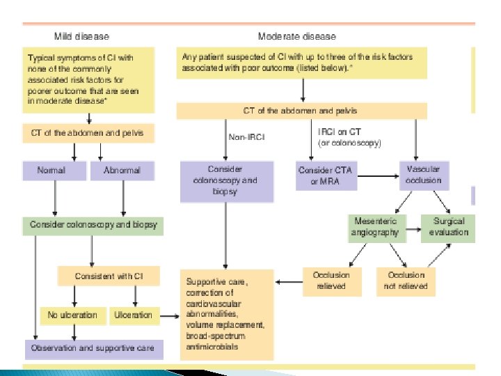
� CT is commonly obtained in the emergency department to assess abdominal pain. � The clinician should consider this modality when patients are classified as having either moderate or severe CI. � CT is useful to exclude serious medical conditions other than CI (e. g. , diverticulitis), can suggest the diagnosis of CI, and reveal which areas of the colon are involved.
� Segmental wall thickening, thumbprinting, and pericolonic fat stranding with or without ascites are signs associated with CI, but are not specific enough to make a definitive diagnosis. � The usual CT findings of colitis are nonspecific and not unique for CI in most patients with abdominal pain.
� “little rose” sign hyperdensity of the mucosa and edema of the submucosa as a sign of “early stage” CI, i. e. , the “wet” appearance. � “Target sign” or “double halo” sign showing different attenuations of the layers of the bowel wall that corresponded to ischemia and reperfusion of the involved segment.
� Further investigation is required to characterize the most common signs of CI from each phase of the ischemic process and course of disease, to describe findings that reliably differentiate CI from other disease entities, and to determine what constellation of findings might be diagnostic of CI. � Adenocarcinoma of the colon is associated with CI and usually the segment of ischemic injury is proximal to the neoplasm. � Tumoral segments can be differentiated from ischemic segments of colon by CT.
� On CT, the segments of CI had significantly less colon thickening (1. 0 vs. 2. 0 cm, P <0. 05) and were longer (10. 1 vs. 5. 9 cm, P <0. 05) than those with malignancy. � In addition, although no statistical comparison was documented, the CI segments appeared to enhance more homogenously(70%), whereas the tumoral segments more frequently enhanced heterogeneously (60%).
� Given the association, although infrequent, between CI and distal malignancy (as well as other potentially obstructing lesions such as strictures and fecalomas), the colon should be screened for cancer with a colonoscopy several weeks following recovery from the ischemic insult, if the patient has not been screened recently. � Screening colonoscopy should not be performed at the time of presentation with CI because a minimum of colon manipulationand distension is recommended to avoid further damage from the reduction in blood flow that occurs with colon insufflation.
� Colonoscopy with biopsy remains the most accurate way to diagnose CI in patients with riskfactors for disease, including vasculitides.
� CI is usually nonocclusive in nature, but CT (or CTA) can be used to identify whether or not vascular occlusions are present. � Unfortunately, the IMA is occluded in up to 10% of asymptomatic patients over 60 years of age and hence the significance of such a finding is uncertain. � With emerging technology, contrast enhanced CT can sometimes assess patency of the celiac, SMA, and IMA without devoted CTA;
� In contrast to AMI in which conventional mesenteric angiography or CTA plays an essential role, vascular imaging studies are not indicated in most patients with suspected CI because by the time of presentation, colon blood flow has usually returned to normal.
� In patients with potentially severe, complicated disease, such as those with IRCI or when the clinician cannot distinguish between AMI and CI (e. g. , in patients with severe abdominal pain and little or no bleeding), a dedicated multiphasic CT is indicated. � In patients with IRCI, CTA should be performed either initially or before discharge, as the colonic episode may be the “heralding” indication of SMA disease and possibly impending AMI.
� Multiphasic CT scanning, including a CT without oral contrast but with intravenous contrast and attention to the angiographic arterial and portal venous phases, has become standard for the diagnosis of AMI. � In any patient in whom the presentation of CI may be a heralding sign of AMI (e. g. , IRCI), and the multiphasic CT is negative for vascular occlusive disease, traditional splanchnic angiography should be considered for further assessment because a negative triple-phase CT does not absolutely exclude vascular obstruction as the cause.
� It is important to note that there are no available published data yet analyzing IRCI as a “heralding” indication of SMA disease. � Select group of patients with IRCI may present again within weeks, months, or even a few years after the heralding presentation of IRCI with catastrophic episodes of AMI, resulting in loss of most of their small bowel or death. �A limited but increasing number of patients with IRCI have undergone angiography and had occluding thrombi found in the SMA; stenting and/or thrombectomy resulted in good outcomes.
� Conversely, some patients with SMA thrombi did not undergo angiography or stenting yet had a positive outcome as well. � Although there are no data to yet support our recommendation, � we believe that the benefit of preventing a potentially catastrophic ischemic insult to the small intestine (AMI) far outweighs the risks of a CTA.
� Pneumoperitoneum, portal venous gas, and pneumatosis are ominous findings in patients suspected of CI. � the presence of pneumatosis and portal venous gas does not necessarily indicate nonviable bowel. � The presence of pneumatosis plus portomesenteric gas showed good specificity (83%) for transmural infarction, but very low sensitivity (17%)
� Patients with CI who also have CT findings of pneumatosis and/or portomesenteric venous gas are likely to have severe disease, but not necessarily with full transmural necrosis. � In the clinical setting of CI, one of these findings is still an indication for prompt operation. � Experience with US in the setting of CI is very limited and it is believed that this technique lacks specificity for bowel wall thickening and has a high false negative rate.
� MRI has been formally studied in only a small number of patients with CI; findings are similar to those of CT, but without the radiation exposure and the need for iodinated contrast agents. � As with CT, such findings usually are not specific enough to make a definitive diagnosis. � MRI may have a role when repeat imaging is required or patients have poor renal function.
� 1 . CT with intravenous and oral contrast should be ordered as the imaging modality of choice for patients with suspected CI, to assess the distribution and phase of colitis (strong recommendation, moderate level of evidence). � 2. The diagnosis of CI can be suggested based on CT findings(e. g. , bowel wall thickening, edema, and thumbprinting)(strong recommendation, moderate evidence). � 3. Multiphasic CT angiography (CTA) should be performed on any patient with suspected IRCI or in any patient in whom the possibility of AMI cannot be excluded (strong recommendation, moderate level of evidence).
� 4 . CT or MRI findings of colonic pneumatosis and portomesenteric venous gas can be used to predict the presence of transmural colonic infarction (strong recommendation, moderate level of evidence). � 5. In a patient in whom the presentation of CI may be a heralding sign of acute mesenteric ischemia (AMI; e. g. , IRCI, severe pain without bleeding, and atrial fibrillation), and the multiphasic CT is negative for vascular occlusive disease, traditional splanchnic angiography should be considered for further assessment (conditional recommendation, low level of evidence).
COLONOSCOPY IN THE DIAGNOSIS OF CI � Colonoscopy offers several advantages over radiologic imaging, including the ability to directly observe the abnormal tissue, to sometimes make a diagnosis just by appearance, and to obtain tissue samples. � The colon single-stripe sign (CSSS) is a highly specific sign of CI described by Zuckerman et al. in 2003. q CSSS is defined as a single inflammatory band of erythema with erosion and/or ulceration along the longitudinal axis of the colon; all CSSSs in the study of Zuckerman et al. were >5 cm in length and 89% of CSSSs were found in the left colon. § This feature had better prognosis than circumferential CI and exhibited a reduced need for surgery (0% vs. 27%) and alesser mortality (4% vs. 41%).
� Colonoscopy enables accurate determination of the anatomic limits of involvement and, therefore, perhaps helps predict prognosis. � It is important to note that the correlation between endoscopic appearance and clinical severity is not absolute and even disease in patients with boggy, purple mucosa may resolve completely without complication. � It is essential to assess the entire clinical picture before making any judgments about the need for surgical intervention.
� colonoscopy enables biopsy specimens to be obtained and, although uncommonly pathognomonic for CI, they often support the diagnosis, but rarely establish etiology. � Risks of colonoscopy are small but must be considered in terms of the pathophysiology of CI.
� Carbon dioxide has been recommended for colonoscopy (and virtual colonography) because it is 10 times more rapidly absorbed from the bowel than is room air, and therefore its use results in a shorter period of distention and a more comfortable examination. � CO 2 increased colonic blood flow in contrast to room air that, at similar pressures, decreased blood flow, thus making CI less likely a complication of colonoscopy when CO 2 rather than room air is used. � CO 2 appears to be the best insufflating agent to use in patients with CI and should be used whenever available.
� Limited colonoscopic evaluation is a safe and useful technique to evaluate patients with CI or suspected of having CI. � CT should be used to characterize the stage of CI and the distribution of disease, but when colonoscopy is performed, the colonoscope should passed only to the distal-most aspect of the affected region. � Biopsies should be taken in all cases, except when there is evidence of gangrene.
� Histopathologic specimens obtained colonoscopically are rarely diagnostic for CI. � Pathognomonic features include infarction and ghost cells, i. e. , preserved individual cellular outlines without cell content, and are infrequently seen � The diagnosis of CI, however, cannot be made conclusively by colonoscopy on just one examination unless mucosal gangrene is observed or infarction or ghost cells are seen at histopathology. � These histopathologic findings are infrequent and of limited value in diagnosing CI.
Recommendations � 1 . Early colonoscopy (within 48 h of presentation) should be performed in suspected CI cases to confirm the diagnosis(strong recommendation, low level of evidence). � 2. When performing colonoscopy on a patient with suspected CI, the colon should be insufflated minimally (conditional recommendation, very low level of evidence). � 3. In patients with severe CI, CT should be used to evaluate the distribution of disease. Limited colonoscopy is appropriate to confirm the nature of the CT abnormality. The endoscopic procedure should be stopped at the distal-most extent of the disease (strong recommendation, low level of evidence).
� 4 . Biopsies of the colonic mucosa should be obtained except in cases of gangrene (strong recommendation, very low level of evidence). � 5. Colonoscopy should not be performed in patients who have signs of acute peritonitis or evidence of irreversible ischemic damage (i. e. , gangrene and pneumatosis) (strong recommendation, very low level of evidence).
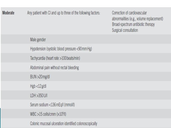
SEVERITY AND TREATMENT OF CI q Various significant predictors of outcome including epidemiologic factors, clinical presentation of disease, vital signs, serologic values, and disease distribution. q Epidemiologic factors § Antimicrobial therapy for CI, § Hepatitis C positivity, § History of cancer, § Male gender, § Warfarin use at the time of diagnosis.
q Presentations of disease § Abdominal pain without rectal bleeding, § Nonbloody diarrhea, § Peritoneal signs on physical examination § Symptom onset after admission q Vital sign § A heart rate of >100 beats per min § Systolic blood pressure of <90 mm Hg at the time of diagnosis
q Serology value § Hgb <12 mg/dl, § hyponatremia (Na <136 m. Eq/l[mmol/l]), § LDH >450 U/l, and § blood urea nitrogen (>28 mg/dl[mmol/l]). q Disease distribution, § A pancolonic distribution of disease and § IRCI also portend a poor outcome
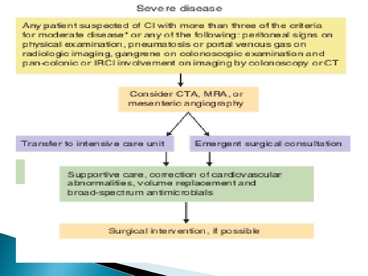
Severe Any patient with CI and more than three of the criteria for moderate disease or any of the following: 1. Peritoneal signs on physical examination 2. Pneumatosis or portal venous gas on radiologic imaging 3. Gangrene on colonoscopic examination 4. Pancolonic distribution or IRCI on imaging or colonoscopy q TREATMENT 1. Emergent surgical consultation (treatment is likely to be surgical) 2. Transfer to intensive care unit 3. Correction of cardiovascular abnormalities (e. g. , volume replacement) 4. Broad-spectrum antibiotic therapy q
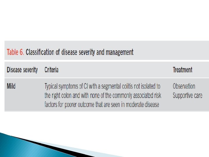
� Treatment of CI varies with the severity of the disease and its presentation. � In general, many patients have a benign, selflimited episode of CI that is neither diagnosed nor treated or is managed in the outpatient setting. � Most cases of CI resolve spontaneously and do not require specific therapy; such patients have reversible ischemic colopathy or transient ischemic colitis.
� Patients with more significant symptoms or findings require hospitalization to enable observation for complications or signs of irreversible disease. � Initial medical management most commonly includes general supportive measures, bowel rest, intravenous hydration, and correction of possible precipitating conditions. � Parenteral nutrition may be indicated if a protracted course is anticipated. � Medical management most commonly included fasting, intravenous fluids, parenteral nutrition, antibiotics, and heparin prophylaxis.
� Indication and timing for antimicrobial use in CI remains untested. � Antibiotics are believed to improve outcome in CI by reducing inflammatory responses stimulated by the normal fecal microbiome, reducing the antigens that prompt innate immune response and minimizing bacterial translocation through compromised colonic mucosa. � This guideline recommends antimicrobial therapy for CI patients who have either “moderate” or “severe”disease.
� Possible antimicrobial regimens include an antianaerobic agent plus a fluoroquinolone or an aminoglycoside or a third-generation cephalosporin. � This guideline recommends that antimicrobials be continued for at least 72 h, at which time the patient’s clinical status should be reevaluated. � If the patient has not clinically improved, one should consider consultation with an infectious disease expert to help define the antimicrobial regimen. � If the patient is symptomatically improved after 72 h, a 7 -day course of therapy should be considered.
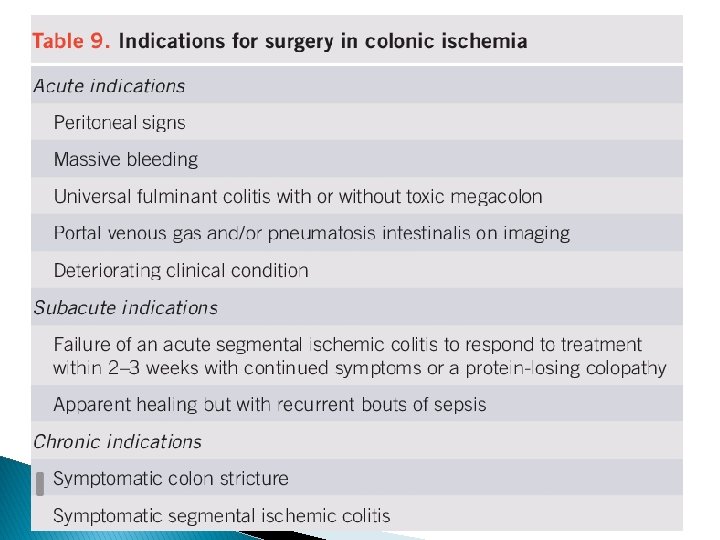
� Glucocorticoids are not recommended for CI, except when it is a complication of a vasculitis. � In such cases, the steroids are used to treat the vasculitis and not the CI. � Surgical intervention for patients with CI is required in its most severe presentations. � There are several indications for surgery in acute CI, but the most common is the presence of signs of necrotic bowel.
� Without surgical intervention, mortality from necrotic bowel approaches 100%. � A less-recognized indication for surgery is the development of recurrent sepsis in a patient who has symptomatically recovered from an acute episode of CI. � Such patients usually have a short segment of unhealed bowel(resection of this segment is curative). � Mortality after surgical intervention for CI is high, ranging from 37 to 47%.