
Diabetic Ketoacidosis Overview o o o Epidemiology presentation
Diabetic Ketoacidosis
Overview o o o Epidemiology, presentation, & diagnosis of type 1 diabetes mellitus (DM) in children and adolescents Clinical features & diagnosis of DKA in children Treatment and complications of DKA in children
o Special situations in children and adolescents with type 1 DM
DKA Epidemiology, presentation, & diagnosis of type 1 DM
Introduction o Type 1 DM n n n one of the most common chronic diseases in childhood insulin deficiency following destruction of the insulin-producing pancreatic beta cells commonly presents in childhood but ¼ of cases are diagnosed in adults
Epidemiology o o Approx. 2/3 of new diagnoses of diabetes in patients 19 years of age Incidence : n n geography, age, gender, and family history environmental factors
Geographical variation o o o Risk appears to rise as the geographical latitude increases Move from low to high incidence, increase risk of developing type 1 diabetes Finland Sardinia vs. Venezuela & China n n 37 to 45 per 100, 000 children younger than the age of 15 years rates that are almost 400 times higher
o o United States - 15 to 17 / 100, 000 Canada – n n Newfoundland - 36 per 100, 000 Quebec - 15 per 100, 000
o o Incidence of childhood type 1 disease is rising worldwide primarily in young children Cause?
Age and gender o Bimodal distribution n n o o o 4 to 6 y/o early puberty - 10 to 14 y/o Gender? Autoimmune – female but not DM Select pop. n n 3: 2 Male vs. female European descent
Genetic susceptibility o Risk is significantly increased in close relatives of a pt with type 1 diabetes n n n n No family history — 0. 4 % Offspring of an affected mother — 2 to 4 % Offspring of an affected father — 5 to 8 % Offspring with both parents affected — as high as 30 % Non-twin sibling of affected patient — 5 % Dizygotic twin — 8 % Monozygotic twin — 50 % lifetime risk, 30 % within 10 years of diagnosis of the first twin
Environmental factors o o o Genetically susceptible individuals Exposure to one or more environmental agents appears to trigger an immune response Identification of environmental factors n n better understanding of the pathogenesis of the disease developing strategies to prevent type 1 diabetes
o o o Viral infections Immunizations Diet n especially exposure of cow's milk at an early age Perinatal factors n maternal age, history of preeclampsia, and neonatal jaundice n Low birth weight decreased the risk of developing type 1 diabetes None of above associations have been verified Many have been contradicted by other studies
o Seasonal variation n n Colder climate has higher risk compared to warmer months Another study – NO seasonal variation in girls Higher incidence in summer months for boys
CLINICAL PRESENTATION o Classic new onset - most common DKA o Silent incidental discovery o
Classic new onset o o Hyperglycemia without acidosis is the most common presentation Symptoms: n n Polyuria & Polydipsia (70%) Weight loss (34%)
Polyuria o o Serum glucose concentration > 180 mg/d. L Exceeding renal threshold for glucose Osmotic diuresis & hypovolemia Presentation: n n Nocturia, bedwetting, or daytime incontinence Not toilet trained: increased frequency of wet diapers and/or unusually heavy (wet)
Polydipsia Hyperglycemia and hypovolemia increased serum osmolality enhanced thirst o
Weight loss o o hypovolemia & increased catabolism Insulin deficiency n n impairs peripheral glucose utilization in skeletal muscle increases fat and muscle breakdown. initially, increased appetite is seen over time, become anorexic, contributing to weight loss
o o Careful Hx is key Classic symptoms of polyuria and polydipsia are often not the initial complaints May initially appear slightly ill and with vague complaints, such as weight loss and lethargy Ask for nocturia and bedwetting, increased frequency and/or unusually wet diapers, and persistent thirst

Visual disturbances o o Changes in refractive index Common due to alterations in the osmotic mileau of the lens Lesser extent - aqueous and vitreous humors Cataract - longstanding hyperglycemia
DKA o o o Similar symptoms but usually more severe Reported frequency as the initial presentation : 15 to 67 % More likely in n n Children who are young (<6 years of age) Low socioeconomic background
o Require : n n n hospitalization, rehydration, and insulin replacement therapy
Silent presentation o o o Least common Typically occurs in children who have another close family member with type 1 diabetes and are being closely monitored Diagnosis is made based upon an elevated blood glucose before symptoms
Frequency of clinical findings o o Polyuria, polydipsia (70%) Weight loss (34 %) Lethargy (14%) DKA n n 25 % of all patients 2/3 of children < 2 years of age
o Age-dependent findings n n enuresis and nocturia (13 & 7 % of children >2 years of age constipation (8 % of patients < 5 years of age first-degree relative - 10% first or second-degree relative - 27%
Young children o o o <6 years of age More likely to present with DKA due to less suspicion More vulnerable due to less ablility to compensate for pathologic processes n n obtaining fluids increasing fluid intake
o o Even more difficult for those <2 years of age Diaper-clad children - unable to communicate their thirst and need for fluid replenishment (Polyuria and polydipsia)
Increasing suspicion o o History or presence of prolonged candidal infection usually of the diaper area Presents for evaluation of dehydration, abdominal pain, or fatigue n consider measuring serum glucose and testing for glycosuria
Diagnosis o o o Fasting plasma glucose 126 mg/d. L on two occasions Symptoms of hyperglycemia and a random venous plasma glucose 200 mg/d. L Abnormal oral glucose tolerance test (OGTT) defined as a plasma glucose 200 mg/d. L measured two hours after a glucose load of 1. 75 g/kg (maximum dose of 75 g)
o Glycosuria : suggestive of diabetes, but not diagnostic n n o renal glucosuria or Fanconi syndrome glycosuria but normoglycemic Glycosylated hemoglobin (A 1 C) n n n 10 to 12 weeks prior measures the % of Hgb bound to glucose in a non-enzymatic reaction Values >6. 5 percent suggest the presence of diabetes, however, levels <6. 5 percent do not exclude diabetes
Type 1 Vs Type 2 DM o Differentiation based upon clinical presentation, history, and laboratory studies
o Body habitus — n n o type 2 diabetes - overweight with body mass index >85 th percentile for age and gender type 1 diabetes - not overweight and often have a recent history of weight loss. Age — n n type 2 diabetes - onset of puberty type 1 disease – at an earlier age and bimodal
o Insulin resistance — n n o type 2 diabetes can have signs and/or symptoms of insulin resistance acanthosis nigricans, and also hypertension, dyslipidemia, and polycystic ovary syndrome Family history — Patients with either type 1 or type 2 diabetes can have an affected close relative
o o Type 1 A diabetes - Insulin deficiency as a result of autoimmune destruction of pancreatic beta cells Type 1 B diabetes – Rare n n n clinical features of type 1 diabetes no circulating autoantibodies no evidence of autoimmune beta-cell destruction and no other cause has been identified
Other causes of diabetes o Diseases of the exocrine system — n o Cystic fibrosis, hereditary hemochromatosis, and chronic pancreatitis Endocrine abnormalities in glucose regulation — n Cushing's syndrome, growth-hormone excess, glucagon-secreting tumors, catecholamine excess in pheochromocytoma
o Drug-induced diabetes — n n glucocorticosteroids, HIV protease inhibitors, cyclosporine, L-asparaginase, and tacrolimus impair glucose tolerance by inhibiting insulin secretion, increasing hepatic glucose production, or causing insulin resistance

DKA Clinical features & diagnosis
DEFINITION o Hyperglycemia n blood glucose of >200 mg/d. L (11 mmol/L) AND o Metabolic acidosis n venous p. H <7. 30 and/or plasma bicarbonate <15 meq/L
o o o Hyperketosis (concentration of total ketone bodies >5 mmol/L) Hyperosmolality The clinical manifestations of DKA are related to the degree of hyperosmolality, volume depletion, and acidosis
Initial presentation of type 1 DM o o Increased risk in Children who are young (<6 years of age) n n o newly diagnosed children with type 1 DM, 26 % presented in DKA incidence of DKA was higher (36 %) in children < five years of age Low socioeconomic background
In established type 1 DM o o o Higher A 1 C levels and higher reported insulin requirements Female adolescents, highest risk > 13 years of age Children > 13 years of age, regardless of gender n o who are underinsured and/or have a history of psychiatric disorders Longer duration of DM
Type 2 diabetes mellitus o Ketosis and DKA can occur in children with type 2 DM n o o o particularly in African-American children 13 % had type 2 DM At presentation, there was no difference in the serum p. H level Type 2 DM had higher blood glucose levels
PRECIPITATING FACTORS o Recurrent episodes n n o Stress n n o underlying poor metabolic control frequently missed insulin injections increases the secretion of cortisol, catecholamines, and glucagon Example: infection preceding DKA Medications
DKA Treatment and complications of DKA in children
TREATMENT o o The approach and principles of management are the same for all children with DKA regardless of the severity However, based upon the severity of presentation, the clinician can ascertain the appropriate clinical setting and needs of the child
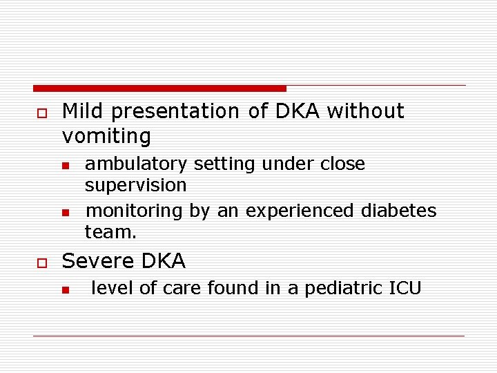
o Mild presentation of DKA without vomiting n n o ambulatory setting under close supervision monitoring by an experienced diabetes team. Severe DKA n level of care found in a pediatric ICU
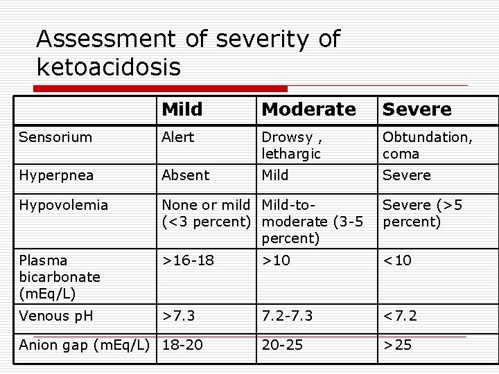
Assessment of severity of ketoacidosis Mild Moderate Severe Sensorium Alert Drowsy , lethargic Obtundation, coma Hyperpnea Absent Mild Severe Hypovolemia None or mild Mild-to(<3 percent) moderate (3 -5 percent) Severe (>5 percent) Plasma bicarbonate (m. Eq/L) >16 -18 >10 <10 Venous p. H >7. 3 7. 2 -7. 3 <7. 2 20 -25 >25 Anion gap (m. Eq/L) 18 -20
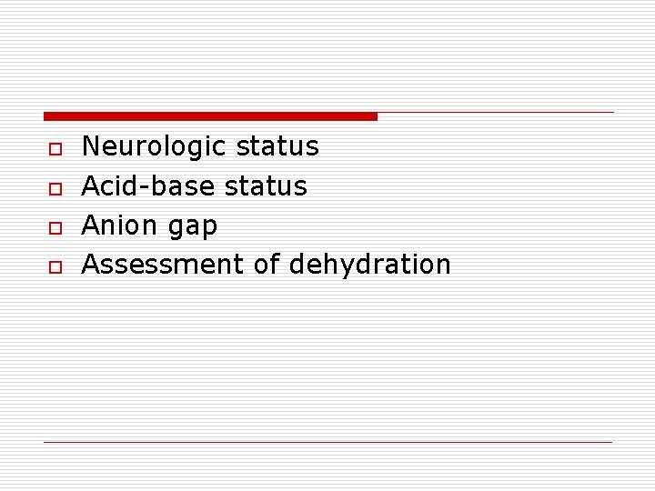
o o Neurologic status Acid-base status Anion gap Assessment of dehydration
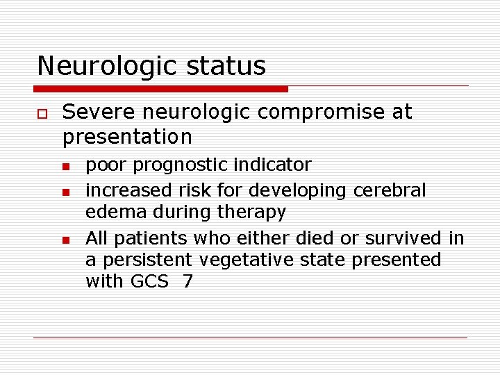
Neurologic status o Severe neurologic compromise at presentation n poor prognostic indicator increased risk for developing cerebral edema during therapy All patients who either died or survived in a persistent vegetative state presented with GCS 7
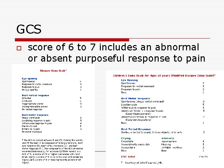
GCS o score of 6 to 7 includes an abnormal or absent purposeful response to pain
Acid-base status o Severity of the acidosis directly reflected by: n n o venous p. H serum [bicarbonate] Respiratory rate also may be helpful
Anion gap o o Another measure of the severity High anion gap n decreased renal perfusion, which limits ketoacid excretion
Moderate and severe DKA o Underlying principles of management n n administer insulin correct the fluid and electrolyte abnormalities in o o o n hyperosmolality, metabolic acidosis, hypovolemia, potassium, and phosphate depletion? During initial therapy, carefully monitored for signs of cerebral edema
Fluid and electrolyte deficits o o o Water — 100 to 125 m. L/kg Sodium — 5 to 13 meq/kg Potassium — 6 to 7 meq/kg
Assessment of dehydration o o o Difficult to clinically assess the degree of dehydration Initial fluid management based upon the of a 7 to 10 % fluid deficit However, treatment should be initiated with a maximal volume of 10 m. L/kg of isotonic solution, unless the patient is hypotensive
o Many of the clinical findings are unusable n n condition of the patient's oropharynx - almost always dry because of hyperventilation and mouth breathing skin turgor is also frequently unreliable because the fluid losses are from both the EC and IC spaces.
o Degree of EC fluid loss is in part masked n n hyperglycemia results in a shift of water from the intracellular to the extracellular fluid compartment effect is rapidly reversed with insulin therapy
o Elevated urine specific gravity n o glucose and, to a lesser degree, ketones raise the specific gravity Elevations in the BUN and Hct n n confirm hypovolemia to follow DKA correction
Fluid repletion o Extracellular volume depletion n n o Glucose osmotic diuresis urinary losses GI losses from vomiting and/or diarrhea Hypovolemic shock is a rare occurrence in DKA and should be evaluated for other causes of shock
Goals o To restore n n o effective circulating volume by replacing sodium and water loss GFR to enhance clearance of ketones and glucose from the blood To avoid cerebral edema
Initial volume expansion o Isotonic solution n o Ringer's lactate n o preferably Ringer's lactate or isotonic saline provides physiologic concentrations of K+ and includes lactate that will be metabolized to bicarbonate The administration of large amounts of isotonic saline can increase the severity of the hyperchloremic metabolic acidosis that
Volume & rate of volume expansion o o o Dependent on effective circulating volume 10 m. L/kg over one hour Additional infusion of 10 m. L/kg can be given over the next hour n If and only if the effective circulating volume is still compromised
o o o Once hemodynamically stable, subsequent volume expansion given more slowly to minimize the risk of cerebral edema. Continued administration of Ringer's lactate, isotonic saline, or one-half isotonic saline Potassium salts are added (since potassium is as osmotically active as sodium).
o Three-quarters isotonic solution n n widely used in children with Reye's syndrome and cerebral edema consists of ½ isotonic saline + 20 meq/L of potassium acetate + 20 meq/L of potassium phosphate (total of 40 meq/L of potassium)
o o Rate calculated to fully replete the patient over 48 hours First 24 hours n n n < 1. 5 to 2 times the maintenance fluid. Based upon BSA, the volume < 2500 m. L/m 2 for 24 hours includes the initial fluid bolus as well as any oral intake
o o Urinary losses should not be added to the calculated total volume of fluids Higher rates of fluid administration have been associated with cerebral edema
Insulin o o Concurrent with volume expansion, an initial insulin infusion rate of 0. 05 to 0. 1 unit/kg per hour Lower dose should be used initially in younger children who are more sensitive to insulin
o Within 60 minutes, steady state serum insulin levels are achieved (100 to 200 mc. U/m. L), n n which offset insulin resistance, suppress glucose and ketone production, stimulate peripheral glucose and ketone metabolism [23, 24]. In addition, volume expansion will lower the serum glucose by dilution
o o The insulin infusion should continue until Venous p. H is >7. 3 and/or the serum HCO 3 is >15 meq/L Then changed to subcutaneous insulin,

o o Almost all patients with DKA develop a normal anion gap acidosis during insulin therapy Normalization of the anion gap is indication for stopping the insulin infusion n If the serum glucose is controlled
o o Hyperglycemia usually corrects before the ketoacidosis Saline infusion D 5 S or 5 percent dextrose in LR when the serum [glucose] < 250 to 300 mg/d. L When < 200 mg/d. L [dextrose] in the IV solution should be increased to 7. 5 or 10% Allows continued administration of insulin to correct the residual ketoacidosis
Pitfall o o o Following the glucose concentration as a measure of improvement of DKA Insulin infusion rate only be reduced when the ketoacidosis is corrected If ketoacidosis persists n n n reassessed for possible mechanisms such as insulin resistance due to infection decreased delivery due to adhesion of insulin to tubing

Anion Gap o o Correction of the ketoacidosis Normalization of the anion gap is manifested by Persistent acidosis without an elevation in anion gap is NOT an indication for continuing the insulin infusion if the serum glucose is controlled
Switch to SC Insulin o 1 st subcutaneous injection should be given at an appropriate interval to allow for absorption prior to stopping insulin infusion n n o rapid-acting insulin (ie, insulin lispro) is approx. 15 minutes regular insulin is 30 to 60 minutes As initial therapy inconsistent absorption n Combination of volume depletion and secondary sympathetic activation decrease local perfusion
Serum sodium Hyperglycemia in uncontrolled DM variable effect on serum [sodium] concentration o Can both lower and raise the measured value. 1. Lowering by dilution – hyperglycemia, by raising the plasma osmolality, results in osmotic water movement out of the cells 2. Glucosuria-induced osmotic diuresis – counteracts above direct effect of hyperglycemia n results in water loss in excess of sodium and potassium raise the serum [sodium] and plasma osmolality n unless there is increase in water intake o
Pseudohyponatremia o Pseudohyponatremia n n hyperlipidemia, will falsely lower the serum sodium since each liter of plasma contains less water and therefore less sodium laboratory artifact
Corrected Na o o “Corrected" serum sodium (Na) concentration Serum [sodium] that should be present if the serum [glucose] were lowered to normal with insulin alone
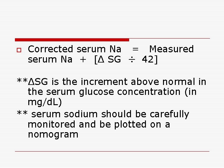
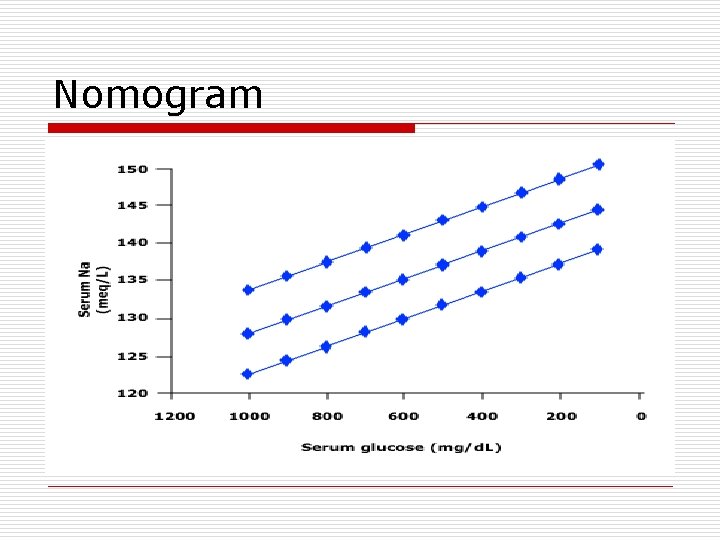
o Corrected serum Na = Measured serum Na + [Δ SG ÷ 42] **ΔSG is the increment above normal in the serum glucose concentration (in mg/d. L) ** serum sodium should be carefully monitored and be plotted on a nomogram
Nomogram
o Risk for cerebral edema n n If doesn’t rise appropriately If the rise falls off the nomogram isobar, o o [sodium] should be increased eg, switching from one-half isotonic to isotonic saline
Serum K o Potassium depletion n o Hyperkalemia n n o Renal and/or Gi losses insulin deficiency - impairs potassium entry into the cells Hyperosmolality - pulls water and potassium out of the cells Ketoacidosis - little effect on transcellular potassium movement
[K] o o o At the time of presentation can be normal, increased, or decreased Regardless, insulin therapy drives potassium into cells Thus, potassium replacement will almost always be required within one to two hours in children without renal failure
Optimal therapy? o o Varies with the initial serum potassium Serum [potassium] should be carefully monitored EKG monitoring in either hypokalemia or hyperkalemia Hypokalemic - K+ needs to start immediately n o o using a [potassium] of 60 to 80 meq/L Normokalemic – K+ at start of insulin therapy (eg, adding 40 meq/L of potassium) Hyperkalemic - K+ when levels normalize
Metabolic acidosis o Insulin promotes n n n metabolism of ketoacid anions, resulting in the generation of bicarbonate stops the ongoing production of new ketoacids improved tissue perfusion corrects any lactic acidosis that might be present
Regeneration of bicarbonate o o Beta-hydroxybutyrate - "potential bicarbonate" Insulin therapy n n o o eliminates the excess ketoacid anions and normalizes the anion gap regenerates bicarbonate from those ketoacid anions that have not been excreted in the urine Urinary loss of these anions represents the loss of potential bicarbonate As a result persistent metabolic acidosis even after anion gap normalized
o o Ketonemia and ketonuria may persist > 36 hours due to the slower removal of acetone (in part via the lungs) However, acetone is biochemically neutral and has no effect on acidbase balance
Bicarbonate therapy o o o Should not be used in DKA Unable to demonstrate any clinical benefit Potential risks
RISK of bicarbonate therapy o Excessive bicarbonate therapy to a near normal p. H n n o rapid rise in PCO 2 (since there is less of an acidemic stimulus to hyperventilation) paradoxical fall in cerebral p. H as lipid-soluble CO 2 rapidly crosses the BBB May slow the rate of recovery of ketosis n Studies in animals suggested that bicarbonate therapy acts by increasing hepatic ketogenesis
o Post-treatment metabolic alkalosis after insulin-induced metabolism of ketoacid anions n o o o regeneration of bicarbonate and spontaneous correction of most of the metabolic acidosi. Risk factor for cerebral edema Hypokalemia Additional sodium load hyperosmolality
When to use? o o Bicarbonate during complications such as cardiac arrest Severe acidemia (arterial p. H <6. 90) n o decreased cardiac contractility and vasodilatation can further impair tissue perfusion Potentially life-threatening hyperkalemia n will drive potassium into the cells
Phosphate o o o Cellular phosphate depletion is a common problem Normal or elevated Phosphate depletion is rapidly unmasked following the institution of insulin therapy
Use or not to use? o Has not been shown to affect tissue oxygenation n o o 2, 3 DPG Prospective studies of phosphate replacement have failed to show clinical benefit May induce hypocalcemia
o o Has been used for concurrent potassium replacement in children with DKA Careful monitoring of the serum calcium is required
Monitoring o Close monitoring for changes n n vital signs, neurologic status, fluid status, and metabolic state (eg, serum electrolytes, glucose, and urea nitrogen, hematocrit, and venous p. H)
o Appropriate documentation n n o easy to follow able to detect clinical changes that require medical attention Flow sheet of laboratory values and clinical parameters allows better visualization and evaluation of the clinical picture
o o o Emphasis should be placed on the hourly evaluation fluid status, changes in osmolality, glucose, electrolytes, and changes in the patient's mental and neurologic status
Hourly monitoring o Blood glucose to avoid hypoglycemia n o In F/S should initially be compared to a venous measurement Electrolytes and venous p. H – n n n Q 1 h for first 3 ~ 4 hours then Q 2 h every two hours When clinically appropriate, frequency can be reduced to every Q 4~Q 6 h
o Clinical parameters n n o Continous HR, RR, BP, oxygen saturation, and neurologic status Severe DKA or altered mental status, frequent neurologic examinations are recommended Warning signs and symptoms of cerebral edema n headache, inappropriate decrease in heart rate, recurrence of vomiting, changes in neurologic status, rising blood pressure, and decreased oxygen saturation
o o ECG monitoring - for evidence of changes characteristic of hypokalemia or hyperkalemia Accurate measurement of I/O n neurologically impaired or it is difficult to ascertain urine output, a urine catheter should be placed
Mild DKA o May be managed in an ambulatory setting n n n o Venous p. H >7. 30, Serum bicarbonate >16 meq/L, No neurologic impairment, and estimated volume deficit less than 3 percent Not vomiting However, children at risk for cerebral edema (eg, those less than five years of age or at initial presentation of diabetes) should be hospitalized
o Rehydralate (75 meq/L sodium and 20 meq/L potassium) can be used alone or in combination with initial parenteral fluid repletion n o o dextrose 2. 5 g/d. L. Short-acting insulin (eg, recombinant modified human insulin) can be given SC and repeated at 3 to 6 hour intervals as needed Dose adjusted depending upon the response
o o Close monitoring of serum electrolytes, glucose, and fluid balance is required The patient should be transferred to an appropriate inpatient setting if there is an inadequate response or the clinical condition deteriorates
DKA COMPLICATIONS AND MORTALITY
o o Reported mortality rates for DKA are consistent in developed countries 0. 15 ~ 0. 51 percent in Canada, U. K. , and the U. S. Cerebral edema - 60 to 90 percent Other causes include n aspiration pneumonia, multiple organ failure, gastric perforation, and traumatic hydrothorax
Cerebral edema o As a complication of therapy n o 0. 4 to 1 percent of patients with DKA Subclinical brain swelling, as detected by CT scan, has been reported
Ventricular width o o o Intercaudate width of the frontal horns of the lateral ventricles was measured by MRI Were significantly smaller during treatment then recovery More likely to have mental status changes
o o o Cerebral expansion occurs commonly Are associated with mental status changes (eg, drowsiness and lethargy) Remains to be determined n n True cerebral edema Potential common finding in any child with severe metabolic disturbance, and dehydration
Pathophysiology o o Generally believed related to our management of DKA Numerous factors implicated but none has been proven n n o May be present before treatment More commonly occurs 4 to 12 hours after the initiation of therapy Thus, therapy may exacerbate but not initiate the pathologic processes
Mechanisms o Vasogenic edema — n n Damage of the brain vascular endothelium breakdown of BBB resulting in interstitial brain edema MRI - there was an increase in water diffusion during the treatment phase
o Osmotic edema during therapy n n o Dilute fluids hyponatremic despite correction of the hyperglycemia water movement from the extracellular fluid into the intracellular compartment Cytotoxic edema — n n Damage to brain cells, particularly astrocytes, cause cellular swelling Brain cell injury can result from ischemic and/or metabolic stresses
Risk factors o o Younger children Children with newly diagnosed diabetes Failure of the serum sodium to rise as predicted following Increased BUN at presentation - represent a greater degree of hypovolemia
o o o The severity of acidosis at presentation - not important in an animal model Bicarbonate therapy After adjusting for the severity of acidosis, a lower initial partial pressure of arterial carbon dioxide
Association with brain edema o o The rate of decrease in plasma glucose concentration The rate of fluid delivery The rate of insulin delivery The initial glucose and sodium concentrations
Signs and symptoms o o Onset of headache is usually the earliest symptom Reduced level of consciousness IICP: hypertension, bradycardia, pupil abnormalities, and decerebrate or decorticate posturing By the time these symptoms develop, cerebral edema is already in an advanced stage
o o Although early diagnosis is important, children with severe DKA may have an altered mental status that can be due to other factors: degree of dehydration, metabolic derangements, and sleep deprivation
Treatment o o o Careful monitoring for changes in mental or neurologic status Reduce rate of fluid administration Mannitol, IV n n n 0. 25 to 1. 0 g/kg over 20 minutes (case report) retrospective study - mannitol did not appear to have either a beneficial or detrimental effect [61] repeated in two hours, if there is no initial response

SICK-DAY MANAGEMENT o o Acute illness varying effects upon insulin requirements. Increased insulin requirements n n o Stress increase in counter-regulatory hormones epinephrine and cortisol, raise peripheral insulin resistance Decreased insulin requirements n reduced oral intake of carbohydrates because of decreased appetite, nausea, or vomiting.
o Thus, a child with type 1 diabetes during an acute illness can develop: n n Hypoglycemia Significant hyperglycemia DKA due to uncontrolled hyperglycemia Ketosis, independent of hyperglycemia
Outside intervention o o Parental involvement is imperative Telephone management with the diabetes care team is helpful
Management o o Increased frequency of blood glucose monitoring (eg, every two to three hours) Checking for urinary ketones with each void, regardless of blood glucose concentration n poor oral intake - ketone formation can occur without hyperglycemia
In hyperglycemia o o o Additional doses of rapid- or veryrapid-acting insulin Insulin at a dose of 0. 05 to 0. 1 units/kg/Q 2 h~Q 4 h based upon blood sugar 0. 1 unit/kg for moderate to large ketones
In decrease intake o o In decrease intake - reduction of insulin dose to avoid hypoglycemia Conventional Tx: n n n fixed schedule intermediate acting-insulin (NPH) daily dose(s) of the intermediate-acting insulin can be reduced by 30 to 50%
Conventional vs. intesive therapy o Under intensive therapy with basal dose (eg, insulin glargine or insulin pump) n o basal dose can be continued at the usual rate or slightly decreased by 10 to 20 % Insulin should NOT be discontinued DKA n insulin requirements, although decreased, are not completely eliminated
o If the usual dose of basal insulin proves too high n n dosing has been incorrect for the child is compensating for food intake without bolus dosing
o With either regimen, increased blood glucose can be treated with rapid or very-rapid-acting insulin
o Poor oral intake ketonuria with hypoglycemia n o o treated with an increased intake of carbohydrate -containing fluids and insulin as necessary Inability to eat and have hypoglycemia Ice cold, sugar-containing liquids and/or glucagon in very low doses (0. 1 -0. 2 mg SC/ every hr for up to two hours)
o o o Evaluate at appropriate medical facility if Persistent vomiting or home therapy cannot correct hypoglycemia Oral hydration - carbohydrates + electrolytes
Other circumstances o o o MEDICAL PROCEDURES TRAVEL SCHOOL AND DAYCARE



DKA Pathogenesis of DKA and NKH
- Slides: 134