ACG Clinical Guideline The Diagnosis and Management of


 or extrahepatic CCA.")


 Ia and III are also at an increased risk")


 is the transformation of normal hepatic")


- Slides: 132
ACG Clinical Guideline: The Diagnosis and Management of Focal Liver Lesions Jorge A. Marrero , MD 1 Joseph Ahn , MD, FACG 2 K. Rajender Reddy , MD, FACG 3 19 August 2014
Dr gavidel � Journal club�
� FLLs are solid or cystic masses or areas of tissue that are identified as an abnormal part of the liver. � Because of the widespread clinical use of imaging modalities such as US, CT, and MRI, previously unsuspected liver lesions are increasingly being discovered in otherwise asymptomatic patients.
�Importantly, the evaluation of liver lesions has taken on greater importance because of the increasing incidence of primary hepatic malignancies, especially hepatocellular carcinoma (HCC) and cholangiocarcinoma (CCA).
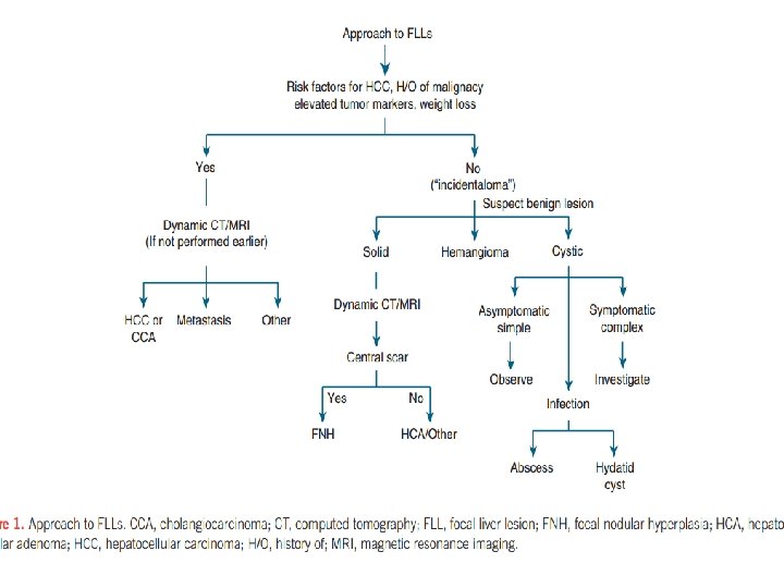
Diagnosis of a liver lesion � The critical components of evaluating an FLL are : � detailed history , physical exam, radiological tests, and pathology. �A history of OCP use in the absence of underlying liver disease suggests a diagnosis of hepatocellular adenoma (HCA), whereas an FLL in the setting of chronic liver disease and portal hypertension should lead to a high suspicion of a diagnosis of HCC.
A radiological test is the most important aspect in the evaluation of a liver lesion.
us � Although US is usually the first imaging test obtained because of its safety and low cost. � It lacks the performance characteristics that CT and MRI have to diagnose and characterize hepatic lesions � Contrast-enhanced US is an emerging modality that has some utility but is not widely available in the United States. � Therefore, we will focus our attention on CT and MRI scans in this guideline.
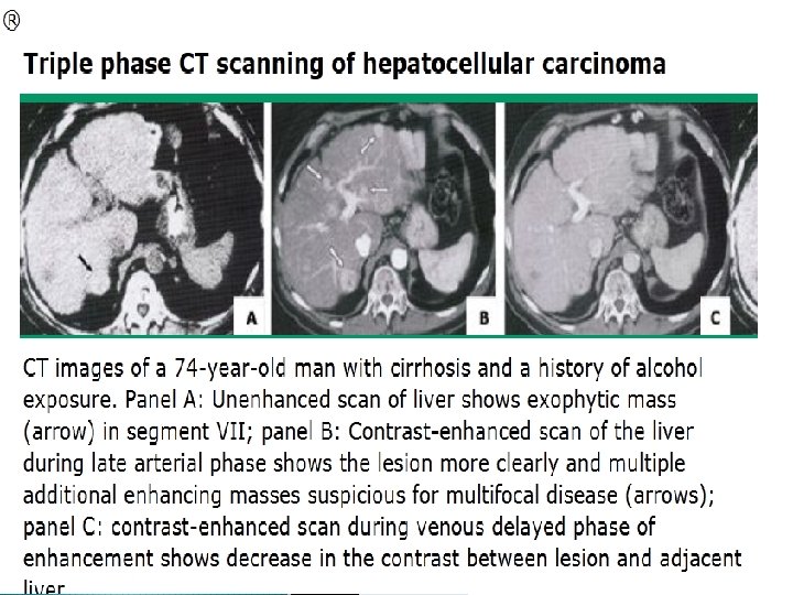
q The most important aspect is the need for : � late arterial phase � portal venous phase � delayed venous phase. � In the context of a CT scan, this is referred to as a “triple-phase” study, distinct from a standard abdominal CT that includes only a portal venous phase and a delayed phase. .
� The evolution of CT and MRI technology has improved their diagnostic capability to often permit making an accurate diagnosis without the requirement for a liver biopsy. � HCC can be diagnosed with ≥ 90 % accuracy with imaging alone when a lesion is 2 cm, thus obviating the need for liver biopsy in nearly all cases under the right clinical circumstances.
� However, if the diagnosis cannot be reliably made radiologically, a biopsy should be performed. � Pathological examination is extremely accurate in making a diagnosis in a patient with an FLL. �A well-sampled biopsy specimen has greater diagnostic accuracy and provides more tissue for ancillary testing (i. e. , immuno histochemistry) when compared with fine-needle aspiration. � A consensus conference on pathology for hepatobiliary malignancies also recommended core biopsies over fine-needle aspiration as it allows for the assessment of both architectural and cytological features
Hepatocellular carcinoma q. Epidemiology � third most common tumor worldwide and the second leading cause of cancer-related deaths. � The incidence of HCC has been rising with chronic hepatitis C virus infection as the main driving force behind this increase
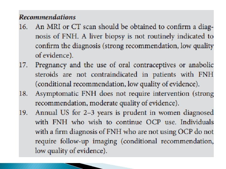
HCC The age-specific incidence rate of HCC starts increasing in the mid 40 s, with those under 60 years of age having the largest increase in incidence, suggesting that this increasing incidence will continue for another 10 to 20 years. � The overall 5 -year survival of patients with HCC is ~ 15 % , indicating its generally poor prognosis. � � � However, 40 % of patients who are diagnosed with disease localized to the liver have improved 5 -year survival rates of 30 %. This indicates that early detection and accurate diagnosis of HCC localized to the liver may improve overall outcomes.
HCC q Risk factors for HCC � In Japan, Europe, and the United States: 1. ~ 60 % of HCV infection 2. 20 % are attributed to HBV infection 3. 20 % to cryptogenic and alcoholic liver disease. In high HBV prevalence areas such as East Asia, China, and Africa � resulting in nearly 80 % of patients with HCC having underlying chronic HBV infection. �
� Cirrhosis HCC. is the most important risk factor for � More than 80 % of the cases of HCC occur in the setting of cirrhosis. � The risk for HCC in individuals with HBV increases from asymptomatic inactive carriers and those with chronic hepatitis without cirrhosis, all with an incidence < 1 per 100 person-years, to 2. 2 – 4. 3 per 100 person-years in cirrhotics.
HCC �~ 20 % of patients with HCC in the setting of HBV infection present without evidence of cirrhosis. q Risk factors for the development of HBV associated HCC include: 1. viral factors such as a high degree of viral replication, 2. viral genotype 3. use of alcohol 4. tobacco.
HCC � The risk for HCC among patients with chronic hepatitis C virus infection occurs almost exclusively in the setting of cirrhosis. Among patients with cirrhosis factors associated with an increase in the risk for the development of HCC are: � alcohol � Tobacco � Obesity � diabetes � older age � male gender �
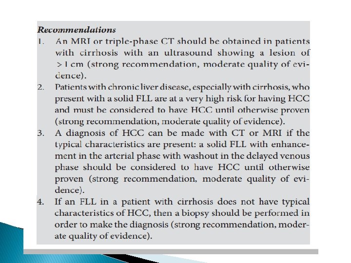
Diagnosis of HCC A CT or MRI should be performed in cirrhotics with: � an ultrasound showing a lesion of > 1 cm � an elevated or rising alpha-fetoprotein in the absence of a liver lesion on US � when there is a clinical suspicion for the presence of HCC. �
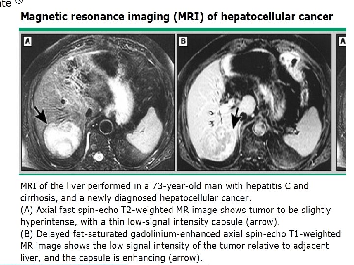
� The choice of MRI versus CT is controversial despite several studies. � These studies have shown that dynamic MRI has a slightly better performance than CT for the diagnosis of HCC. � One should utilize the locally available expertise, whether MRI or CT. �An essential characteristic of HCC is that it is an arterially hypervascular tumor.
� However, only using hypervascularity on the arterial phase as the sole criterion for the diagnosis of HCC has poor specificity � During the portal venous phase of a patient with HCC, the previously arterially enhanced mass lacks contrast and appears hypodense compared with the rest of the liver that is now enhanced in the portal venous phase, a term labeled “washout. ” � These characteristic findings of arterial hyperenhancement with “ washout ” in the portal venous or delayed phase are highly specific and sensitive for a diagnosis of HCC. � These criteria have been validated and accepted in the guidelines for the diagnosis of HCC
� It has been estimated that ~ 85 % of patients with HCC have arterial enhancement and washout. � In those who do not have these characteristic features on radiological examination, a directed biopsy of the mass may be needed in order to confirm a diagnosis of HCC.
� This strategy of biopsying atypical lesions on imaging has been validated. � The decision to proceed with a directed biopsy must be made weighing the risk of tumor seeding and potential bleeding versus the impact of the results on the patient ’ s treatment options and prognosis. � The use of the coaxial biopsy technique, in which the actual needle is introduced percutaneously into the tumor inside a sheath, can mitigate this risk of tumor seeding by insulating the needle inside the sheath.
CHOLANGIOCARCINOMA � CCA can be divided further into intrahepatic CCA (ICCA) or extrahepatic CCA. � This guideline will focus on ICCA. � The incidence of ICCA has been shown to be increasing � The 1 -year survival for ICCA remains low at 27. 6 % and the 5 -year survival is < 10% because of the diagnosis often being made at advanced stages.
Risk factors for CCA � patients with PSC have a risk of developing CCA at ~ 1. 5 % per year after diagnosis. � Among patients with PSC who progress to CCA, ~ 30 % are diagnosed with CCA within 2 years after the initial diagnosis of PSC. � CCA should be strongly suspected in patients with PSC who present with an FLL.
Other important risk factors 1. 2. 3. 4. 5. 6. 7. 8. 9. 10. smoking alcohol use older age ( > 65 years of age) liver fluke infestation Caroli ’ s disease choledochal cyst bile duct adenoma chronic intrahepatic stones chemical agents (such as vinyl chloride) cirrhosis
� The current evidence does not support routine screening for CCA in patients with underlying PSC, despite the increased incidence of CCA in this population.
Diagnosis of CCA � Patients with ICCA may present with nonspecific symptoms including abdominal pain, diminished appetite, weight loss, malaise, and night sweats. � Laboratory tests are usually nonspecific. � CT and MRI can greatly assist in the diagnosis of CCA.
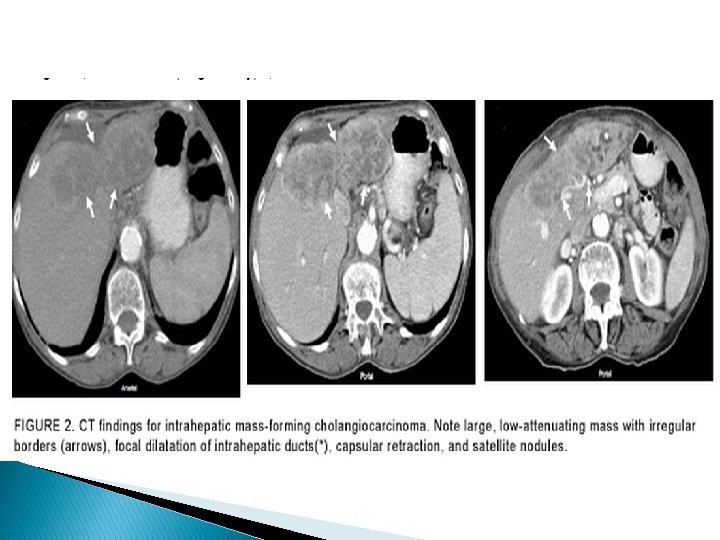
� ICCA takes up contrast agent progressively during the arterial and venous phases of studies — especially if the lesion is > 2 cm, because of its extensive desmoplastic reaction. � Other 1. 2. 3. associated findings may include : hepatic capsular retraction vascular encasement that may lead to lobar atrophy dilatation of peripheral bile ducts
� ICCA may be difficult to differentiate from a metastatic lesion (especially a metastasis from a foregut adenocarcinoma) by imaging and histology.
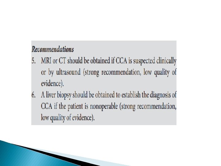
�A diagnosis of CCA cannot be confidently made with radiological imaging alone. � If surgery is indicated for a patient with an FLL suspected of having underlying primary hepatic malignancy, a diagnostic biopsy may not be required because its results will not change the management strategy and may lead to seeding � In all other cases, a biopsy specimen should be obtained to confirm the diagnosis if CCA is suspected.
� Either CT or MRI is appropriate for the evaluation � Multidetector CT may be more accurate than MRI for predicting resectability, with an accuracy of 85 % to 100 % , and may be better for identifying extrahepatic metastases.
� The utility of PET in the diagnosis of CCA is limited and PET should not be used routinely; � PET detects ICCA with sensitivity values ranging from 18 % (for infiltrating types) to > 80 % (for mass-forming types) � Carbohydrate antigen 19 -9 is a serum marker that can be measured to identify patients with ICCA, with 62 % sensitivity and 63 % specificity
Treatment of CCA � When surgical resection can be offered for ICCA, the median survival time is 36 months, with a recurrence rate of 62. 2 % after a median of 26 months of follow-up However, even in centers with expertise, < 30 % of all patients undergo curative resections � Liver transplantation is contraindicated in ICCA because of its poor results. � � For inoperable tumors, combination chemotherapy with gemcitabine plus cisplatin is the standard therapy.
Hepatocellular adenoma � Hepatocellular adenoma is rare with 0. 007 – 0. 012 % of the population developing these lesions.
Risk factors for hepatocellular adenoma �a marked increase in hepatocellular adenomas has been noted in women taking oral contraceptive (OCP) therapy. � The incidence of these lesions in women not taking OCPs is 1 – 1. 3 per million, whereas in those taking OCPs the frequency has been higher at 34 per 1 million. � Strengthening this causal relationship, hepatocellular adenomas tend to regress after the discontinuation of OCP therapy
Other risk factors � Anabolic androgen steroids Males taking androgens are not the only individuals at risk from hormonal abnormalities. � Males and females with high levels of endogenous androgens or estrogens are also at risk of developing hepatocellular adenomas.
� glycogen storage disease (GSD) Ia and III are also at an increased risk for hepatocellular adenomas. ( male , patients > 25 years of age ) � Managing hepatocellular adenomas in patients with GSD requires treatment strategies unique to this population. � Tumor size has been shown to decrease as a result of continuous nocturnal feeding. � Despite higher morbidity in GSD patients, surgical resection to prevent tumor progression is a feasible intermediate step until definitive treatment with liver transplantation can be achieved.
� Obesity � features of the metabolic syndrome such as diabetes mellitus, insulin resistance, hypertension, and dyslipidemia
� Obese patients who use OCP are likely at an increased risk for hepatocellular adenoma. � the prevalence of transformation of hepatocellular adenomas to HCC is 10 times more likely in males, with metabolic syndrome the most frequently associated condition for this transformation.
Clinical variants of hepatocellular adenoma � Liver adenomatosis : � Multiple adenomas, defined as between > 3 and ≥ 10 lesions, are collectively referred to as liver adenomatosis � These multiple lesions have identical clinical, histological, and radiographic features as hepatocellular adenomas and are managed in the same manner. Their precise pathogenesis is unclear. � Of interest, preliminary retrospective analysis has noted stability or regression of these lesions with weight loss. �
� THCA � OCP is asubtype of inflammatory adenoma. use, hormonal therapy, and obesity are frequently associated with the development of THCA. � Up to 40 % of patients with THCA typically present concomitantly with another benign liver lesion. � THCA should be managed as aggressively as hepatocellular adenomas as they are likely to be symptomatic, prone to hemorrhage, and may contain focal areas of necrosis. � The high likelihood of hemorrhage combined with an unknown potential of transformation to HCC makes surgery the recommended treatment.
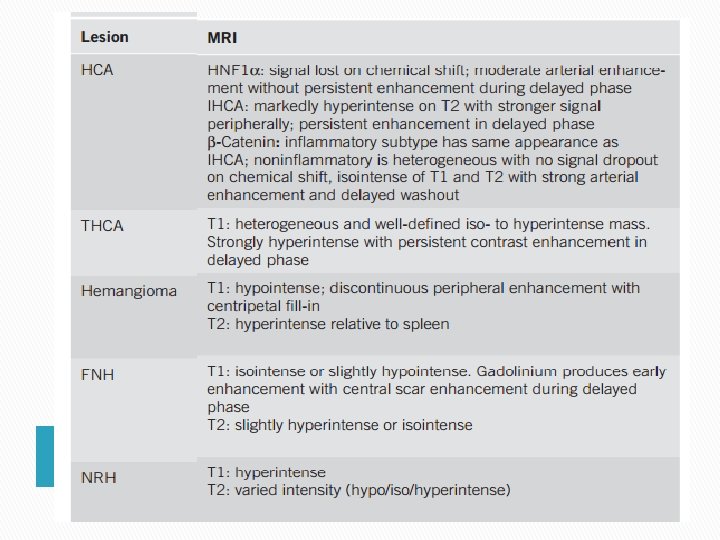
Diagnostic characteristics of hepatocellular adenoma Although CT can be used to diagnose hepatocellular adenomas, recent findings suggest that not only can MRI be used to diagnose hepatocellular adenomas, but it can also identify the subtypes of hepatocellular adenomas based on the imaging patterns, obviating the need for biopsy to distinguish these subtypes � MRI enhanced with gadobenate dimeglumine or gadoxetate disodium can be very effective in differentiating hepatocellular adenomas from FNH and other lesions �
� Liver biopsy can aid in identifying the subtype of hepatocellular adenomas as each category has specific genetic and molecular markers. � However, the vascular nature of hepatic lesions coupled with their propensity to hemorrhage can make biopsy risky. � Thus, biopsy should be reserved for cases in which imaging is inconclusive and the results will have an impact on treatment decisions
q Hepatocellular adenoma and pregnancy: � increase in size during pregnancy � Instead of advocating contraindication to pregnancy in all cases of hepatocellular adenomas, an individualized approach is advocated in which pregnancy is not discouraged when lesions are < 5 cm.
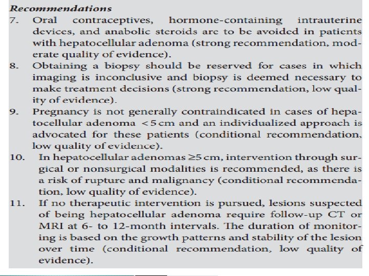
q. Management adenoma � more of hepatocellular aggressive than that for most other benign hepatic lesions because of the potential for hepatocellular adenomas to hemorrhage or progress to HCC. � Hemorrhage has been reported in 11 – 29 % of hepatocellular adenomas cases, with nearly all instances of spontaneous rupture occurring in lesions ≥ 5 cm. � Thus, resection should be considered when hepatocellular adenomas are found to be ≥ 5 cm.
� Nonsurgical modalities such as embolization can be pursued as an alternative to resection in high surgical risk patients or in patients with lesions in anatomically challenging locations.
� In cases where hemorrhage does occur, conservative management: � using blood products is a temporary approach to achieve hemodynamic stability and avoid emergent liver resection � Packing the liver, performing an emergency hepatectomy, embolizing the hepatic artery, or even liver transplantation can be used to control hemorrhage
� Hepatocellular adenomas of the -β catenin subtype should be considered for early referral for resection as malignant transformation occurs most frequently in this subtype, occurring in up to 5 – 10 % of cases
� Hepatocellular adenomas < 5 cm can be managed conservatively as these lesions are rarely observed to rupture or undergo malignant transformation � Nevertheless, some hepatocellular adenomas have been reported to increase in size despite the discontinuation of OCP or anabolic steroids, and the development of HCC has been reported despite regression in size. � Thus, follow-up imaging should be conducted once every 6 months for at least 2 years to establish any growth patterns and monitor for malignant transformation. � Annual imaging can be performed after this period based on the growth patterns and stability of the lesion.
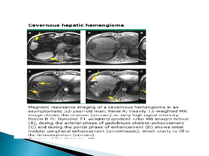
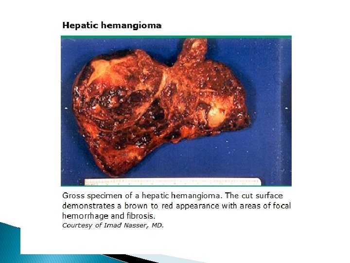
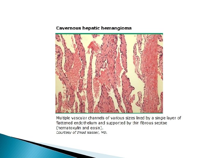
HEPATIC HEMANGIOMA �A benign vascular liver lesions of unknown etiology that are thought to arise from congenital hamartomas. � Alternatively, could result from dilation of existing blood vessels in tissues that developed normally. � The observed increase in the size of the lesions is thought to result from progressive ectasia rather than hyperplasia or hypertrophy. � Hepatic hemangiomas are the most common benign hepatic tumors with a prevalence of 0. 4 – 20 % identified during autopsy
� earlier, larger, and are more often found in women than in men, with a 5 to 1 female to male preponderance. � Female sex hormones may have a role in their pathogenesis � There is no causal link between hepatic hemangiomas and pregnancy or OCP use
q Diagnostic characteristics of hepatic hemangioma � Hepatic hemangiomas can be found in all age groups, typically discovered in those between the ages of 30 and 50 years. � Most of these lesions are asymptomatic and are discovered incidentally during imaging studies. � Hemangiomas occur with symptoms in ~ 11 – 14 % of all hepatic hemangioma cases � The most common presentations are right upper quadrant pain or a mass felt in the epigastrium. � Other common symptoms include severe pain, nausea, dyspepsia, early satiety, vomiting, weight gain, and hepatomegaly
� In rare cases, giant hemangiomas may cause consumptive coagulopathy known as Kasabach – Merritt syndrome that manifests as thrombocytopenia, disseminated intravascular coagulation, and systemic bleeding.
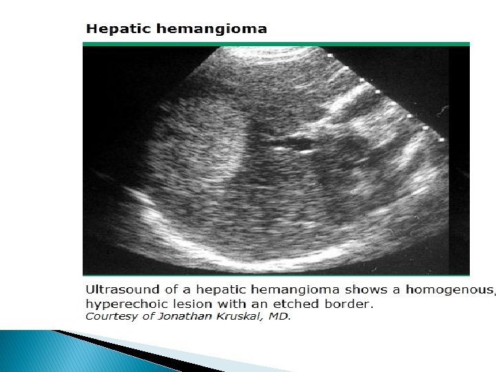
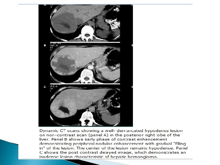
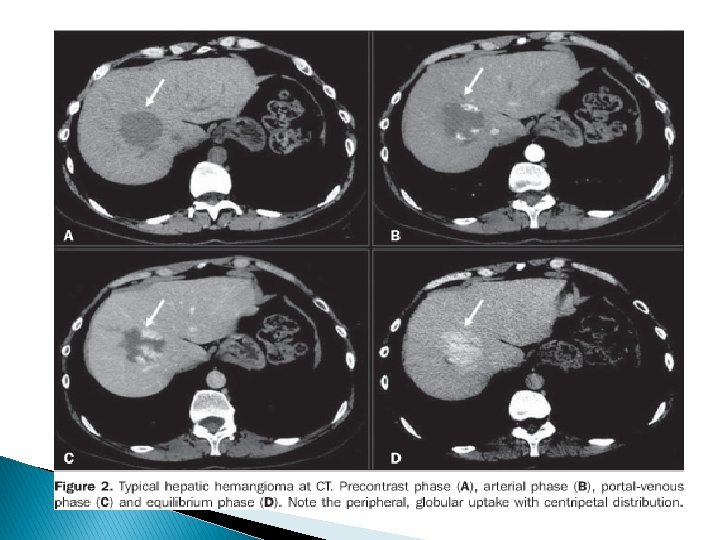
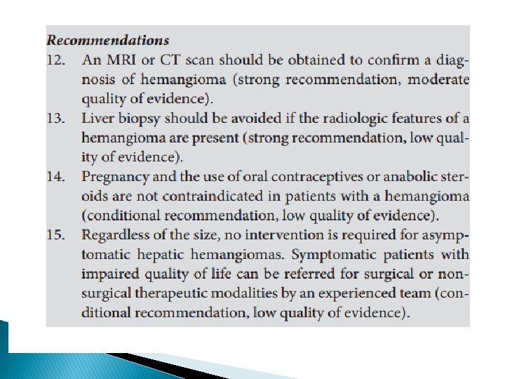
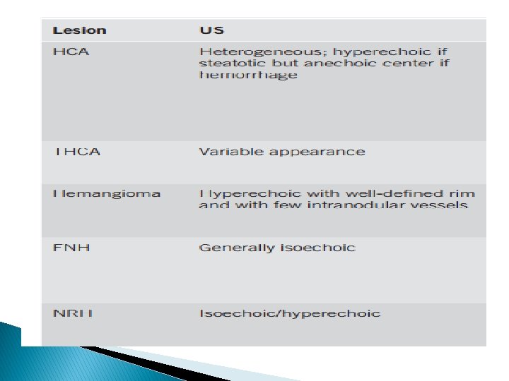
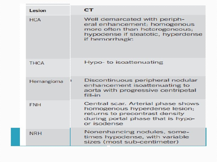
� CT, MRI, and US studies are reliable in establishing a diagnosis of hepatic hemangioma as this lesion displays unique features upon imaging with peripheral nodular enhancement and progressive centripetal fill-in. MRI is preferred in cases where the lesion is < 3 cm or found close to the heart or intrahepatic vessels � Contrast-enhanced US, if available, can increase both the sensitivity and specificity of US and is effective in diagnosing hepatic hemangioma �
� Spontaneous bleeding of hemangiomas is rare. � However, owing to its highly vascular nature, biopsy should be avoided because of the risk of potential bleeding. Furthermore, the high sensitivity and specificity of radiologic studies in the diagnosis of hepatic hemangioma obviates the need for a biopsy. � In cases of smaller lesions where there is uncertainty in the diagnosis, a follow-up imaging study may be more prudent than a biopsy. �
q Management of hepatic hemangioma � The majority of hemangiomas are asymptomatic and remain stable over time. � Thus, preventing rare complications with surgical intervention is not needed, and instead a conservative approach is advocated. � Surgical intervention can be considered in cases where the lesion grows very large ( > 10 cm) or the patient begins to report symptomatic compression or recurrent pain � Follow-up imaging is not required in cases of classical hemangioma.
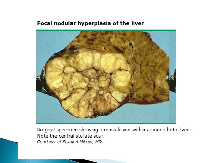
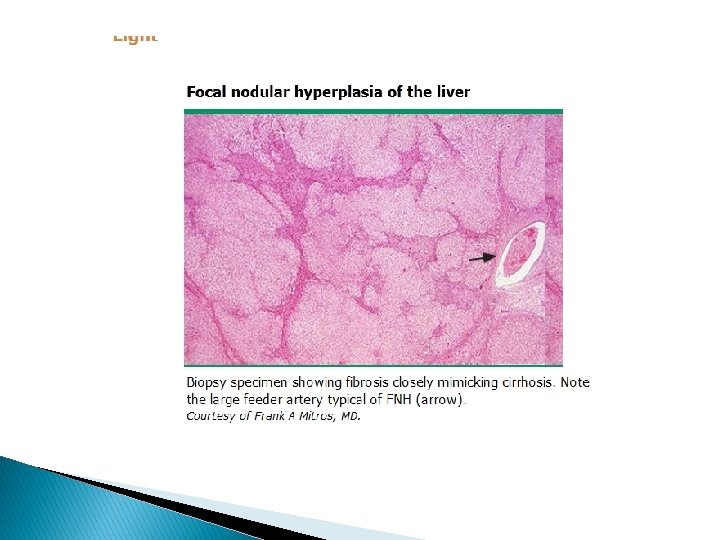
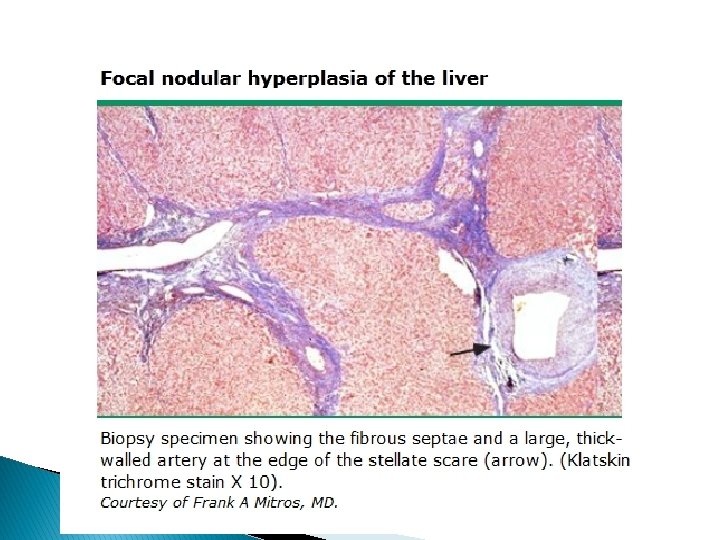
FOCAL NODULAR HYPERPLASIA Second most common hepatic lesion and is found at autopsy with a prevalence of 0. 3 – 3 %. � Clinically relevant cases of FNH are rare with a reported prevalence in US studies of 0. 03 %. � � The development of focal nodular hyperplasia is caused by an injury to the portal tract resulting in the formation and enlargement of arterial to venous shunts. � This in turn causes hyperperfusion in local arteries resulting in oxidative stress that triggers a response from hepatic stellate cells to produce the central scar typically seen in cases of FNH
q Diagnostic characteristics of FNH � Although 20 – 40 % of cases of FNH may present with symptoms, most are discovered incidentally. � Up to 20 % of cases are associated with a concomitant diagnosis of hepatic hemangioma. � other hypervascular tumors such as hepatocellular adenomas and HCC have been observed concurrently with FNH.
� FNH is noted primarily in women in their 40 s and 50 s � Female sex hormones were suspected to have a role in FNH development as not only is the prevalence higher in women but also women tend to develop larger and earlier lesions compared with men. � However, changing OCP use over time has not led to alterations in the prevalence of FNH, and pregnancy has not been associated with an increase in tumor size, making this association less likely.
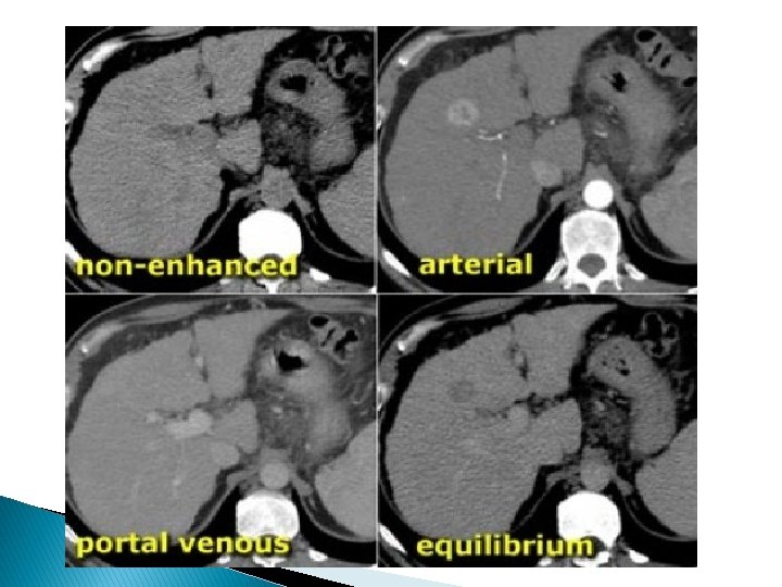
� Identification of classic FNH by way of its “ spoke-wheel ”central scar on crosssectional imaging is relatively straightforward. � Ambiguous cases that cannot exclude hepatocellular adenomas must be differentiated accurately as FNH or hepatocellular adenomas as their management differs significantly � The diagnostic accuracy of MRI for FNH has improved because of the improvement in hepatobiliary contrast agents, such as gadobenate dimeglumine � In addition, adherence to the technical specifications of triphasic and multisection spiral CT has been reported to accurately diagnose FNH.
� Improvements in imaging have been made with the use of contrast enhanced US that has been shown to be similarly accurate as MRI and CT in identifying FNH. � In cases where FNH cannot be distinguished from hepatocellular adenomas, immunohistochemical analysis performed on biopsy specimens can discriminate FNH from hepatocellular adenomas with good performance characteristics.
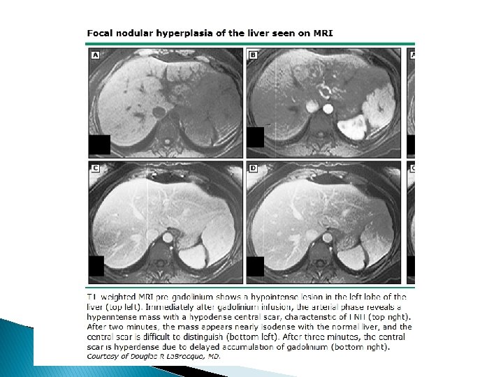
well defined showing arterial enhancing lesion in the left lobe of liver with central non enhancing scar. On venous phase image the lesion shows homogeneous contrast enhancement. In delayed phase image there is enhancement of scar.
Management of FNH q § § q A conservative approach should be taken when managing FNH due to: Most cases of FNH are asymptomatic and stable over time. The occurrence of HCC and spontaneous rupture are rare. However, further evaluation of symptomatic lesions in which a diagnosis of FNH cannot be firmly established is recommended. � Partial hepatic resection is the most common intervention, embolization and radiofrequency ablation have more recently been utilized as they are associated with fewer complications and lower morbidity.
�Follow- up annual US for 2 – 3 years is prudent in women diagnosed with FNH who wish to continue OCP use. �Individuals with a firm diagnosis of FNH who are not using OCP do not require follow up imaging.
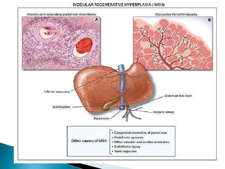
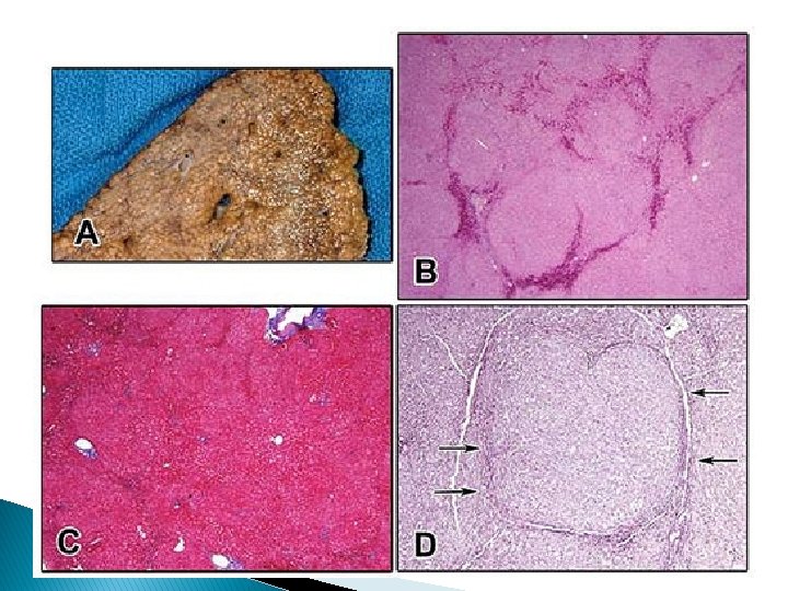
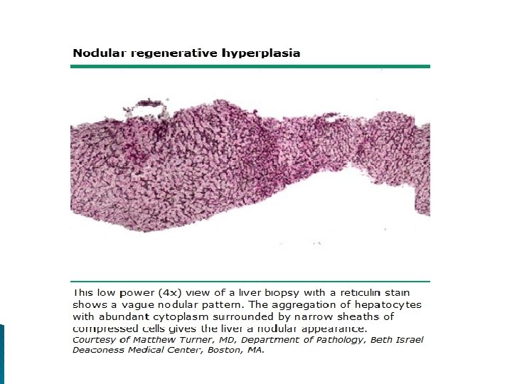
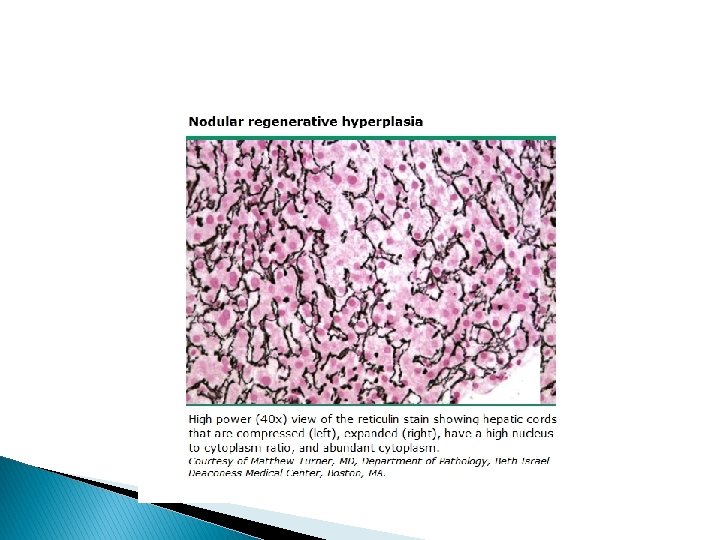
NODULAR REGENERATIVE HYPERPLASIA � Nodular regenerative hyperplasia (NRH) is the transformation of normal hepatic parenchyma into small regenerative nodules. �A secondary consequence of altered blood flow in which obstructive portal venopathy, due to thrombosis or phlebitis, causes ischemia. � This, in turn, leads to hyperplasia of hepatic acini to maintain adequate blood flow to compensate for atrophied hepatocytes.
� This process forms nodules that are separated by atrophic areas with little to no fibrosis � prevalence of over 5. 3 % in individuals > 80 years old � The general population presents with NRH at a lower frequency of 2. 1 – 2. 6 %. � No apparent relationship is found between NRH and gender.
q. A number of conditions do seem to be associated with NRH, including : 1. immunological and hematological disorders 2. cardiac and pulmonary disorders 3. several drugs and toxins 4. Neoplasias 5. organ transplantation
Diagnostic characteristics of NRH � NRH is most commonly discovered incidentally. � Symptomatic cases are rare and most often present with features of portal hypertension, such as ascites, splenomegaly, hepatomegaly, and esophageal varices. � Imaging studies are insufficient in establishing a definitive diagnosis of NRH. � The lesions are rountinly too small to observe radiographically and, when visualized, too difficult to distinguish from the regenerating nodules of cirrhosis.
� The definitive method for establishing a conclusive diagnosis of NRH is biopsy. � Although NRH shares common features with micronodular cirrhosis, three histological criteria — 1. nodules of regenerative hepatocytes separated by atrophic parenchyma 2. absence of fibrous septa between nodules 3. curvilinear compression of the central lobule — � can be used to distinguish NRH from cirrhosis � There are no reliable tumor markers known to be useful for diagnosing NRH.
Management of NRH � Treating NRH requires addressing the underlying etiological condition. � The B-blocker prophylaxis and / or endoscopic therapy for esophageal varices, pharmacologic therapy of ascites, and TIPS are possible forms of treatment when NRH is complicated by portal hypertension.
� Although very rare, NRH can lead to liver failure that may require treatment with liver transplantation. � As NRH is commonly associated with other underlying disease processes, follow-up care requires determining and managing the underlying disease. � There are no absolute contraindications to pregnancy or the use of OCPs.
HEPATIC CYSTS
� Hepatic cysts typically present for evaluation upon being found incidentally on imaging studies. � Early laparotomy series reported a prevalence of 0. 2 to 1 %. � US � CT series have reported a 3 – 5 %. series have reported the prevalence to be as high as 15 – 18 %.
� The presence which not characteristic of simple hepatic cysts and should prompt further diagnostic evaluation. : 1. multiple cysts ( > 20) 2. large cysts ( > 4 – 5 cm) 3. septations, 4. Calcifications 5. fenestrations 6. loculations 7. Heterogeneity 8. daughter cysts 9. symptoms on presentation
q. Simple � Simple hepatic cysts are postulated to be congenital exclusions of hyperplastic bile duct rests that lack a communication with biliary ducts. � They are composed of an outer layer of fibrous tissue and are lined by a cuboidal, columnar epithelium that continually produces cystic fluid. � Simple hepatic cysts are usually < 1 cm and can grow up to 30 cm. � Simple hepatic cysts are uncommon before the age of 40 years and have a female predilection of 1: 4.
� There is no clear correlation with OCP use or pregnancy. � The presence of symptoms or a rapid increase in size on follow-up imaging should lead to consideration of alternative diagnosis such as biliary cystadenoma (BC) or BCA.
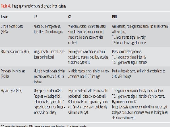
� Diagnostic � characteristics of simple hepatic cysts Ultrasound typically reveals an anechoic, homogenous, fluidfilled lesion with smooth margins. CT shows a well-demarcated, water-attenuated, smooth lesion without an internal structure, and no enhancement with contrast. � MRI shows a well-defined, homogeneous lesion with low signal intensity on T 1 weighting, and high intensity on T 2, without contrast enhancement. � The differential diagnosis includes : � BCA, � PCLD, � hydatid cysts, � cystic metastases from primary cystic tumors � cystic necrosis of large solid neoplasms. q
q Cysts that have : � internal septations � fenestrations � calcifications, � irregular walls � daughter cysts on US q should be evaluated with CT or MRI for features of BCA or hydatid cysts. � Aspiration of fluid contents is not needed to diagnose simple hepatic cysts and is not recommended. � if done, the findings should show normal fluid CA 19 -9 levels and negative testing for cytology.
Management of simple hepatic cysts �A hepatic cyst identified on US with septations, fenestrations, calcifications, irregular walls, or daughter cysts should prompt further evaluation with CT or MRI (strong recommendation, low quality of evidence). � Asymptomatic simple hepatic cysts should be observed with expectant management (strong recommendation, moderate quality of evidence).
� Aspiration of asymptomatic, simple hepatic cysts is not recommended (strong recommendation, low quality of evidence). � Symptomatic simple hepatic cysts may be managed with laparoscopic deroofing rather than aspiration and sclerotherapy, dictated based on availability of local expertise (conditional recommendation, low quality of evidence).
BILIARY CYSTADENOMAS AND BILIARY CYSTADENOCARCINOMAS � Biliary cystadenomas are congenitally derived, aberrant bile duct remnants composed of three layers of tissue. � Early pathophysiologic suppositions regarding an origin from heterotopic ovarian tissue have been disproven by recent immunohistochemistry and electron microscopy studies. � The outer layer of thick collagen and mixed connective tissue surrounds a middle layer of mesenchymal smooth muscle cells and fibroblasts, and an inner lining of cuboidal / columnar epithelium that typically secretes mucin
� Biliary cystadenomas are reported to constitute up to 1 – 5 % of total hepatic cysts, and up to 10% of cysts > 4 cm.
� There are no known associations with the use of oral contraceptives, although the 1: 4 female predilection suggests a possible hormonal involvement � Although rare, BC is the most common form of a primary hepatic cystic neoplasm.
The presence of calcifications along with mixed solid and cystic components on imaging as well as constitutional symptoms has been reported to be associated with BCA. � Diagnostic characteristics of biliary cystadenomas US typically shows irregular walls and internal septations forming loculi. � US is most sensitive in identifying these internal septations. � If a complex cyst is found on US, crosssectional imaging with CT and MRI should be obtained.
� � Aspiration and biopsy is not recommended for focusing the differential because it has limited sensitivity and can disseminate malignancy if there is underlying BCA. Although imaging can suggest the possibility of BC or BCA, surgical resection is ultimately necessary to confirm and treat the suspected BC or BCA.
q Management of biliary cystadenomas � Routine fluid aspiration is not recommended when BCA is suspected because of limited sensitivity and the risk of malignant dissemination (strong recommendation, low quality of evidence). � Imaging characteristics suggestive of BC or BCA, such as internal septations, fenestrations, calcifications, or irregular walls, should lead to referral for surgical excision (strong recommendation, low quality of evidence). � Complete surgical excision, by an experienced team, is recommended if BC or BCA is suspected (strong recommendation, low quality of evidence).
POLYCYSTIC LIVER DISEASE � PCLD is thought to be a part of a clinical spectrum of ciliopathies including congenital hepatic fibrosis, choledochal cysts, microhamartomas, and Caroli ’ s disease that are associated with mutations that impair cholangiocyte ciliary function. � PCLD is characterized by the presence of extensive hepatic cysts that are microscopically similar to simple hepatic cysts but more numerous (usually > 20) and larger.
Diagnostic characteristics of PCLD � There is a female predilection and a noted increase in the size of hepatic cysts and symptoms with age. � Pregnancy and female hormones are also thought to be risk factors for an increase in the size and number of hepatic cysts. � PCLD tends to be asymptomatic until the size and number of cysts increase to a critical level. � Patients are typically more likely to have hepatomegaly and symptoms from mass effect, such as abdominal bloating, pain, fullness, and shortness of breath, when the cyst to hepatic parenchyma ratio becomes > 1.
� Patients may also present with complications such as traumatic rupture, infection, bleeding, extrinsic compression of the biliary tree or gastrointestinal tract, and compression of the inferior vena cava. � In advanced cases, patients may develop portal hypertension with relatively preserved hepatic synthetic function. � The diagnosis of PCLD is supported by the presence of multiple hepatic cysts on US, CT, or MRI. � CT or MRI may provide additional information to exclude other disease processes and to evaluate for the presence of concomitant renal cysts.
Management of PCLD � Routine medical therapy with mammalian target of rapamycin inhibitors or somatostatin analogs is not recommended(strong recommendation, low quality of evidence). � Aspiration, deroofing, resection of a dominant cyst can be performed based on the patient ’ s clinical presentation and underlying hepatic reserve (conditional recommendation, low quality of evidence). � Liver transplantation with or without kidney transplantation can be considered in patients with refractory symptoms and significant cyst burden (conditional recommendation, low quality of evidence).
HYDATID CYSTS � Hydatid cysts, or cystic echinococcosis, are most common in patients from sheepgrazing areas such as the Mediterranean, South America, Australia, and East Africa. � Hydatid cysts are due to Echinococcus granulosus infection in which humans serve as accidental intermediate hosts when they eat food contaminated with echinococcus eggs or eat organ meat from infected animals such as sheep or cows.
� The eggs hatch in the human host small intestine and penetrate into the vasculature and then into the liver and lungs. � The cysts become visible in the liver at 3 to 4 weeks and grow into a mature cyst that has a germinal layer surrounding a fluid-filled central hydatid cavity. � The cysts develop an ectocyst or pericyst in response to the compressive forces of the host ’ s liver parenchyma. � The cyst can have high pressure from fluid production that can lead to rupture after trauma or operative manipulation.
� Most small cysts, < 5 cm, are asymptomatic. � Larger cysts can elicit an inflammatory reaction and may lead to abdominal pain. � An acute presentation with pain should lead to consideration of rupture or secondary infection of the cyst. � The incidental rupture or iatrogenic puncture of the cyst with spillage of its antigenic contents can lead to a severe allergic reaction, leading to ascites, peritonitis, and shock. � Rarely, the cysts can extrude into the biliary tree, leading to jaundice and cholangitis
�Diagnostic characteristics of hydatid cysts � On US, small hydatid cysts may appear similar to simple hepatic cysts as a unilocular collection. � With progression, the lesions may develop a thick, often calcified wall and daughter cysts in the periphery of the main cyst.
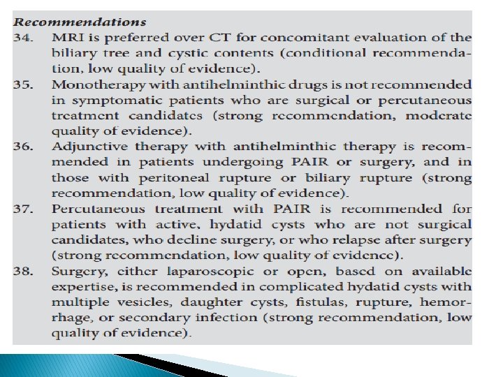
� CT and MRI should be obtained if hydatid cysts are suspected. � MRI is preferred for pre surgical evaluation to look for cystobiliary communication and evaluate cystic content characteristics.
Management of hydatid cysts � Treatment of hydatid cysts depends on the size, location, and symptoms of the cysts as well as the availability of clinical expertise and patient preferences. � Asymptomatic, inactive, calcified hydatid cysts can be managed expectantly. � Chemotherapy alone with antihelminthic drugs is generally not utilized unless the patient is not a candidate for primary percutaneous or surgical treatment, has multiorgan dissemination, or declines other intervention. � Chemotherapy as an adjunct treatment to other modalities is recommended before and after percutaneous treatment or surgery to prevent relapse.
� chemotherapy be started before the procedure and at least 1 month to 6 months afterward. � PAIR (puncture, aspiration, injection, and reaspiration) is a percutaneous treatment alternative to surgery. � PAIR is recommended for patients with active hydatid cysts > 5 cm who are not candidates for or decline surgery, or who relapse after surgery.
PAIR is not recommended in patients with biliary fistulas or communications with the biliary tree because of the risk of biliary sclerosis. � PAIR is also contraindicated in patients with inaccessible cysts, or with complicated, multivesiculated cysts. � The risk of lethal anaphylaxis related to percutaneous treatment was extremely rare at 0. 03 % of reported procedures. � Surgical treatment, either radical pericystectomy or conservative deroofing, has been reserved for complicated cysts that have fistulas, multiple daughter vesicles, rupture, hemorrhage, or secondary infection. � Surgery also remains an option when percutaneous treatment such as PAIR is not available. �
� The size of the liver lesion is extremely important in guiding the evaluation. � Lesions < 1 cm are commonly benign incidental findings.
� Certain solid FLLs such as FNH and hemangiomas can often precisely be diagnosed by a quality imaging modality alone. � In many benign lesions such as hemangiomas and hepatocellular adenomas, liver biopsy carries a high risk of bleeding and is not of any additional value to the radiologic diagnosis.
THANK YOU