A silicon strip detector for a novel 2


 γ photons Water-equivalent phantom Radiotherapy energies photon interactions : Compton Effect")

 Siemens PRIMUS linac dual energy machine")


- Slides: 42
A silicon strip detector for a novel 2 D dosimetric method for complex radiotherapy treatment verification Alessio Bocci DITANET Experienced Researcher DITANET Workshop 08 November 2011 Alessio Bocci, CNA 1
Outline Ø Complex radiation therapy treatments - IMRT Ø Detectors for dosimetric applications Ø RADIA 2 project : experimental set-up Ø Measurements and results Ø Conclusions DITANET Workshop 08 November 2011 Alessio Bocci, CNA 2
Motivation and Objective Ø Motivation: Cancer is the second most frequent cause of death in developed countries. At present, although surgery is the most effective way to remove the malignant tissue, when it is combined with radiation therapy improves the cure rate by 40% approximately. Ø Objective Characterization of a silicon strip detector dedicated to 2 D dose measurements in the axial plane of a phantom for the verification of complex radiation therapy treatment plans (i. e. IMRT) DITANET Workshop 08 November 2011 Alessio Bocci, CNA 3
Intensity Modulated Radiation Therapy Many beam directions and entrance points for conformal doses distributions modulating in space the fluence of each radiation field DITANET Workshop 08 November 2011 Alessio Bocci, CNA 4
Dosimetry for radiotherapy verification 2 D Detectors dedicated to radiotherapy verification plans Ø Ionization chambers – “gold standard” detectors Ø Radiographic and radiochromic films Ø Semiconductor dosimeters: Silicon diodes DITANET Workshop 08 November 2011 Alessio Bocci, CNA 5
Dose distribution verification Film dosimeters: traditional detectors for verifying treatment plans dose distribution Film dosimetry used as 2 D dosimeters : Advantages: high spatial resolution (sub-mm), good uniformity, axial plane Disadvantages: Unusable as on-line detectors and are unreliable and time consuming detectors DITANET Workshop 08 November 2011 Coronal plane (ventral and dorsal) Axial plane (inferior and superior) Sagittal plane Alessio Bocci, CNA 6
2 D Digital Detectors in radiotherapy treatments Single silicon detectors and ionization chambers assembled for 2 D detectors IMRTLog (ONCOlog Medical) 2 D array silicon diodes spatial resolution 0. 5 to 1. 0 cm I’MRT Matri. XX (Scanditronix/Wellhöfer) array of ionizations chambers Map. CHECK (Sun Nuclear) silicon diodes Spatial resolution is still poor with respect to film dosimeters These devices are useful only for one irradiation angle and they can measure dose distribution only in the coronal plane of the patient DITANET Workshop 08 November 2011 Alessio Bocci, CNA 7
New detection systems Traditional detectors New detection systems Radiographic films Film dosimeters On-line 2 D commercial digital detectors New detection systems no ü ü Spatial resolution ü poor ü 2 D detectors ü ü yes Not monolithic! ü Axial plane ü no ü DITANET Workshop 08 November 2011 It is necessary to develop new detection systems that enhance the traditional ones, and that are able to verify in a simple and accurate way complex treatment planning inexpensive radiation hard easy to use Alessio Bocci, CNA 8
From Nuclear Physics to Medical Applications Medical applications can benefit from the developments on nuclear and high energy physics detector technology Silicon strip detectors mounted @ CNA in a telescope configuration Silicon strip detectors mounted @ Virgen Macarena Hospital in Seville silicon tracking detectors silicon detectors for medical applications DITANET Workshop 08 November 2011 Alessio Bocci, CNA 9
RADIA 2 Project Si-DETECTOR • Commercial silicon detector • Low cost Normally used on particle detection (W 1 type from Micron Semiconductor Ltd) • Single sided 16 strips (3. 1 mm pitch) • Active area 50 x 50 mm² & 500 µm thick DITANET Workshop 08 November 2011 Alessio Bocci, CNA 10
Why Silicon detectors ? Advantages: Linear relationship between photon energy and e/h pairs ; High sensitivity (Si - 3. 6 e. V/pair – sensitivity per unit volume: 640 n. C Gy-1/mm 3 ) (about 18000 times the sensitivity per unit volume of an ionization chamber) ; Small active volume that allows to obtain high spatial resolution ; Well developed technology for the production of segmented monolithic planar detectors ; Radiation hard material ; C. Talamonti et al, NIM-A 658 (2011) 84– 89 DITANET Workshop 08 November 2011 Alessio Bocci, CNA 11
Single-sided-Si-strip detectors (SSSSD) γ photons Water-equivalent phantom Radiotherapy energies photon interactions : Compton Effect Secondary electrons from the phantom interacts centemeters away Signals from each strip are converted to the absorbed dose DITANET Workshop 08 November 2011 Alessio Bocci, CNA 12
RADIA 2 Project – Collaboration Z. Abou-Haidar 1, M. A. G. Alvarez 1 , R. Arrans 3, A. Bocci 1, M. A. Cortes-Giraldo 2 , J. M. Espino 2 , M. I. Gallardo 2, A. Perez Vega-Leal 4 F. J. Perez Nieto 5, J. M. Quesada 2 1. DITANET group @ National Accelerator Centre (CNA) 2. Department of Atomic, Molecular and Nuclear Physics (FAMN), University of Seville 3. Virgen Macarena University Hospital, Seville 4. School of Engineering, University of Seville 5. Instalaciones Inabensa S. A. A. Bocci et. al. , Empirical characterization of a silicon strip detector for a novel 2 d mapped method for dosimetric verification of radiotherapy treatments, Radiotherapy and Oncology, Volume 99, Supplement 1, May 2011, Page S 172 A. Bocci et. al. , A silicon strip detector for a novel 2 D dosimetric method for radiotherapy treatment verification submitted to NIM-a (October 2011) M. A. Cortes-Giraldo et al. , "Geant 4 Simulation to Study the Sensitivity of a MICRON Silicon Strip Detector Irradiated by a SIEMENS PRIMUS Linac", Progress in Nuclear Science and Technology, in press (2011) DITANET Workshop 08 November 2011 Alessio Bocci, CNA 13
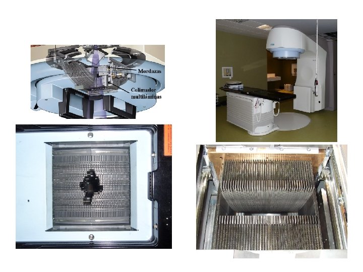
LINAC accelerator University Hospital Virgen Macarena (Seville, Spain) Siemens PRIMUS linac dual energy machine operating at 6 MV photon mode A treatment planning system TPS (Philips Pinnacle) was used to calculate dose distributions. Calculations were compared to experimental data DITANET Workshop 08 November 2011 Alessio Bocci, CNA 14
Experimental Set-up Two phantoms prototypes were designed and built Polyethylene slab material 1. A slab phantom for: detector characterization (sensitive area perpendicular to the beam direction) DITANET Workshop 08 November 2011 Cylindrical phantom 2. A cylindrical phantom for: angular response measurements & 2 D treatment plans verification in the axial plane Alessio Bocci, CNA 15
Detector in the axial plane Set-up 2: The active area of the detector is parallel to the beam direction (axial plane) plane Since common ways to present dose distributions is a dose map in the patient axial plane, as an innovation the detector was placed in the cylindrical phantom in the axial plane, parallel to the beam axis This work is part of a more ambitious project aiming to: Employ this prototype for obtaining 2 D dose maps by an in-house developed algorithm dedicated to verify IMRT treatment plans. This system is patent pending DITANET Workshop 08 November 2011 Alessio Bocci, CNA 16
Electronics discrete electronics developed in-house School of Engineering, University of Seville Charge integrators for each strip (electrometers) digitized (12 bits) and analyzed by a Digital signal processor (DSP) Virgen Macarena University Hospital, Seville A PC allows to control and to retrieve data via an RS-232 serial bus based on a Lab. VIEW software DITANET Workshop 08 November 2011 Alessio Bocci, CNA 17
Measurements Detector Set-up 1: characterization • Linearity • Uniformity • Calibration • Percent Depth Dose (PDD) • Penumbra Angular response Set-up 2: • TPS and Geant 4 Simulations • Angular measurements • Final calibration DITANET Workshop 08 November 2011 Alessio Bocci, CNA 18
Measurements Detector Set-up 1: characterization ü Linearity • Uniformity • Calibration • Percent Depth Dose (PDD) • Penumbra Angular response Set-up 2: • TPS and Geant 4 simulations • Angular measurements • Final calibration DITANET Workshop 08 November 2011 Alessio Bocci, CNA 19
Linearity Set-up 1: SSSSD perpendicular to the beam direction Linearity with dose better than 0. 1 % for all channels DITANET Workshop 08 November 2011 Alessio Bocci, CNA 20
Measurements Detector Set-up 1: characterization • Linearity ü Uniformity • Calibration • Percent Depth Dose (PDD) • Penumbra Angular response Set-up 2: • TPS and Geant 4 simulations • Angular measurements • Final calibration DITANET Workshop 08 November 2011 Alessio Bocci, CNA 21
Uniformity Set-up 1: SSSSD perpendicular to the beam direction Non-uniformities depend by the different strip efficiency and gain of the electronics Uniformity better than 0. 5 % for all channels DITANET Workshop 08 November 2011 Alessio Bocci, CNA 22
Measurements Detector Set-up 1: characterization • Linearity • Uniformity ü Calibration • Percent Depth Dose (PDD) • Penumbra Angular response Set-up 2: • TPS and Geant 4 simulations • Angular measurements • Final calibration DITANET Workshop 08 November 2011 Alessio Bocci, CNA 23
Calibration Set-up 1: SSSSD perpendicular to the beam direction Monitor Units –> Voltage -> c. Gy Calibration in standard condition radiation field 10 × 10 cm 2 source-to-surface distance (SSD) = 100 cm 1. 5 cm of water-equivalent solid slabs DITANET Workshop 08 November 2011 Alessio Bocci, CNA 24
Measurements Detector Set-up 1: characterization • Linearity • Uniformity • Calibration ü Percent Depth Dose (PDD) • Penumbra Angular response Set-up 2: • TPS and Geant 4 simulations • Angular measurements • Final calibration DITANET Workshop 08 November 2011 Alessio Bocci, CNA 25
Percent Depth Dose Set-up 1: SSSSD perpendicular to the beam direction Dose at different depth measured using different water-equivalent solid slabs The difference between SSSSD and ionization chamber is: 0. 68 % at 10 cm and 0. 73 % at 15 cm DITANET Workshop 08 November 2011 Alessio Bocci, CNA 26
Measurements Detector Set-up 1: characterization • Linearity • Uniformity • Calibration • Percent Depth Dose (PDD) ü Penumbra Angular response Set-up 2: • TPS and Geant 4 simulations • Angular measurements • Final calibration DITANET Workshop 08 November 2011 Alessio Bocci, CNA 27
Penumbra Region between 20 % and 80 % of the maximum dose levels at 1. 5 cm water depth SSSSD: 6. 17 ± 0. 56 mm - single silicon diode: 3. 92 ± 0. 20 mm Set-up 1: SSSSD perpendicular to the beam direction SSSSD penumbra value larger than the one obtained when using a single silicon detector This was mainly due to the SSSSD strips pitch of 3. 1 mm Geant 4 simulations gave compatible results Improvements with a future prototype (next project) DITANET Workshop 08 November 2011 Alessio Bocci, CNA 28
Measurements Detector Set-up 1: characterization • Linearity • Uniformity • Calibration • Percent Depth Dose (PDD) • Penumbra Angular response Set-up 2: ü Geant 4 simulations and TPS calculations • Angular measurements • Final calibration DITANET Workshop 08 November 2011 Alessio Bocci, CNA 29
Geant 4 simulations and TPS calculations Treatment head Geometry Model Phantom SSSSD Detector The geometry of the Siemens treatment head at 6 MV nominal energy photons, was reproduced in detail The geometric model of the phantoms and of the SSSSD was also reproduced The absorbed dose in each strip was calculated Geant 4 Simulations were also performed for calculating the dose-to-water case for comparisons with TPS calculations M. A. Cortés Giraldo, Ph. D. Thesis, 2011 DITANET Workshop 08 November 2011 M. A. Cortes-Giraldo et al. , "Geant 4 Simulation to Study the Sensitivity of a MICRON Silicon Strip Detector Irradiated by a SIEMENS PRIMUS Linac", Progress in Nuclear Science and Technology, in press (2011) Alessio Bocci, CNA 30
Measurements Detector Set-up 1: characterization • Linearity • Uniformity • Calibration • Percent Depth Dose (PDD) • Penumbra Angular response Set-up 2: • Geant 4 simulations and TPS calculations ü Angular measurements • Final calibration DITANET Workshop 08 November 2011 Alessio Bocci, CNA 31
Angular response 00 450 Set-up 2: SSSSD parallel to the beam direction Two experimental measurements detector gantry 3150 DITANET Workshop 08 November 2011 Alessio Bocci, CNA 32
Angular Response Steps of 450 Comparison between exp. data, TPS and Geant 4 Set-up 2: SSSSD parallel to the beam direction The agreement between the tendency of experimental data at different angles and the TPS is notable This implies that the new calibration factors are independent of the irradiation angle Constant calibration factors DITANET Workshop 08 November 2011 Alessio Bocci, CNA 33
Measurements Detector Set-up 1: characterization • Linearity • Uniformity • Calibration • Percent Depth Dose (PDD) • Penumbra Angular response Set-up 2: • Geant 4 simulations and TPS calculations • Angular measurements ü Final calibration DITANET Workshop 08 November 2011 Alessio Bocci, CNA 34
Final Calibration Constant calibration factors Calibration factors: Experimental/TPS Set-up 2: SSSSD parallel to the beam direction detector gantry Calibration factors independent of angular irradiation and of strip number DITANET Workshop 08 November 2011 Alessio Bocci, CNA 35
Final Calibration Calibrated dose Set-up 2: SSSSD parallel to the beam direction detector gantry Relative difference between the calibrated dose and TPS calculations are better than 2 % DITANET Workshop 08 November 2011 Alessio Bocci, CNA 36
Conclusions Ø Main Objective: Characterize and benchmark a new detection system based on a Si-strip detector dedicated to 2 D dose measurements in the axial plane of a cylindrical phantom Ø SSSSD characterization: results shows that the prototype is suitable for IMRT verification plans (remarkable linearity, uniformity, PDD) Ø The angular response of the detector in the axial plane was independent of the irradiation angle and of the strip number Ø Geant 4 simulations gave compatible results with respect to TPS and to experimental data Ø Final calibration with respect to TPS in the axial plane gives differences smaller than 2 % for all the strips Ø This system is patent pending OEMP PATENT number P 201101009 DITANET Workshop 08 November 2011 Alessio Bocci, CNA 37
Future developments Ø Future: work is in progress in order to obtain 2 D dose maps from experimental data in the axial plane using an in-house developed algorithm based on the Radon Transform Ø A new SSSSD prototype and a new experimental set-up has been designed to improve the spatial resolution, coupling the data acquisition system with a reconstruction algorithm and with an on-line graphical interface software DITANET Workshop 08 November 2011 Alessio Bocci, CNA 38
RADIA 2 Project – Collaboration Z. Abou-Haidar 1, M. A. G. Alvarez 1 , R. Arrans 3, A. Bocci 1, M. A. Cortes-Giraldo 2 , J. M. Espino 2 , M. I. Gallardo 2, A. Perez Vega-Leal 4 F. J. Perez Nieto 5, J. M. Quesada 2 1. DITANET group @ National Accelerator Centre (CNA) 2. Department of Atomic, Molecular and Nuclear Physics (FAMN), University of Seville 3. Virgen Macarena University Hospital, Seville 4. School of Engineering, University of Seville 5. Instalaciones Inabensa S. A. A. Bocci et. al. , Empirical characterization of a silicon strip detector for a novel 2 d mapped method for dosimetric verification of radiotherapy treatments, Radiotherapy and Oncology, Volume 99, Supplement 1, May 2011, Page S 172 A. Bocci et. al. , A silicon strip detector for a novel 2 D dosimetric method for radiotherapy treatment verification submitted to NIM-a (October 2011) M. A. Cortes-Giraldo et al. , "Geant 4 Simulation to Study the Sensitivity of a MICRON Silicon Strip Detector Irradiated by a SIEMENS PRIMUS Linac", Progress in Nuclear Science and Technology, in press (2011) DITANET Workshop 08 November 2011 Alessio Bocci, CNA 39
Thank you for your attention!!!
Single Strip and 2 D monolithic silicon detectors Research is directed towards silicon microstrip technology to improve spatial resolution 1. DOSI Single crystal n-Si 128 channels 32 mm x 0. 2 mm I. Redondo-Fernandez et al, NIM-a, (2007) 141– 144 2. CMRP DMG 128 phosphor implanted n+ strips on a p-type silicon wafer J. H. D. Wong et al. , Medical Physics 37 (2010) 427– 439 3. European project MAESTRO Pixellated monolithic silicon detectors such as the 2 D array 441 Si n+p diodes 50 µm epi layer growth on MCz p. Active area: 6. 29 x 6. 29 cm 2. D. Menichelli et al. , Nucl. Instr. and Meth. A, 583, 109 (2007) DITANET Workshop 08 November 2011 Alessio Bocci, CNA 29