VTE in malignancy and palliative care setting Dr


. \"Thrombose und Embolie. Gefässentzündung und septische Infektion\". Gesammelte Abhandlungen")


 • asthma (33%)")

![• NICE guidance [CG 144] Published date: June 2012 Objective diagnosis of DVT](https://slidetodoc.com/presentation_image_h2/6b0e98bd8e641943569be1da6f080747/image-16.jpg "• NICE guidance [CG 144] Published date: June 2012 Objective diagnosis of DVT")
![• NICE guidance [CG 144] Published date: June 2012 CTPA Imaging test of](https://slidetodoc.com/presentation_image_h2/6b0e98bd8e641943569be1da6f080747/image-17.jpg "• NICE guidance [CG 144] Published date: June 2012 CTPA Imaging test of")

 LMWH (mostly Xa) Fondaparinux")
 Rapidly acting parenteral anticoagulant (LMWH / UFH/")


")


- Slides: 61
VTE in malignancy and palliative care setting Dr Amin Islam MB, MRCPUK, FRCPath UK Consultant Haematologist Southend University NHS FT Palliative care Study Day, 19 th July 2017 Robina parker room
Background • Cancer is associated with a 4 fold greater risk of thrombosis. • Approximately 1 in 5 cancer patients develop a blood clot at some point in their lives. • Occurrence of VTE has been proven to increase the likelihood of death in cancer patients by between two to six fold • However, cancer patients are also associated with a higher annual risk of bleeding (13%) versus 4% in non-cancer patients.
MOST COMMON CAUSES OF DEATH IN CANCER PATIENTS • • Sepsis Thromboembolism Organ failure Cachexia
Aims • Why is there increased frequency of blood clots in cancer patients? • Which agents are most effective for therapy and prophylaxis of VTE? Can we use NOACs? • How long should we treat patients with VTEs for? • Should we give high-risk cancer patients prophylactic treatment even in the absence of VTEs?
1. Why is there an increased VTE risk? • Stasis of blood caused by tumour compression • Vascular injury caused by intravasation of cancer cells, drug, or therapeutic devices • Blood hypercoagulability due to the release of cancer cell proagulant factors
VTE – some figures • • • The 3 rd most common cardiovascular disorder Annual incidence 1 -2 : 1000 (<40 y 1: 10000, >80 y 1: 100) Affects 2 -5 % of population during their lifetimes 20 % of patients with PE will die before diagnosis or on the first day PE mortality 30% without treatment , 2 -8% with adequate treatment Long term complications: PTS (up to 40%) and chronic thromboembolic pulmonary hypertension (1 -4%) Nearly two thirds of all VTE events result from hospitalization VTE is the single, most common, preventable cause of death in hospital patients More than 25000 deaths in the UK every year from preventable hospital acquired VTE (House of Commons Health Committee Report, 2005) Partly as a result of our ageing population and partly due to increased risk factors the number of cases DVT and PE is expected to rise
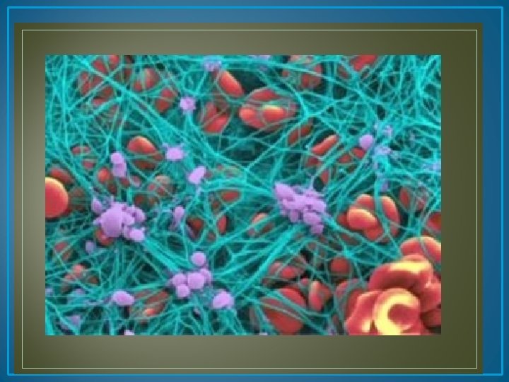
VTE Pathogenesis Virchow, R. (1856). "Thrombose und Embolie. Gefässentzündung und septische Infektion". Gesammelte Abhandlungen zur wissenschaftlichen Medicin (in German). Frankfurt am Main: Von Meidinger & Sohn. pp. 219– 732. Matzdorff AC, Bell WR (1998). Thrombosis and embolie (1846 -1856). endothelial injury hypercoagulability stasis of blood flow Virchow’s triad
VTE Pathogenesis Trauma or surgery Heart valve disease or replacement Indwelling catheters AS AF Left ventricular dysfunction Immobility or paralysis Venous insufficiency, varicose veins Venous obstruction Malignancy Pregnancy, peri-partum period Oestrogen therapy. I Inflammatory bowel disease Nephrotic syndrome Sepsis Thrombophilia
VTE – pathophysiology PE DVT
VTE diagnosis Presenting symptoms and signs are not specific • Only 10 -20% of patients investigated for DVT actually have the disease • DVT impersonators - Cellulitis (6 -9% probability) - Muscle injury (6 -7%) - Superficial Thrombosis (6 -7%) - Chronic Oedema or Venous Insufficiency (5 -7%) - Pelvic tumours compressing lymphatic or venous drainage (1%) - Ruptured Baker's cyst (3%)
VTE diagnosis • • PE presentation is extremely varied and vague PE suspected when dyspnoea, pleuritic chest pain Massive PE ? ---syncope, collapse PE involving segmental or subsegmental arteries ? --minimal or no symptoms
VTE diagnosis PE impersonators Common causes of dyspnoea: (PE – 2%) • asthma (33%) • heart failure (31%) • COPD (9%) • arrhythmia (7%) • infection (5%) • interstitial lung disease (4%) • Anaemia (2%) Common causes of pleuritic pain (PE – 18%) • viral or idiopathic (46%) • pneumonia (8%) • chest wall trauma /cancer/other (25%)
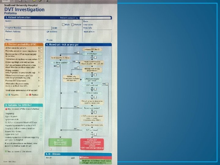
VTE diagnosis Clinical prediction tools for VTE diagnosis
• NICE guidance [CG 144] Published date: June 2012 Objective diagnosis of DVT depends on diagnostic imaging (proximal leg US scan) Patients with low pretest probability and negative D-dimer can have DVT excluded without need for imaging D-dimer test -93 -95% sensitivity 44 -46% specificity Threshold for positive result is determined locally
• NICE guidance [CG 144] Published date: June 2012 CTPA Imaging test of choice for PE diagnosis Allergy to contrast Renal impairment Risk from irradiation is high V/Q spect scan V/Q planar scan Patients with low pretest probability and negative D-dimer can have PE excluded without need for imaging
VTE MANAGEMENT • Anticoagulants are the cornerstone of VTE treatment • Goal: Ø Prevent propagation of thrombus Ø Relieve symptoms Ø Prevent recurrent events
Anticoagulants = Depleted by VKAs = Heparins: UFH (all factors) LMWH (mostly Xa) Fondaparinux (Xa) = Heparinoids Danaparoid Oral anti-Xa: Rivaroxaban Apixaban etc = Direct thrombin inh. Dabigatran Argatroban
VTE MANAGEMENT Initial phase (5 -7 d) Rapidly acting parenteral anticoagulant (LMWH / UFH/ Fondaparinux) overlapped with VKA or rivaroxaban (? other NOACs) Long term phase (7 d-3 m) VKA / rivaroxaban / ? NOAC Extended phase (> 3 -6 months) VKA / rivaroxaban / ? NOAC
VTE MANAGEMENT- initial phase - Current standard for initial VTE treatment LMWH - 200 u/kg, 120 u/kg if GFR<10 -20 ml/min (unlicenced) - SC, once daily, weight adjusted dose - Until INR from concomitant VKA is therapeutic (at least 5 days or until INR >2 for at least 24 h, whichever is longer) • PE with haemodynamic instability • Increased risk of bleeding UFH Creatinine clearance < 30 ml/min → LMWH (anti. Xa monitoring) or → UFH (APTT monitoring) NICE CG 144
VTE MANAGEMENT- long term phase VKA LMWH NOACs Target INR 2. 5 (range 2 -3) VTE on warfarin? Limited evidence to suggest target INR 3. 5 (3 -4) Load with 5 mg (5 -5 -5 -INR) Cancer patients (labile INRs, drug interactions) 150 u/kg sc od from month 2 onwards Fast acting, no need for bridging with LMWH Possible all- oral treatment Replicable dosing effect, monitoring NOT required Rivaroxaban 15 mg bd for 3 weeks→ 20 mg od, no dose adjustments Dabigatran 150 mg bd Apixaban 10 mg bd for 7 days → 5 mg bd
VTE MANAGEMENT- thrombolysis NICE Consider catheter directed thrombolysis for patients with symptomatic iliofemoral DVT + : CG 144 Symptoms >14 days Good functional status Low risk of bleeding >1 year life expectancy Consider pharmacological thrombolysis in patients with PE and haemodynamic instability, do not offer to patients with PE and haemodynamic stability ? PE + haemodynamic stability + right ventricular dysfunction ? ?
VTE MANAGEMENT Temporary inferior vena caval filters NICE CG 144 Offer to patients with proximal DVT or PE who cannot have anticoagulation treatment, and remove the inferior vena caval filter when the patient becomes eligible for anticoagulation treatment. Consider inferior vena caval filters for patients with recurrent proximal DVT or PE despite adequate anticoagulation treatment only after considering alternative treatments such as: increasing target INR to 3– 4 for long-term high-intensity oral anticoagulant therapy or switching treatment to LMWH Ensure that a strategy for removing the inferior vena caval filter at the earliest possible opportunity is planned and documented when the filter is placed, and that the strategy is reviewed regularly
VTE MANAGEMENT Outpatient management of DVT • Safe and effective in most patients • Improved quality of life, cost savings Admit the following patients: • • • Co- existent serious medical pathology Severe acute venous obstruction Patients in significant pain Renal impairment creatinine > 200 µmol/l Liver disease Communication problems Poor social background Limited mobility Active bleeding High risk of bleeding
VTE MANAGEMENT Outpatient management of PE NICE CG 144 Evidence update 55 Selected patients who are at low risk of adverse effects could safely receive anticoagulation on an outpatient basis or be discharged within 3 days PESI or s. PESI could be used to select
RISK ASSESSMENT FOR VTE RECURRENCE • Recurrences cluster in the first 2 -3 months after discontinuation of anticoagulation, then decline reaching a plateau of: - <3% every year (provoked VTE) - 5 -10% (unprovoked VTE) • Prandoni et al: Risk after cessation of anticoagulation - 6 months 8. 6% - 2 years 17. 5% - 5 years 24. 8% - 8 years 30. 3%
RISK ASSESSMENT FOR VTE RECURRENCE NICE CG 144 • Offer a VKA to patients with confirmed proximal DVT or PE within 24 hours of diagnosis. Continue for 3 months. At 3 months consider risks and benefits of continuing VKA. • Offer LMWH to patients with active cancer and confirmed proximal DVT or PE, and continue the LMWH for 6 months. At 6 months, assess the risks and benefits of continuing anticoagulation. • Offer a VKA beyond 3 months to patients with an unprovoked PE, taking into account the patient's risk of VTE recurrence and whether they are at increased risk of bleeding. Discuss with the patient the benefits and risks of extending their VKA treatment. • Consider extending the VKA beyond 3 months for patients with unprovoked proximal DVT if their risk of VTE recurrence is high and there is no additional risk of major bleeding. Discuss with the patient the benefits and risks of extending their VKA treatment.
RISK ASSESSMENT FOR VTE RECURRENCE ACCP recommendations, Chest, 2012 Patient categories Drug Duration (months) Comments First episode of DVT/ PE secondary to a transient (reversible) risk factor VKA 3 Recommendation applies to both proximal and calf vein thrombosis First episode of idiopathic DVT / PE VKA 6 -12 Continuation of anticoagulant therapy after 6– 12 months may be considered First episode of DVT/ PE and cancer LMWH 6 Continuation of LMWH is recommended indefinitely or until the cancer is resolved First episode of DVT/ PE with a documented thrombophilic abnormality VKA 6 -12 Continuation of anticoagulant therapy after 6– 12 months may be considered First episode of DVT/ PE with documented antiphospholipid antibodies or two or more thrombophilic abnormalities VKA 12 Continuation of anticoagulant therapy after 12 months may be considered Second episode of DVT/PE VKA Consider indefinite duration anticoagulation
RISK ASSESSMENT FOR VTE RECURRENCE Factors NOT predictive of recurrence • • • VTE in pregnancy, OCP and gynae surgery Recent surgery, trauma or fracture. Recent immobilzation Hormonal therapy (Tamoxifen) Failed prophylaxis Distal DVT, deep muscular DVT Short term oral anticoagulation 3/12 considered
Hybrid approach for optimizing the duration of anticoagulation in patients with VTE. Goldhaber S Z , and Piazza G Circulation. 2011; 123: 664 -667 Copyright © American Heart Association, Inc. All rights reserved.
NOACs • • For more than 50 years VKAs were the only available anticoagulants September 2008 - NICE TA 157→ Dabigatran for primary VTE prevention (THR, TKR) TA 170, TA 1245 → Rivaroxaban, Apixaban for primary VTE prevention (THR, TKR) TA 249, TA 256, TA 275 → Dabigatran, Rivaroxaban, Apixaban for prevention of stroke and systemic embolism in non valvular AF • July 2012, TA 261 → Rivaroxaban, DVT treatment, prevention of recurrent VTE and DVT • June 2013, TA 287 → Rivaroxaban, PE treatment, prevention of recurrent VTE • Dabigatran for DVT, PE treatment and prevention (December 2014) • Apixaban (June 2015) • Edoxaban (October 2015)
NOACs Comparison of the designs of the trials evaluating NOACs for extended VTE treatment Dabigatran Trial Comparator Design Number of patients Noninferiority margin Duration of prior anticoagulation treatment (mo) Treatment protocol Duration (mo) Rivaroxaban Apixaban RE-SONATE RE-MEDY EINSTEIN-Ext AMPLIFY-Ext Placebo Warfarin Placebo Double-blind 1343 2856 1197 2486 — 2. 85 — — 6 -18 3 -12 6 -12 3 -12 150 mg BID 20 mg OD 2. 5 or 5 mg BID 6 6 -36 6 -12 12 Ø All 3 superior to placebo for extended VTE treatment Ø RE- MEDY - Dabigatran non inferior to warfarin - Less bleeding with dabigatran - MI more common with dabigatran
NOACs Dosing information
Choosing the right anticoagulant for the right VTE patient Characteristic Drug choice Rationale Extensive DVT or massive PE Heparin Such patients often require advanced therapy and were excluded from trials with the NOACs High initial risk of bleeding Heparin Enables dose titration; rapid offset Protamine as an antidote Active cancer LMWH No trials comparing NOACs with LMWH Pregnancy LMWH Warfarin and NOACs cross the placenta Liver dysfunction with increased PT/INR at baseline Warfarin NOACs undergo hepatic metabolism Limited access to anticoag clinic (impaired mobility or geographical inaccessibility) All-oral therapy Creatinine clearance <30 m. L/min NOAC Rivaroxaban or apixaban Warfarin Given in fixed doses without monitoring Only NOACs to be evaluated in all-oral regimens Such patients were excluded from trials with NOACs Creatinine clearance 30 -50 m. L/min Rivaroxaban, apixaban, or edoxaban Less affected by renal impairment than dabigatran; if edoxaban is chosen, the 30 -mg OD dose should be used Dyspepsia or upper gastrointestinal symptoms Rivaroxaban, apixaban, or edoxaban Dyspepsia in as much as 10% given dabigatran Apixaban More gastrointestinal bleeding with dabigatran, rivaroxaban, and edoxaban than with warfarin Recent gastrointestinal bleed Recent acute coronary syndrome Poor compliance with long-term bd dosing Rivaroxaban, apixaban or edoxaban Rivaroxaban or edoxaban Small myocardial infarction signal with dabigatran OD regimens for long-term use
Special situations: Brain Metastases • Brain tumours per se is not a contraindication to anticoagulation • As to prophylaxis, benefits and risks have to be weighted individually using predictive scores such as the Khorana model • In practice, we would give prophylactic treatment to patients with high Khorana scores.
NOACs Potential limitations… • Lack of specific antidotes APART FROM DABIGATRAN PRAXBIND as reversal available NICE approved 2016 • Adherence difficult to assess
NOACs Suggestions for further research… • • NOACs in cancer NOACs in patients with thrombophilia NOACs in HIT NOACs in patients requiring concomitant antiplatelet therapy • NOACs in thrombocytopenic patients
Case 1 • • • 73 yrs. old Admitted with SOB HB 34 Blood test confirmed AIHA Transfused CT showed left axillary node Biopsy : AITL/CD 20 positive Prednisolone started Readmitted with SOB and pulseless legs CT PA : confirmed PE on LMWH CT ANGIO: left legs Arterial clots
• • • Had embolectomy Failed Underwent above knee amputation Had milder chemotherapy Ritux and pred AW for RCHOP Addmitted with pyrexia and SOB Blood test WCC 45 90% plasma cells : plasma cell leukaemia Palliative dexamethasone CT PA : massive bilateral PE expanding
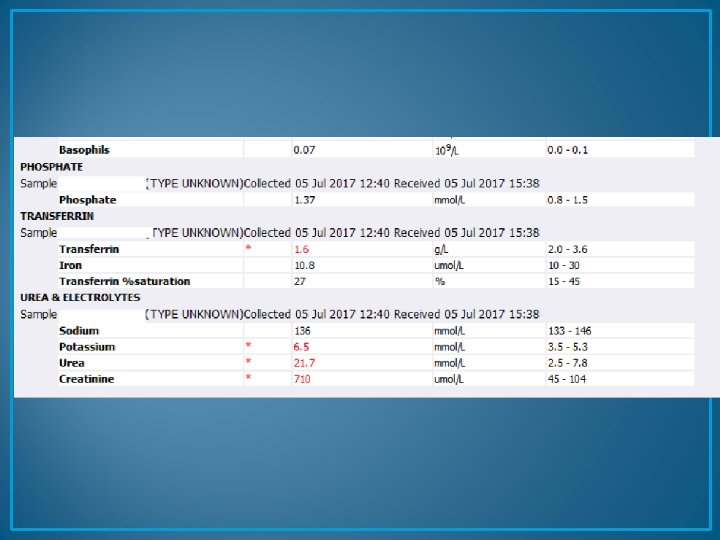
bloods
Initial treatment of PE • • Transfuse platelets Keep PLT>50 full dose LMWH Refractory thrombocytopenia needing daily transfusion DW/Carer and palliative care team Stop treatment and transfer to hospice RIP day 4
Case 2 81 years old • • 6 weeks history of SOB Wt loss Night sweat Left leg swelling Previously IVDU and on Methadone replacement HEP C positive for watch and wait
CT
Had emergency • • • Chest drain Pleural fluid flow: Confirmed DLBCL BMAT: Consistent DLBCL Urgent; Dexamethasone 40 mg Emergency RT for SVC obstructions Had Rituxmab and Prednisolone Stable and escalate to RCHOP X 6 Was in CR Had IT MTX X 4
Readmitted with pleuritic chest pain and fits • CT PA : PE • MRI: brain lymphoma • Dex started and initiated RT to brain as palliative approach • Deteriorated with sepsis and further fits • Stopped all medication • RIP as inpatient day 8
Case 3 55 years old • • • AML Treated with intensive chemo at Barts Had allo SCT Severe GVHD On long term immunosuppressives Admitted with legs cellulittis Spread rapidly and became Necrotising fasiatis Emergeny debridelent HL : blocked US: extensive clots
• Had intermediate dose LMWH • Plan for HL removal Day 3 then line removal • Patient deteriorated day 15 and RIP in south borne ward
Case 4 57 yrs. old • • Abdominal pain and distension for 6 weeks Non specific B symptoms Left leg swealling Bloods: normal Reviewed in OG opd and admitted Had US and CT Us doppler : extensive DVT LMWH started
CT
O&G admission • • For complex abdominal open surgery Haematology advise Stop LMWH and insert IVC filter TEDs stocking Monitoring Day 10 post surgery: biopsy DLBCL for CHEMO Removal of IVC filter and LMWH started
Case 5 56 yrs. old lady • • • Metastatic ovarian cancer Left leg DVT and PE Bloods Stable: PLT 600 Clotting Normal LMWH started as treatment dose Patient find SC too painful and extensive abdominal wall bruising What to give ? Opted for Dabigatran We have reversal in case of bleeding Patient was transgffered to Hospice and died 5 weeks after transfer
Case 6 60 yrs. old known T cell lymphoma • • • CHOEP chemotherapy X 6 - PR ASCT and PD Failed Brentuximab SOB CT PA: Bilateral PE
IV heparin started • patient deteriorated • RIP day 5
Few more points • Prophylaxis with LMWH if PLT>30 and normal renal function and clotting • Careful monitoring essential • Risk benefit must be discussed with patient/relatives and careers • NOACs not licensed but should be consider on case by case basis
• If needed then DABIGATRAN preferred as we have now reversal: Praxbind • IVC filter is temporary >3 months at best if palliative and prognosis is poor any way then leave in situ • In emergency and multiorgan failure with VTE: IV heparin preferable
Thank you !