ULTRASOUND EVALUATION OF THE RENAL ARTERIES AND THE


 is")


 • Observe morphology bilaterally")


 AND NORMAL SYSTOLIC NOTCH (ARROW) AND NORMAL")


 PSV – EDV PSV Normal = <. 8 Abnormal = >.")
. Example Data:")
 of >100 msec is considered abnormal. • Proximal")

- Slides: 46
ULTRASOUND EVALUATION OF THE RENAL ARTERIES AND THE KIDNEY 2017 Technique and Interpretation Holdorf RVT
• The renal arteries arise within 1. 5 cm of the origin of the superior mesenteric artery. • They arise from the lateral sides of the aorta and enter each kidney. • The right renal artery courses behind the IVC to enter the right kidney. • Approximately 22% of patients have two renal arteries. A small percentage have three or more.
Patient History: • Many patients who are given a prescription to have a renal artery Doppler examination will present with hypertension, controlled or not. • Many patients who have hypertension have renovascular hypertension, usually caused from renal artery stenosis. • Renal artery stenosis can be secondary to • Atherosclerosis • Fibromuscular dysplasia • A condition that causes stenosis or aneurysm of the medium-sized arteries (Usually of the kidneys) • Occlusion
• Mechanism of hypertension: • Renin regulates the body's mean arterial blood pressure. • Renin's primary function is to eventually cause an increase in blood pressure, leading to restoration of perfusion pressure in the kidneys. Renin is secreted from kidney cells, which sense changes in renal perfusion pressure, via stretch receptors in the vascular walls. Hypertension: Abnormally high blood pressure and especially arterial blood pressure. Normal Blood Pressure: 120/80 The top number refers to Cardiac output The bottom number refers to peripheral resistance Blood pressure measures the ARTERIAL blood pressure.
NORMAL RENAL ARTERIES
NORMAL RENAL ARTERIES TWO ON THE RIGHT
RENAL STENOSIS
RENAL STENOSIS
RENAL ARTERY ULTRASOUND TECHNIQUE • Technique: Obtain Celiac artery and SMA velocity data Obtain Aorta PSV (Peak Systolic velocity)
NORMAL FLOW RESISTANCES • Aorta high • Renal artery low • Celiac artery low • SMA High (Pre-prandial) LOW (Post Prandial)
MESENTERIC ISCHEMIA • Mesenteric ischemia is a medical condition in which injury of the small intestine occurs due to not enough blood supply. • It can come on suddenly, known as acute mesenteric ischemia, or gradually, known as chronic mesenteric ischemia. Acute disease often presents with sudden severe pain.
CELIAC ARTERY VELOCITY
SMA VELOCITY
SMA VELOCITY
AORTA PSV-PEAK SYSTOLIC VELOCITY
• In transverse: • Locate renal arteries • Left renal vein (LRV) is a landmark for identifying the left renal artery
LEFT RENAL ARTERY IN TRANSVERSE VIEW
RIGHT RENAL ARTERY IN TRANSVERSE VIEW
• Obtain kidney size (length) • Observe morphology bilaterally
KIDNEY MEASUREMENT FOR LEFT AND RIGHT
NORMAL LEFT KIDNEY
OBTAIN PEAK SYSTOLIC VELOCITY AND END DIASTOLIC VELOCITY BILATERALLY OF: • Renal artery • Proximal, mid and distal bilaterally • Upper and lower pole of the kidneys in the segmental arteries
SEGMENTAL ARTERIES
RENAL ARTERY PEAK SYSTOLIC VELOCITY ALSO SHOWING RENAL ARTERY END DIASTOLIC FLOW
SEGMENTAL ARTERIES PSV: BRISK SYSTOLIC UPSTROKE (ARROWHEAD) AND NORMAL SYSTOLIC NOTCH (ARROW) AND NORMAL RI OF. 64
DUPLEX RENAL ARTERIES: OBSERVE FOR MULTIPLE RENAL ARTERIES
INTERPRETATION • Renal arteries and kidney arteries are normally LOW RESISTANT in quality as are: • Celiac arteries • Hepatic arteries • Splenic arteries • The aorta is usually higher resistant in quality, as are a fasting SMA and IMA
LOW RESISTANT RENAL ARTERY WAVEFORM
HIGH RESISTANT AORTIC WAVE FORM
RAR: RENAL TO AORTIC RATIO Highest Renal Artery PSV Aorta PSV* * Taken by the SMA
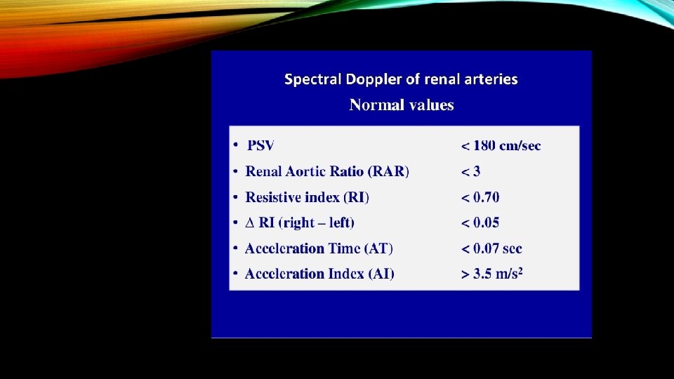
RAR INTERPRETATION • Normal = < 3. 5 • Abnormal = > 3. 5 * • Suggests > 60% diameter reduction
CANNOT USE RAR IF THE FOLLOWING IS PRESENT If AAA is detected If aortic PSV > 90 cm/s If aortic PSV < 40 cm/s
RAR Example: Highest Renal artery PSV = 260 Aortic PSV = 55 260/55 = 4. 7 • Normal = < 3. 5 • Abnormal = > 3. 5 Range is Abnormal Renal artery
AORTA WITH A PSV OF 3. 7 M/S OR 368. 7 CM/S REASON: STENOSIS OF THE MIDDLE AORTIC SEGMENT
ABNORMAL RENAL ARTERY WITH PSV = 326. 6 CM/S
DOPPLER ULTRASOUND INVESTIGATION OF THE KIDNEY • Kidney arteries are normally low resistant in quality. • Observe the kidney for morphologic abnormalities (cysts, cortex thinning, other defects). • Normal pole to pole length varies from 10 – 12 cm, depending on patient size.
RENAL VASCULATURE
RENAL SEGMENTAL ARTERY FIRST BRANCH OFF THE MAIN RENAL ARTERY
Resistive Index (RI) PSV – EDV PSV Normal = <. 8 Abnormal = >. 8 • RI = (peak systolic velocity - end diastolic velocity ) / peak systolic velocity • the normal value is ≈ 0. 60 • with 0. 70 being around the upper limits of normal
Abnormal calculations indicate an increase in distal resistance (e. g. nephron-sclerotic disease). Example Data: • PSV of 45 cm/sec: EDV of 5 cm/sec. End Diastolic Velocity EDV 5 Peak Systolic Velocity PSV 45 PSV – EDV 45 – 5 PSV 45 • The RI 40 45 = 0. 88 Normal value is <0. 8
• An acceleration time (AT) of >100 msec is considered abnormal. • Proximal high grade stenosis/occlusion of the renal artery may result in dampened, weak, Doppler signals distally, but still of low resistant quality. • Another term for this is TARDUS-PARVUS. • The stenosis is located proximal to the waveform • The waveform is distal to the stenosis.
RENAL ARTERY TARDUS PARVUS