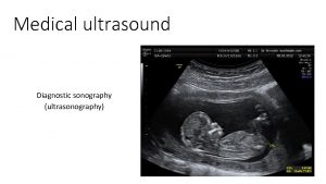
Medical Ultrasound Image documentation and reporting Ultrasound Documentation


- Slides: 13

Medical Ultrasound: Image documentation and reporting

Ultrasound Documentation � Ultrasound requests/examinations should be justified and performed to answer a specific clinical question � “An ultrasound report is a public document and part of the patient’s medical record” (SCo. R & BMUS 2015) The report should be written and issued by a suitably qualified practitioner who performs the examination

Imaging Report – RCR Standards The Royal College of Radiologists (RCR) state that investigations must only be reviewed by individuals who have the relevant knowledge and qualifications to do so. A written account of the findings from said review is required in order to influence patient management and treatment where necessary.

Imaging Report – RCR Standards Standard 1 - Every imaging investigation must be reported within an agreed time by an individual qualified to interpret that particular investigation. Prompt feedback is critical in the management of patients. Standard 2 - All imaging investigations must be accompanied by a formal permanently recorded written report. A verbal report can be given to the patient at the time of scan depending on individual preference, however, a separate written account must be documented

Image documentation: why and how? Any kind of patient documentation should be stored in an appropriate way. Data stored should clearly reflect the information gathered during the ultrasound examination. The patients’ data should be kept safe.

Reports should: Comply with local protocol Be concise Be easy to understand Be unambiguous Use ultrasound terminology Explain significance of findings Only use common abbreviations Be audited regularly to ensure satisfactory standards and facilitate improvements

The Written Report In essence the written report should answer the clinical question The author is responsible for the accuracy of the report

Report writing -What Should be Included? Reports should include: Patient’s full details (Name, Do. B, Unique identifier/CHI) Examination date and time Clinical history and reason for examination Examination performed/Structures assessed/Probe employed A record of any relevant measurements taken Description of relevant findings Interpretation of findings/diagnosis/differentials Actions taken on account of findings Recommendations for follow up where appropriate Limitations of examination Details of the individual performing the procedure

Reporting: Exam findings � Location (area, tissue) � Size � Type (cystic, solid) � Echogenicity (anechoic, hypoechoic, isoechoic, hyperechoic) � Echo-structure (homogenous, heterogenous) � Borders (well defined, regular, irregular) � Relation to surrounding structures (attached to…, displacing…) � Response to compression (compressible, movable, floating) � Vascularity

How to document the pathologies? � Images: most clearly showing the pathology, its size, vascularity and relation to surrounding tissues. should include common landmarks. MUST be recorded in two perpendicular planes. � Description: should support documented images, additional information on location of the pathology, relation to other structures, compressibility, displacement, and response to the dynamic examination (i. e. flexion/extension of the related joint).

Image Management All Ultrasound service providers should have facility to store whole examination's (RCR, 2014)

References Royal College of Radiologists, 2014, Standards for the provision of an ultrasound service [online] https: //www. rcr. ac. uk/sites/default/files/publication/BFCR%2814%2917_Standard s_ultrasound. pdf Society and College of Radiographers and British medical Ultrasound Society, 2015, Guidelines for Professional Ultrasound practice [online] https: //www. bmus. org/tatic/uploads/resources/GUIDELINES_FOR_PROFESSIONAL_ULTR ASOUND_PRACTICE. pdf

Thank you
 Score de ferriman
 Rbt documentation and reporting
 Medical ultrasound awareness month
 Amplitude mode
 Ultrasound image optimisation
 Cheddar method medical documentation
 Types of medical documentation
 معنى medication reconciliation
 Difference between medical report and medical certificate
 Analog image and digital image
 California medical license application
 Gbmc infoweb
 Torrance memorial transitional care unit
 Cartersville medical center medical records
