ULTRASOUND SCANNING Introduction to Ultrasound Ultrasound Scanning Physics


 �Hydronephrosis. �Infections and abscess.")


 �Mass – Fibroids and")


 Spleen (nodules and")


 selection done by a procedure called “gating” Gating +beam dimensions, define “sample")
 B-mode imaging + Doppler")

- Slides: 36
ULTRASOUND SCANNING
Introduction to Ultrasound
Ultrasound Scanning Physics �Characterized by sound waves of high frequency Higher than the range of human hearing �Sound waves are measured in Hertz (Hz) Diagnostic U/S = 1 -20 MHz �Sound waves are produced by a transducer
Instrumentation - Ultrasound Probes B C
Transducers/Probes • Sector scanner – Fan-shaped beam – Small surface required for contact – Cardiac imaging • Linear scanner – Rectanglular beam – Large contact area required • Curvi-linear scanner – Smaller scan head – Wider field of view
Indications • As a compliment to abdominal radiographs – To determine the origin of an abdominal mass • Spleen, Liver – To facilitate fine needle aspiration/cystocentesis – To evaluate organ parenchyma – To rule in/out intestinal obstruction (foreign body) – ***If clinical signs or history indicate abdominal ultrasound, then it should be performed even if radiographs are normal!!!
Indications for USG �Indications: abdomen, OBG, & Gynec. �Abdomen: Liver- Size, configuration, texture and contour. �To R/o ascites, abscess, cystic disease etc. �Growth – Primary and secondaries. �Jaundice: Viral Obstructive – Calculi, growth and annular pancreas.
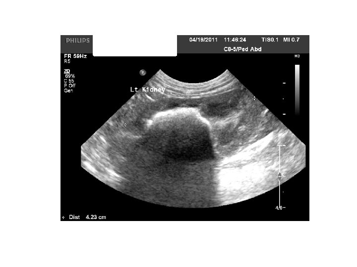
Renal US �Kidney: size and trauma. �Congenital: Agenesis �Calculi (Radiolucent) �Hydronephrosis. �Infections and abscess. �Tumors �
Pancreas �Pancreas: �Infections – calcification, duct dilatation �Complications of pancreatitis. �Pseudo cyst and abscess. �Spleen: �Splenomegaly- Fever and abscess. �Portal hypertension �RTA – Splenic injury
Abdominal US �Abdominal mass, Lymphadenopathy �G. I tract: Mass Obstruction Volvulus Worms Omental secondaries
OBG � OBG: � Missed abortion � Ectopic pregnancy � 5 th week – Gestation sac � 6 th week - Fetal pole �Cardiac activity � 12 th week – Head (BPD) � 18 -20 week- Fetal anomaly scan. � Placenta: Praevia and abruption. � Hydramnios, twins, molar pregnancy. � Position of fetus - Cephalic, breech. � Gestational age: BPD, HC, AC and FL
Gynec �Gynec: �Uterus – Size, congenital anomaly (Bifid, septate etc) �Mass – Fibroids and growth. �Infertility. �Ovaries: Ovulation, mass – cysts and growth. �P. I. D: Free fluid in POD.
Why do you need both X-ray and USG? �Examples Prostatic adenocarcinoma seen on ultrasound ▪ Has it spread to the lumbar vertebrae? Coughing patient with mitral regurgitation on echocardiogram ▪ Does the patient have pulmonary edema? Enlarged liver on radiographs ▪ Can get a guided FNA with ultrasound
Mirror Image Artifact
Comet Tails
Artifacts • Acoustic shadowing – U/S beam does not pass through an object because of reflection or absorption – Black area beyond the surface of the reflector – Examples: cystic calculi, bones • Acoustic enhancement – Hyperintense (bright) regions below objects of low U/S beam attenuation – AKA Through transmission – Examples: cyst or urinary bladder
Acoustic Shadowing
Acoustic Enhancement
Artifacts �Refraction: Occurs when the sound wave reaches two tissues of differing acoustic impedances U/S beam reaching the second tissue changes direction May cause an organ to be improperly displayed
Ultrasound-Guided FNA/ Biopsies � Routinely aspirate: Liver (masses and diffuse disease) Spleen (nodules and diffuse disease) Gastrointestinal masses Enlarged lymph nodes Enlarged prostate Pulmonary/ mediastinal masses (usually don’t biopsy due to risk of pneumothorax � Occasionally aspirate: Kidneys (esp. if enlarged) Pancreas Urinary bladder masses � Never aspirate: Adrenal glands Gall bladder
Ultrasound-Guided FNA/Biopsies � Non-aspiration Technique 22 g 1. 5 in needle 10 cc syringe Short jabs into organ Spray onto slide, smear, and check abdomen for hemorrhage
DOPPLER
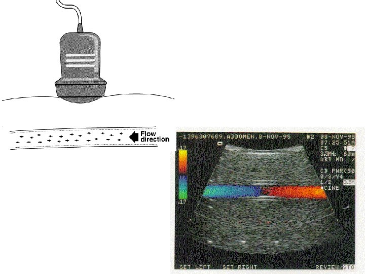
Waves from stationary and moving sources Stationary Moving
Doppler Effect �Shift in perceived frequency when either source or listener are moving relative to one another �Familiar occurrence in audible sounds �Also occurs in medical ultrasound
Please note: the ‘squishing’ of the wave-fronts in the middle diagram and the stretching in the lower diagram are exaggerated. Realistically the Doppler shifts are so small in ultrasound you would hardly see any difference in the wave-fronts compared to the unshifted one on top. These artists’ diagrams.
Doppler shift � Doppler shift is the difference between the transmitted and received frequencies. � Transmitted and received frequencies are in the MHz range � Doppler shift frequencies often in audible range
Scattering from blood � Source of signals for flow imaging: Red blood cells Much smaller than l Rayleigh scatterers � Scattering increases with frequency � Scattering increases with number of targets Double the number of scatterers, scattered intensity doubles!
Cosine Function h cos q = a/h q a h a cos 2 o = 0. 999 h cos 60 o = 0. 5 a
The frequency of the Doppler shift is proportional to the cosine of the Doppler angle Angle formed by the ultrasound beam and the direction of flow � Doppler frequency varies with the cosine of the angle. Cosine = 1 for 0 o Cosine = ½ for 60 o Cosine = 0 for 90 o For angles between 0 and 10 o, cosine is close to 1. � � The larger the angle (up to 90 o), the smaller the cosine
Angle Correct Error
Range (depth) selection done by a procedure called “gating” Gating +beam dimensions, define “sample volume”
Duplex Mode (Duplex Doppler) B-mode imaging + Doppler
THANK YOU