Endocrine System Overview Endocrine system nervous system controls


 valves . 1 Blood returning to the heart")
 valves. Aorta Pulmonary trunk As ventricles contract and")
 valves. As ventricles relax and intraventricular pressure falls,")
 Inferior vena cava (IVC) Coronary sinus Tricuspid valve Right atrium")


 tracing. Ventricular depolarization Ventricular repolarization Atrial depolarization Sinoatrial")
 The relationship of blood vessels to each other")


- Slides: 29
Endocrine System Overview • Endocrine system + nervous system controls and coordinates metabolic activites • Regulates metabolic activities via hormones - transported in blood • Slow acting but longer lasting than nervous system responses Endocrine system controls and integrates: • • • Maintenance of electrolyte, water, and nutrient balance of blood Reproduction Growth and development metabolism and energy utilization body defenses
Pineal gland Hypothalamus Pituitary gland Thyroid gland Parathyroid glands (on dorsal aspect of thyroid gland) Thymus Adrenal glands Pancreas Gonads • Ovary (female) • Testis (male) © 2016 Pearson Education,
Hypothalamus Anterior lobe of pituitary Anterior Pituitary Hormones: Hypothalamic neurons synthesize releasing and inhibiting hormones (GHRH, GHIH, TRH, CRH, Gn. RH, PIH). Superior hypophyseal artery Growth hormone (GH) Thyroid-stimulating hormone (TSH) Adrenocorticotropic hormone (ACTH) Follicle-stimulating hormone (FSH) Luteinizing hormone (LH) Prolactin (PRL) © 2016 Pearson Education, Posterior Pituitary Hormones: Oxytocin Anti-Diuretic Hormone
ADH – Anti- Diuretic Hormone Water Homeostasis • High solute concentration causes its release from the posterior pituitary • Helps reabsorb water in the kidneys • Increases blood volume & hence BP
Thyroid Gland Thyroid Hormones: T 3, T 4 and Calcitonin T 3 & T 4 – Helps regulate metabolism Oxidize glucose to make ATP Regulate HR, BP Calcitonin – Helps regulate high blood calcium levels back to normal Figure 16. 8
Pancreas Location & Function Secretes two major hormones 1. Insulin - Lowers blood glucose to normal levels - Converts excess glucose to glycogen in the liver - Helps cells utilize glucose to make ATP 2. Glucagon - increases dropping blood glucose levels to normal
Regulation of Blood Glucose Levels • The hyperglycemic effects of glucagon & the hypoglycemic effects of insulin Figure 16. 18
Adrenal Cortex Zona Glomerulosa – Mineralocorticoids – Aldosterone - increases blood concentrations of Na+ and decreases K+ concentrations; increases blood volume & BP Zona Fasciculata - Glucocorticoids – Cortisol - Glucose homeostasis Zona Reticularis – Androgens - Sex hormones Adrenal Medulla – Epinephrine & Nor- epinephrine “Fight or flight” hormones Increase HR and blood flow to muscles, heart and brain
Blood – Composition and Function Blood is the only fluid tissue in body Physical Characteristics - Blood is a viscous, opaque fluid with metallic taste - Color varies with O 2 content - High O 2 levels show a scarlet red - Low O 2 levels show a dark red - p. H 7. 35– 7. 45 - Average volume: Males: 5– 6 L Females: 4– 5 L Composition of Blood Type of connective tissue Matrix is nonliving fluid called plasma – Temperature & p. H homeostasis, transport of nutrients and wastes Cells are living blood cells called formed elements Cells are suspended in plasma Formed elements Erythrocytes (red blood cells, or RBCs) - Transports oxygen and carbon dioxide Leukocytes (white blood cells, or WBCs) - defense and immunity Platelets – clot formation
Figure 18. 2 a Location of the heart in the mediastinum. Midsternal line 2 nd rib Sternum Diaphragm © 2016 Pearson Education, Inc. Location of apical impulse
The systemic and pulmonary circuits . Capillary beds of lungs where gas exchange occurs Pulmonary Circuit Pulmonary arteries Aorta and branches Venae cavae Right atrium Pulmonary veins Left atrium Left ventricle Right ventricle Heart Systemic Circuit Oxygen-rich, CO 2 -poor blood © 2016 Pearson Education, Inc. Oxygen-poor, CO 2 -rich blood Capillary beds of all body tissues where gas exchange occurs
Gross anatomy of the heart. Left common carotid artery Brachiocephalic trunk Left subclavian artery Aortic arch Superior vena cava Ligamentum arteriosum Right pulmonary artery Left pulmonary artery Ascending aorta Left pulmonary veins Pulmonary trunk Auricle of left atrium Right pulmonary veins Circumflex artery Right atrium Right coronary artery (in coronary sulcus) Left coronary artery (in coronary sulcus) Anterior cardiac vein Left ventricle Right marginal artery Great cardiac vein Small cardiac vein Anterior interventricular artery (in anterior interventricular sulcus) Inferior vena cava Apex Anterior view © 2016 Pearson Education, Inc.
Gross anatomy of the heart. Aorta Left pulmonary artery Superior vena cava Left atrium Right pulmonary artery Left pulmonary veins Pulmonary trunk Right atrium Mitral (bicuspid) valve Right pulmonary veins Fossa ovalis Aortic valve Pectinate muscles Pulmonary valve Tricuspid valve Right ventricle Left ventricle Chordae tendineae Papillary muscle Interventricular septum Trabeculae carneae Epicardium Myocardium Inferior vena cava Endocardium Frontal section © 2016 Pearson Education, Inc.
Gross anatomy of the heart. Aorta Superior vena cava Right pulmonary artery Left pulmonary artery Right pulmonary veins Left pulmonary veins Right atrium Auricle of left atrium Left atrium Inferior vena cava Great cardiac vein Coronary sinus Posterior vein of left ventricle Right coronary artery (in coronary sulcus) Left ventricle Posterior interventricular artery (in posterior interventricular sulcus) Middle cardiac vein Right ventricle Apex Posterior surface view © 2016 Pearson Education, Inc.
Left subclavian artery Left common carotid artery Aortic arch Brachiocephalic trunk Superior vena cava Ligamentum arteriosum Pulmonary trunk Gross anatomy of the heart. Pulmonary valve Right auricle Pulmonary vein Left auricle Tricuspid valve Chordae tendineae of mitral valve Papillary muscle Myocardium of left ventricle Myocardium of right ventricle Trabeculae carneae Interventricular septum Internal aspect of ventricles; dissection of view © 2016 Pearson Education, Inc. similar to (e)
Label the parts for review
The function of the atrioventricular (AV) valves . 1 Blood returning to the heart fills atria, pressing against the AV valves. The increased pressure forces AV valves open. Direction of blood flow Atrium Cusp of atrioventricular valve (open) 2 As ventricles fill, AV valve flaps hang limply into ventricles. Chordae tendineae 3 Atria contract, forcing additional blood into ventricles. Ventricle AV valves open; atrial pressure greater than ventricular pressure © 2016 Pearson Education, Inc. Papillary muscle
The function of the semilunar (SL) valves. Aorta Pulmonary trunk As ventricles contract and intraventricular pressure rises, blood is pushed up against semilunar valves, forcing them open. Semilunar valves open © 2016 Pearson Education, Inc.
The function of the semilunar (SL) valves. As ventricles relax and intraventricular pressure falls, blood flows back from arteries, filling the cusps of semilunar valves and forcing them to close. Semilunar valves closed © 2016 Pearson Education, Inc.
Superior vena cava (SVC) Inferior vena cava (IVC) Coronary sinus Tricuspid valve Right atrium Right ventricle Pulmonary semilunar valve Oxygen-poor blood Pulmonary trunk Pulmonary arteries SVC Coronary sinus Pulmonary trunk Right atrium Tricuspid valve Right ventricle IVC The heart is a double pump, each side supplying its own circuit. Pulmonary semilunar valve To heart Oxygen-poor blood is carried in two pulmonary arteries to the lungs (pulmonary circuit) to be oxygenated. Oxygen-poor blood returns from the body tissues back to the heart. Systemic capillaries To body To lungs Pulmonary capillaries Oxygen-rich blood returns to the heart via the four pulmonary veins. Oxygen-rich blood is delivered to the body tissues (systemic circuit). Aorta To heart Pulmonary veins Aortic semilunar valve Left atrium Mitral valve Left ventricle Aorta Aortic semilunar valve Left ventricle Mitral valve Left atrium © 2016 Pearson Education, Inc. Four pulmonary veins Oxygen-rich blood
Pathway of Blood Through Heart • Right side of the heart • Superior vena cava (SVC), inferior vena cava (IVC), and coronary sinus • Right atrium • Tricuspid valve • Right ventricle • Pulmonary semilunar valve • Pulmonary trunk • Pulmonary arteries • Lungs © 2016 Pearson Education, Inc.
Pathway of Blood Through Heart • Left side of the heart • • Four pulmonary veins Left atrium Mitral valve Left ventricle Aortic semilunar valve Aorta Systemic circulation © 2016 Pearson Education, Inc.
Slide 5 Intrinsic cardiac conduction system Superior vena cava Right atrium 1 The sinoatrial (SA) node (pacemaker) generates impulses. Internodal pathway 2 The impulses pause (0. 1 s) at the atrioventricular (AV) node. Left atrium 3 The atrioventricular (AV) bundle connects the atria to the ventricles. Subendocardial conducting network (Purkinje fibers) 4 The bundle branches conduct the impulses through the interventricular septum. Interventricular septum Anatomy of the intrinsic conduction system showing the sequence of electrical excitation © 2016 Pearson Education, Inc.
QRS complex R An electrocardiogram (ECG) tracing. Ventricular depolarization Ventricular repolarization Atrial depolarization Sinoatrial node T P Atrioventricular node Q P-R Interval 0 S 0. 2 © 2016 Pearson Education, Inc. S-T Segment Q-T Interval 0. 4 Time (s) 0. 6 0. 8
Venous system Large veins (capacitance vessels) The relationship of blood vessels to each other and to lymphatic vessels Arterial system Heart Large lymphatic vessels Lymph node Lymphatic system Small veins (capacitance vessels) Elastic arteries (conducting arteries) Muscular arteries (distributing arteries) Arteriovenous anastomosis Lymphatic capillaries Sinusoid Postcapillary venule Arterioles (resistance vessels) Terminal arteriole Precapillary Thoroughfare Capillaries sphincter channel (exchange vessels)Education, Inc. © 2016 Pearson Metarteriole
Tunica intima • Endothelium • Subendothelial layer • Internal elastic membrane Artery Vein Tunica media (smooth muscle and elastic fibers) • External elastic membrane Tunica externa (collagen fibers) Generalized structure of arteries, veins, and capillaries • Vasa vasorum Valve Capillary network Lumen Basement membrane Capillary Endothelial cells © 2016 Pearson Education, Inc.
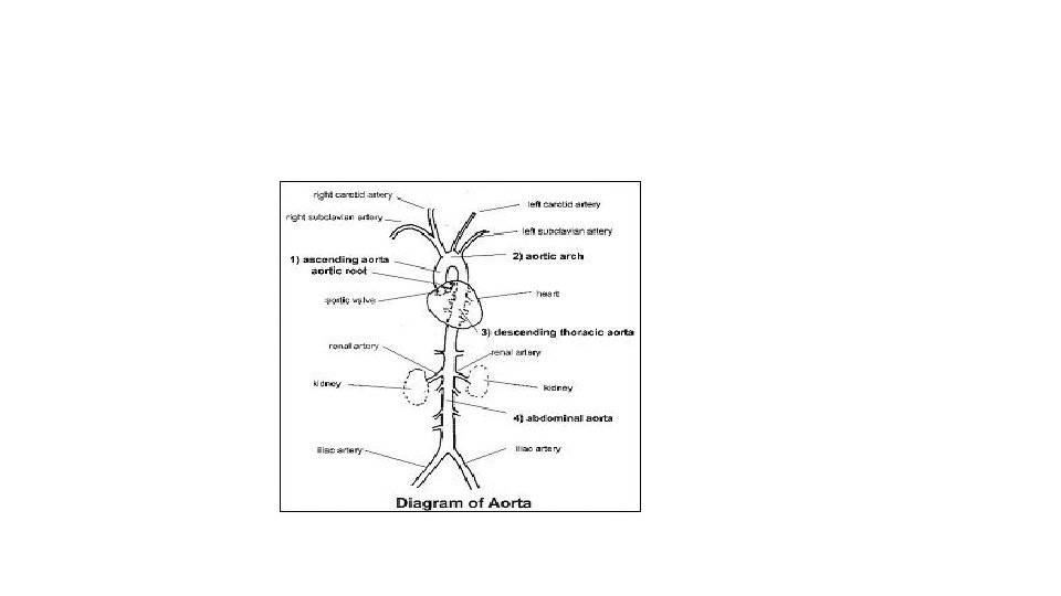
Arteries of the head and trunk Internal carotid artery External carotid artery Common carotid arteries Arteries that supply the upper limb Subclavian artery Vertebral artery Subclavian artery Brachiocephalic trunk Aortic arch Ascending aorta Axillary artery Coronary artery Celiac trunk Brachial artery Abdominal aorta Superior mesenteric artery Renal artery Radial artery Gonadal artery Ulnar artery Inferior mesenteric artery Common iliac artery Internal iliac artery Deep palmar arch Superficial palmar arch Digital arteries Arteries that supply the lower limb External iliac artery Femoral artery Popliteal artery Anterior tibial artery Posterior tibial artery Arcuate artery Illustration, anterior view © 2016 Pearson Education, Inc.
Veins of the head and trunk Dural venous sinuses External jugular vein Vertebral vein Veins that drain the upper limb Internal jugular vein Right and left brachiocephalic veins Subclavian vein Superior vena cava Cephalic vein Axillary vein Brachial vein Great cardiac vein Basilic vein Hepatic veins Splenic vein Hepatic portal vein Renal vein Median cubital vein Superior mesenteric vein Ulnar vein Inferior mesenteric vein Radial vein Inferior vena cava Common iliac vein Internal iliac vein Digital veins Veins that drain the lower limb External iliac vein Femoral vein Great saphenous vein Popliteal vein Posterior tibial vein Anterior tibial vein Small saphenous vein Dorsal venous arch Dorsal metatarsal veins © 2016 Pearson Education, Inc. Illustration, anterior view. The vessels of the pulmonary circulation are not shown.