ULTRASONOGRAPHY IN DENTISTRY Dentistry in the modern era


 Soft tissue Mass, patients are exposed to")


 Disease The ultrasound probe may offer an important alternative to traditional")


- Slides: 29
ULTRASONOGRAPHY IN DENTISTRY
Dentistry in the modern era is emerging with the use of advanced imaging techniques such as computed tomography(CT), Magnetic Resonance Imaging (MRI), Nuclear Medicine (NM), and Ultrasound (US), of which MRI and Ultrasound are the only imaging techniques, which operate without causing radiation hazards to the patient. Ultrasound is one of the advanced imaging techniques which uses sound waves for viewing the normal and Pathological conditions involving bone and soft tissue of the oral and Maxillofacial region. `
In Dentistry for detecting a bony (or) Soft tissue Mass, patients are exposed to multiple radiographs which might cause radiation effects at the tissue and organ level. So this valuable technique can be used prior to use of X-ray radiation.
Sonography
Medical Ultrasound uses the frequency of 1 -15 MHz (2. 5, 3. 5, 7. 5 and 10 MHz). The transducer has a special property called piezoelectric effect i. e. they can convert sound waves in to electrical waves and 1 vice versa. Image Display- Ultrasound signals may be displayed in several ways 1. A-Mode ( Amplitute mode – not in use) 2. B-Mode ( Brightness Mode – Producing different echogenicity) 3. TM Mode Motion mode – used in foetal (Time Motion) ECG and Doppler study Special Imaging Modes 1. Harmonic imaging 2. Spatial Compounding 3. 3 -D Ultrasound
From sound to image The creation of an image from sound is done in three steps: 1 -Producing a sound wave. 2 -Receiving the echoes. 3 -Forming the image. During the ultrasound examination, a machine called a transducer is used to view the target organ and produce pictures for study. The transducer emits sound and detects the returning echoes when it is placed on or over the body part being studied. When the emitted sound encounters a border between two tissues that conduct sound differently, some of the sound waves bounce back to the transducer, creating an echo. The echoes are analyzed by a computer in the ultrasound machine and transformed into moving pictures of the organ or tissue being examined.
Ultrasonography is one method of imaging which lacks radiation hazards, this imaging technique can be used for bone and soft tissue examination, either Normal (or) Pathological lesions, detection of calculi in major salivary glands, TMJ imaging, Detection of fractures & Vascular lesions. So proper application of this method can be of great importance in dentistry.
Application of US in Dentistry Depending on the application and ultrasonic intensities it is divided into two 1. Diagnostic ultrasound – the ultrasonic intensities used are typically 5 to 500 m. W/cm 2 and it includes : • Ultrasonic echography has been used as an instant, non-invasive method for the observation of relatively deep areas, Recently, however high frequency echography has been developed that can provide detail. investigation of more superficial regions • Ultrasound in dentistry is used for detection of fractures of the Maxillo facial region i. e Nasal bone fractures, Orbital rim fractures, Maxillary fractures, Mandibular fractures, Zygomatic arch fractures and for locating the position of Mandibular condyles. And Post operative view can be done instantly to view the reduction & healing of fractures.
• Ultrasound can be used to detect parotid lesions where solid and cystic lesions are reliably differentiated and diffuse enlargement of the parotid gland (or) focal disease is readily shown by Ultrasound. Sonographically, benign lesions usually look well defined, homogeneous and hypoechoic, while Malignant lesions tend to be ill defined and hypoechoic with heterogeneous internal architecture and enlarged cervical lymph node may be visible and reactive intra parotid lymph nodes may also be readily assessed.
• inflammatory soft tissue conditions of the head and neck region and superficial tissue disorders of the maxillofacial region. Ultra sound can provide the content of the lesion before any surgical procedure, both solid and cystic contents could be identified in ultrasound. The mixed lesions should be considered neoplastic and should be. biopsied before surgical procedure Ultrasound with aid of high resolution transducer, can demonstrate the internal Muscle structures more clearly than CT. hyper echoic bands, which corresponds to the internal fascia are usually observed on US Image of normal, Muscles and are sometimes referred to as septa. These bands diminish or disappear with inflammation; hence this is an important structural Index of Masseteric Infection.
Transverse sonogram of the left parotid gland. Echogenic swelling of both parotid glands was present. The deep parts of the gland are not visualized due to strong acoustic attenuation. Sialoadenosis was diagnosed
• Ultrasound is also an accurate Modality for measuring the thickness of Muscles, data regarding thickness may provide information useful in diagnosis and treatment especially in. follow up examination. • Ultrasound can also be used for detecting Sialoliths in Parotid, submandibular and sublingual salivary glands, which appear as echodense spots with a. characteristic acoustic shadow • In Ultrasound, color Doppler sonography has been developed to identify vasculatures and to enable evaluation of the blood flow, velocity and vessel resistance together with surrounding Morphology. It can be used for detecting the coarse of the facial artery and for detecting hemangioma.
• So the use of ultrasound is unlimited, so proper application of this Imaging can be of use in detecting various normal & Pathological lesion in the Maxillofacial region. • Swellings in orofacial region • Periapical lesions • Lymph nodes – benign/malignant • Intraosseous lesions • Temporomandibular disorders • Assessment of masticatory muscles in temporomandibular dysfunction • Congenital vascular lesions of head and neck • Primary lesions of the tongue
• Detecting the foreign bodies • Distracted mandibular bone • Miscellaneous lumps and bumps of the head and neck • Submandibular gland injection of botulinum toxin for hypersalivation in cerebral palsy 2. Therapeutic ultrasound – use intensities of 1 to 3 W/cm 2 and it includes • Myofascial pain • Temporomandibular joint dysfunction • Bone healing and oestointegration • Oral Cancer • Healing of full thickness excised skin lesion.
• Other applications 1. 2. 3. 4. Ultrasonic scaling Modifications of the ultrasonic scaling Endodontics Surgical applications
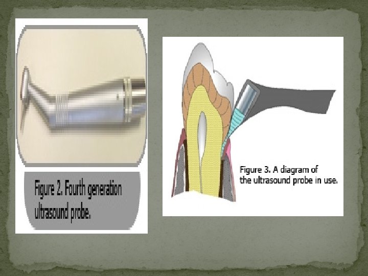
Dental ultrasound technology An innovative device is currently being developed that uses ultrasound waves for dental diagnostic purposes. The advantage of ultrasound is that it is very safe and does not emit any cancer causing ionizing radiation. Dental ultrasound will be able to detect tooth decay in a new, more accurate way, the exact location and size of the cavity even if the cavity is between teeth (interproximal) or under and existing filling made from a metal, which is impenetrable to dental x-rays. Software is under development for accurately detecting Periodontal (gum) Disease. dental ultrasound software will be able automatically measurement of the space between the gum and tooth to determine the health status of the gums.
Detecting Periodontal (gum) Disease The ultrasound probe may offer an important alternative to traditional manual periodontal probing because 1 -it is non-invasive 2 - painless 3 - potentially more sensitive 4 may give additional histological information. *more research is needed to validate these claims
Ultrasonographic picture of the swelling showing oval-shaped lesion with anechoic internal echo pattern and unchanged posterior wall echo. These features are suggestive of a dentigerous cyst
Ultrasonography picture showing anechoic lesion in the region of buccal space suggesting of buccal space abscess. (USG, 8 M 8 H 2 probe
ADVANTAGES • particularly useful in the examination of superficial structures. • widely available and relatively inexpensive. • non-invasive • tolerated by the patient. • not interfere with normal function. • Artifacts are few. • highly acceptable to most patients. • Images are rapidly acquired. • Images are simple to store and retrieve. • Images obtained are easy to read once the observer is trained. • performed without heavy sedation. • no known cumulative biological effects. • easily accessible and painless. • absolute non ionizing nature. • Equipments are relatively cheap. • possibility of real time imaging. • helps to distinguish between solid and cystic lesions.
DISADVANTAGES • cortex can be visualized in intact bone due to ultrasound frequencies. • be performed by experienced investigators. • Images when archived they may be difficult to orientate and to interpret unlike CT and MRI scans, which have acquired in standard reproducible scans. • The difficulty of picturing the TMJ using ultrasounds depends on the limited accessibility of the deep structures, especially the disc, due to absorption of the sound waves by the lateral portion of the head of the condyle and the zygomatic process of the temporal bone. • Ultrasonography waves do not visualize bone or pass through air, which acts as an absolute barrier during both emission and reflection.
Summary Ultrasound is an inexpensive, non-invasive and readily available imaging technique, that can be used as an primary investigative Imaging technique So as to avoid radiation hazards caused by X-ray radiation (or) MRI which may be highly economical to the patients. So proper application and Utilization of this technique can be of great use in dentistry.