Lecture on Ultrasonography www Assignment Point com Ultrasonography


")


 image of a fetus (sagital view).")

- Slides: 28
Lecture on Ultrasonography www. Assignment. Point. com
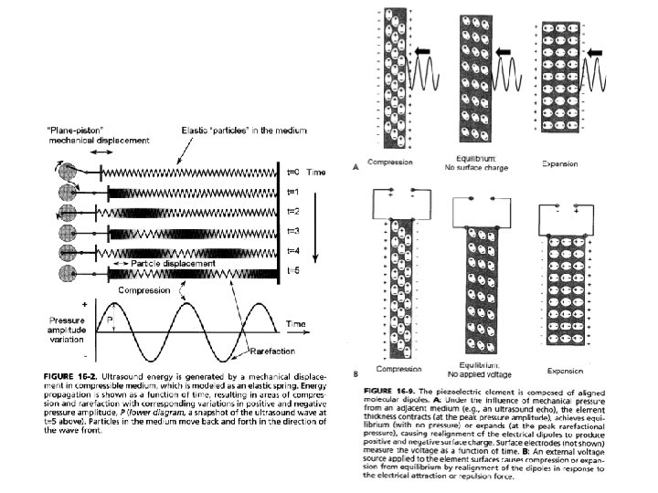
Ultrasonography • Ultrasound in the frequency range of 1 − 20 MHz is used in diagnostic ultrasonography. • The velocity of propagation of ultrasound through a medium depends upon its compressibility. • Lower compressibility results in higher velocity. Typical velocities in human tissues: 330 m/s in air (the lungs); 1, 540 m/s in soft tissue; and 3, 300 m/s in bone.
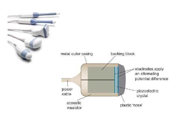
• A wave of ultrasound may get reflected, refracted, scattered, or absorbed as it propagates through a body. • Most modes of diagnostic ultrasonography are based upon the reflection of ultrasound at tissue interfaces. • A gel is used to minimize the presence of air between the transducer and the skin to avoid reflection at the skin surface.
• Typically, pulses of ultrasound of about 1 μs duration at a repetition rate of about 1, 000 pps (pulses per second) are applied, and the resulting echoes are used for locating tissue interfaces and imaging. • Large, smooth surfaces in a body cause specularreflection, whereas rough surfaces and regions cause nonspecularreflection or diffuse scatter.
• The normal liver, for example, is made up of clusters of parenchyma that are of the order of 2 mm in size. • Considering an ultrasound signal at 1 MHz and assuming a propagation velocity of 1, 540 m/s, the wavelength is 1. 54 mm: of the order of the size of parenchymal clusters. • For this reason, ultrasound is scattered in all directions by the liver, which appears with a speckled texture.
• Absorption of ultrasound by bone causes shadowing in images: tissues past bones and dense objects along the path of propagation of the beam are not imaged accurately.
• Fluid-filled regions such as cysts have no internal structure, generate no echoes except at their boundaries, and appear as black regions on ultrasound images.
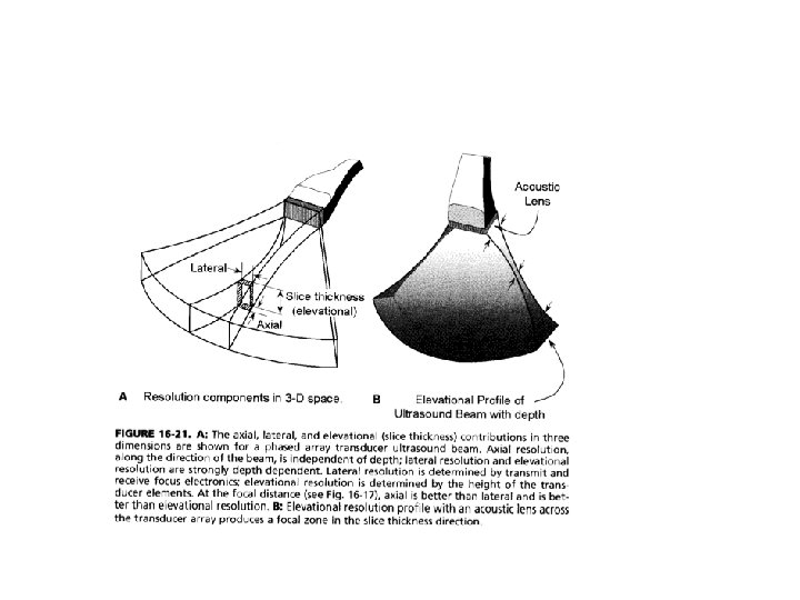
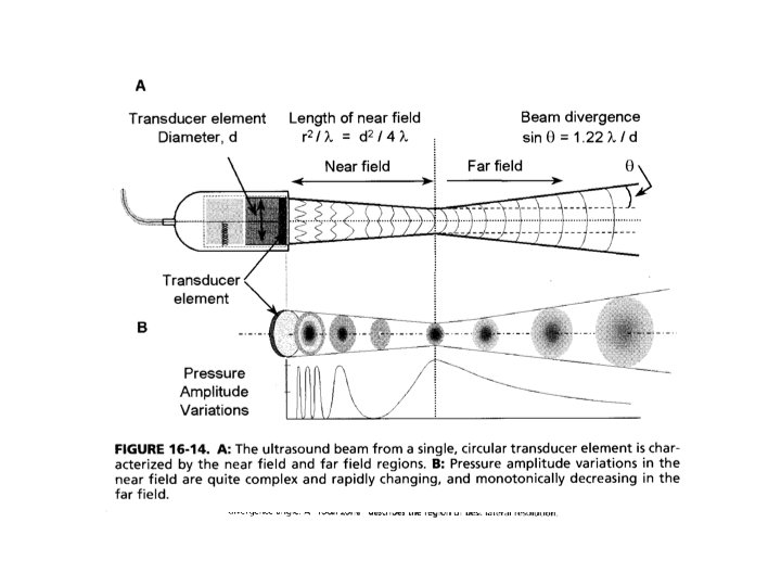
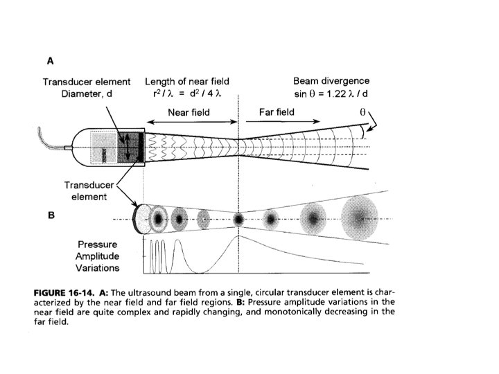
Image Quality • The quality of ultrasonographic images is affected by —multiple reflections, —speckle noise due to scattering, and —spatial distortion due to refraction. • Spatial resolution of ultrasound images: 0. 5 − 3 mm.
Modes of Operation A mode: • A single transducer is used in this mode. • The amplitude (A) of the echoes is displayed on the vertical axis, with the corresponding depth (related to the time of arrival of the echo) on the horizontal axis. • The A mode is useful in distance measurement (ranging), with applications in the detection of retinal detachment and the detection of shift of the midline of the brain.
M mode: • This mode produces a display with time on the horizontal axis and echo depth on the vertical axis. • The M mode is useful in the study of movement or motion (M), with applications in cardiac valve motion analysis.
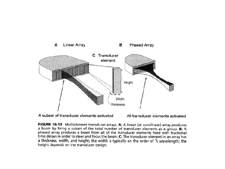
B mode: • An image of a 2 D section or slice of the body is produced by using a single transducer to scan the region of interest or by using an array of sequentially activated transducers. • Real-time imaging is possible at 15 − 40 fps. • The B mode is useful in studying large organs, such as the liver, and in fetal imaging.
Doppler ultrasound: • Based upon the change in frequency of the investigating beam caused by a moving target (the Doppler effect). • Useful in imaging blood flow. • Detection of turbulence and retrograde flow: useful in the diagnosis of stenosis or insufficiency of cardiac valves and plaques in blood vessels. • Doppler imaging may be used to obtain a combination of anatomic information with Bmode imaging and flow information obtained using pulsed Doppler.
Mitral Valve (valve between left atrium and left ventricle)
Umbilical cord
Echocardiography
Special probes: • A variety of probes have been developed for ultrasonography of specific organs and for special applications: – transrectal probes for imaging the prostate, – endovaginal probes for fetal imaging, – transesophageal probes for imaging the heart via the esophagus, and – intravascular probes for the study of blood vessels.
Examples: • Echocardiography —ultrasonography for the assessment of the functional integrity of heart valves. • An array of ultrasound transducers is used in the B mode to obtain a video illustrating the opening and closing activities of the valve leaflets. • Useful in the detection of stenosis and loss of flexibility of the cardiac valves due to calcification.
a b Two frames of the echocardiogram of a subject with normal function of the mitral valve. (a) Mitral valve in the fully open position. (b) Mitral valve in the closed position.
• M-mode ultrasound image of a subject with normal function of the mitral valve. The horizontal axis represents time. The echo signature of the mitral valve leaflets as they open and close is illustrated.
• B-mode ultrasound (3. 5 MHz) image of a fetus (sagital view).
• In spite of limitations in image quality and resolution, ultrasonography is an important medical imaging modality due to the nonionizing nature of the medium. • Ultrasonography is particularly useful in fetal imaging. • Ultrasonography is also useful in tomographic imaging, discriminating between solid masses and fluid filled cysts in the breast, and tissue characterization.