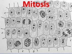
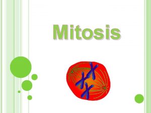
This Week 1116 Mitosis overview Major Vocab and

This Week § 11/16 – Mitosis overview Major Vocab and Steps § 11/17 – Regulation of the Cell Cycle § 11/18 - Loss of Regulation Cancer § 11/19 – Chapter 9 Quiz § 11/20 – Steps of Mesiosis Bozeman video 28 Khan academy is high quality on this!

11/16 Today – Mitosis Overview §Prequiz § 1. ) Why Mitosis? § 2. ) Vocab § 3. ) Steps of Mitosis http: //www. nature. com/scitable/t opicpage/p 53 -the-mostfrequently-altered-gene-in 14192717

Overview: The Key Roles of Cell Division § The ability of organisms to produce more of their own kind best distinguishes living things from nonliving matter § The continuity of life is based on the reproduction of cells, or cell division © 2014 Pearson Education, Inc.

Figure 9. 1 © 2014 Pearson Education, Inc.

§ In unicellular organisms, division of one cell reproduces the entire organism § Cell division enables multicellular eukaryotes to develop from a single cell and, once fully grown, to renew, repair, or replace cells as needed § Cell division is an integral part of the cell cycle, the life of a cell from formation to its own division © 2014 Pearson Education, Inc.

Figure 9. 2 100 m 200 m (a) Reproduction (b) Growth and development 20 m (c) Tissue renewal © 2014 Pearson Education, Inc.

Figure 9. 2 a 100 m (a) Reproduction © 2014 Pearson Education, Inc.

Figure 9. 2 b 200 m (b) Growth and development © 2014 Pearson Education, Inc.

Figure 9. 2 c 20 m (c) Tissue renewal © 2014 Pearson Education, Inc.

Cellular Organization of the Genetic Material § Genome § Chromosomes § Chromatin § Sister Chromatid § Centriole © 2014 Pearson Education, Inc. Kinetochore Somatic Cell Microtubule Aster Centrosome

Figure 9. 3 20 m Eukaryotic chromosomes © 2014 Pearson Education, Inc.

§ Eukaryotic chromosomes consist of chromatin, a complex of DNA and protein § Every eukaryotic species has a characteristic number of chromosomes in each cell nucleus § Somatic cells (nonreproductive cells) have two sets of chromosomes § Gametes (reproductive cells: sperm and eggs) have one set of chromosomes © 2014 Pearson Education, Inc.

Distribution of Chromosomes During Eukaryotic Cell Division § In preparation for cell division, DNA is replicated and the chromosomes condense § Each duplicated chromosome has two sister chromatids, joined identical copies of the original chromosome § The centromere is where the two chromatids are most closely attached © 2014 Pearson Education, Inc.

Figure 9. 4 Sister chromatids Centromere © 2014 Pearson Education, Inc. 0. 5 m

§ During cell division, the two sister chromatids of each duplicated chromosome separate and move into two nuclei § Once separate, the chromatids are called chromosomes © 2014 Pearson Education, Inc.

Figure 9. 5 -1 Chromosomes 1 Chromosomal DNA molecules Centromere Chromosome arm © 2014 Pearson Education, Inc.

Figure 9. 5 -2 Chromosomes 1 Chromosomal DNA molecules Centromere Chromosome arm Chromosome duplication 2 Sister chromatids © 2014 Pearson Education, Inc.

Figure 9. 5 -3 Chromosomes 1 Chromosomal DNA molecules Centromere Chromosome arm Chromosome duplication 2 Sister chromatids Separation of sister chromatids 3 © 2014 Pearson Education, Inc.

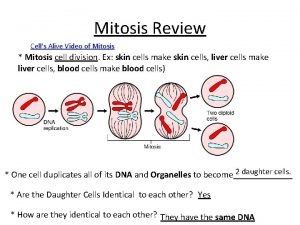
§ Eukaryotic cell division consists of § Mitosis, the division of the genetic material in the nucleus § Cytokinesis, the division of the cytoplasm § Gametes are produced by a variation of cell division called meiosis § Meiosis yields nonidentical daughter cells that have only one set of chromosomes, half as many as the parent cell © 2014 Pearson Education, Inc.

Concept 9. 2: The mitotic phase alternates with interphase in the cell cycle § In 1882, the German anatomist Walther Flemming developed dyes to observe chromosomes during mitosis and cytokinesis © 2014 Pearson Education, Inc.

Phases of the Cell Cycle § The cell cycle consists of § Mitotic (M) phase, including mitosis and cytokinesis § Interphase, including cell growth and copying of chromosomes in preparation for cell division © 2014 Pearson Education, Inc.

§ Interphase (about 90% of the cell cycle) can be divided into subphases § G 1 phase (“first gap”) § S phase (“synthesis”) § G 2 phase (“second gap”) § The cell grows during all three phases, but chromosomes are duplicated only during the S phase © 2014 Pearson Education, Inc.

Figure 9. 6 S (DNA synthesis) G 1 is s e © 2014 Pearson Education, Inc. ito C M yt si s n i ok G 2

§ Mitosis is conventionally divided into five phases § Prophase § Prometaphase § Metaphase § Anaphase § Telophase § Cytokinesis overlaps the latter stages of mitosis © 2014 Pearson Education, Inc.

Video: Animal Mitosis Video: Microtubules Mitosis Animation: Mitosis Video: Microtubules Anaphase Video: Nuclear Envelope © 2014 Pearson Education, Inc.

10 m Figure 9. 7 a G 2 of Interphase Centrosomes (with centriole pairs) Nucleolus Chromosomes Early mitotic Centromere (duplicated, spindle Aster uncondensed) Nuclear envelope © 2014 Pearson Education, Inc. Prophase Plasma membrane Two sister chromatids of one chromosome Prometaphase Fragments of nuclear envelope Kinetochore Nonkinetochore microtubules Kinetochore microtubule

10 m Figure 9. 7 b Metaphase Anaphase Metaphase plate Spindle Centrosome at one spindle pole © 2014 Pearson Education, Inc. Telophase and Cytokinesis Cleavage furrow Daughter chromosomes Nuclear envelope forming Nucleolus forming

Figure 9. 7 c G 2 of Interphase Centrosomes (with centriole pairs) Chromosomes (duplicated, uncondensed) Nucleolus Nuclear envelope © 2014 Pearson Education, Inc. Plasma membrane Prophase Early mitotic Centromere spindle Aster Two sister chromatids of one chromosome

Figure 9. 7 d Prometaphase Fragments of nuclear envelope Kinetochore © 2014 Pearson Education, Inc. Metaphase Nonkinetochore microtubules Kinetochore microtubule Metaphase plate Spindle Centrosome at one spindle pole

Figure 9. 7 e Telophase and Cytokinesis Anaphase Cleavage furrow Daughter chromosomes © 2014 Pearson Education, Inc. Nuclear envelope forming Nucleolus forming

10 m Figure 9. 7 f G 2 of Interphase © 2014 Pearson Education, Inc.

10 m Figure 9. 7 g Prophase © 2014 Pearson Education, Inc.

10 m Figure 9. 7 h Prometaphase © 2014 Pearson Education, Inc.

10 m Figure 9. 7 i Metaphase © 2014 Pearson Education, Inc.

10 m Figure 9. 7 j Anaphase © 2014 Pearson Education, Inc.

10 m Figure 9. 7 k Telophase and Cytokinesis © 2014 Pearson Education, Inc.

The Mitotic Spindle: A Closer Look § The mitotic spindle is a structure made of microtubules and associated proteins § It controls chromosome movement during mitosis § In animal cells, assembly of spindle microtubules begins in the centrosome, the microtubule organizing center © 2014 Pearson Education, Inc.

§ The centrosome replicates during interphase, forming two centrosomes that migrate to opposite ends of the cell during prophase and prometaphase § An aster (radial array of short microtubules) extends from each centrosome § The spindle includes the centrosomes, the spindle microtubules, and the asters © 2014 Pearson Education, Inc.

§ During prometaphase, some spindle microtubules attach to the kinetochores of chromosomes and begin to move the chromosomes § Kinetochores are protein complexes that assemble on sections of DNA at centromeres § At metaphase, the centromeres of all the chromosomes are at the metaphase plate, an imaginary structure at the midway point between the spindle’s two poles Video: Mitosis Spindle © 2014 Pearson Education, Inc.

Figure 9. 8 Aster Sister chromatids Centrosome Metaphase plate (imaginary) Kinetochores Microtubules Chromosomes Overlapping nonkinetochore microtubules Kinetochore microtubules 1 m 0. 5 m © 2014 Pearson Education, Inc. Centrosome

Figure 9. 8 a Microtubules Chromosomes 1 m Centrosome © 2014 Pearson Education, Inc.

Figure 9. 8 b Kinetochores Kinetochore microtubules 0. 5 m © 2014 Pearson Education, Inc.

§ In anaphase, sister chromatids separate and move along the kinetochore microtubules toward opposite ends of the cell § The microtubules shorten by depolymerizing at their kinetochore ends § Chromosomes are also “reeled in” by motor proteins at spindle poles, and microtubules depolymerize after they pass by the motor proteins © 2014 Pearson Education, Inc.

11/17 Regulating the Cell Cycle §. )Cleavage Furrows §. )Regulation at Different Steps §. ) CDK §. ) p 53

Figure 9. 9 Experiment Results Kinetochore Spindle pole Conclusion Mark Chromosome movement Motor protein Chromosome Microtubule © 2014 Pearson Education, Inc. Kinetochore Tubulin subunits

Figure 9. 9 a Experiment Kinetochore Spindle pole Mark © 2014 Pearson Education, Inc.

Figure 9. 9 b Results Conclusion Chromosome movement Motor Microtubule protein Chromosome © 2014 Pearson Education, Inc. Kinetochore Tubulin subunits

Cytokinesis: A Closer Look § In animal cells, cytokinesis occurs by a process known as cleavage, forming a cleavage furrow § In plant cells, a cell plate forms during cytokinesis Animation: Cytokinesis Video: Cytokinesis and Myosin © 2014 Pearson Education, Inc.

Figure 9. 10 (a) Cleavage of an animal cell (SEM) Cleavage furrow Contractile ring of microfilaments 100 m (b) Cell plate formation in a plant cell (TEM) Vesicles forming cell plate Wall of parent 1 m cell Cell plate New cell wall Daughter cells © 2014 Pearson Education, Inc.

Figure 9. 10 a (a) Cleavage of an animal cell (SEM) Cleavage furrow Contractile ring of microfilaments © 2014 Pearson Education, Inc. 100 m Daughter cells

Figure 9. 10 aa Cleavage furrow © 2014 Pearson Education, Inc. 100 m

Figure 9. 10 b (b) Cell plate formation in a plant cell (TEM) Vesicles forming cell plate Wall of parent 1 m cell Cell plate New cell wall Daughter cells © 2014 Pearson Education, Inc.

Figure 9. 10 ba Vesicles forming cell plate © 2014 Pearson Education, Inc. Wall of parent cell 1 m

Figure 9. 11 Nucleus Chromosomes Nucleolus condensing Chromosomes 10 m 1 Prophase 2 Prometaphase Cell plate 3 Metaphase © 2014 Pearson Education, Inc. 4 Anaphase 5 Telophase

Figure 9. 11 a Chromosomes Nucleus Nucleolus condensing 1 Prophase © 2014 Pearson Education, Inc. 10 m

Figure 9. 11 b Chromosomes 10 m 2 Prometaphase © 2014 Pearson Education, Inc.

Figure 9. 11 c 3 Metaphase © 2014 Pearson Education, Inc. 10 m

Figure 9. 11 d 4 Anaphase © 2014 Pearson Education, Inc. 10 m

Figure 9. 11 e Cell plate 5 Telophase © 2014 Pearson Education, Inc. 10 m

Binary Fission in Bacteria § Prokaryotes (bacteria and archaea) reproduce by a type of cell division called binary fission § In E. coli, the single chromosome replicates, beginning at the origin of replication § The two daughter chromosomes actively move apart while the cell elongates § The plasma membrane pinches inward, dividing the cell into two © 2014 Pearson Education, Inc.

Figure 9. 12 -1 Origin of replication E. coli cell 1 Chromosome replication begins. © 2014 Pearson Education, Inc. Two copies of origin Cell wall Plasma membrane Bacterial chromosome

Figure 9. 12 -2 Origin of replication E. coli cell 1 Chromosome replication begins. 2 One copy of the origin is now at each end of the cell. © 2014 Pearson Education, Inc. Two copies of origin Origin Cell wall Plasma membrane Bacterial chromosome Origin

Figure 9. 12 -3 Origin of replication E. coli cell 1 Chromosome replication begins. 2 One copy of the origin is now at each end of the cell. 3 Replication finishes. © 2014 Pearson Education, Inc. Two copies of origin Origin Cell wall Plasma membrane Bacterial chromosome Origin

Figure 9. 12 -4 Origin of replication E. coli cell 1 Chromosome replication begins. 2 One copy of the origin is now at each end of the cell. 3 Replication finishes. 4 Two daughter cells result. © 2014 Pearson Education, Inc. Two copies of origin Origin Cell wall Plasma membrane Bacterial chromosome Origin

The Evolution of Mitosis § Since prokaryotes evolved before eukaryotes, mitosis probably evolved from binary fission § Certain protists (dinoflagellates, diatoms, and some yeasts) exhibit types of cell division that seem intermediate between binary fission and mitosis © 2014 Pearson Education, Inc.

Figure 9. 13 Chromosomes Microtubules Intact nuclear envelope (a) Dinoflagellates Kinetochore microtubule Intact nuclear envelope (b) Diatoms and some yeasts © 2014 Pearson Education, Inc.

A. What would you expect to happen if you fuse a cell in S with a cell in G 1? Why? B. What would you expect to happen to a nucleus from a G 1 cell if it is introduced into an enucleated M cell? Why? © 2014 Pearson Education, Inc.

Figure 9. 14 Experiment 1 S G 1 Experiment 2 M G 1 Results S S G 1 nucleus immediately entered S phase and DNA was synthesized. M M G 1 nucleus began mitosis without chromosome duplication. Conclusion Molecules present in the cytoplasm control the progression to S and M phases. © 2014 Pearson Education, Inc.

Checkpoints of the Cell Cycle Control System § The sequential events of the cell cycle are directed by a distinct cell cycle control system, which is similar to a timing device of a washing machine § The cell cycle control system is regulated by both internal and external controls § The clock has specific checkpoints where the cell cycle stops until a go-ahead signal is received © 2014 Pearson Education, Inc.

Figure 9. 15 G 1 checkpoint Control system G 1 M G 2 M checkpoint G 2 checkpoint © 2014 Pearson Education, Inc. S

§ For many cells, the G 1 checkpoint seems to be the most important § If a cell receives a go-ahead signal at the G 1 checkpoint, it will usually complete the S, G 2, and M phases and divide § If the cell does not receive the go-ahead signal, it will exit the cycle, switching into a nondividing state called the G 0 phase © 2014 Pearson Education, Inc.

Figure 9. 16 G 1 checkpoint G 0 G 1 Without go-ahead signal, cell enters G 0. (a) G 1 checkpoint S M G 1 G 2 With go-ahead signal, cell continues cell cycle. G 1 M G 2 M checkpoint Prometaphase Without full chromosome attachment, stop signal is received. (b) M checkpoint © 2014 Pearson Education, Inc. Anaphase G 2 checkpoint Metaphase With full chromosome attachment, go-ahead signal is received.

Figure 9. 16 a G 1 checkpoint G 0 G 1 Without go-ahead signal, cell enters G 0. (a) G 1 checkpoint © 2014 Pearson Education, Inc. G 1 With go-ahead signal, cell continues cell cycle.

Figure 9. 16 b G 1 M G 2 M checkpoint Prometaphase Without full chromosome attachment, stop signal is received. (b) M checkpoint © 2014 Pearson Education, Inc. Anaphase G 2 checkpoint Metaphase With full chromosome attachment, go-ahead signal is received.

§ The cell cycle is regulated by a set of regulatory proteins and protein complexes including kinases and proteins called cyclins © 2014 Pearson Education, Inc.

§ An example of an internal signal occurs at the M phase checkpoint § In this case, anaphase does not begin if any kinetochores remain unattached to spindle microtubules § Attachment of all of the kinetochores activates a regulatory complex, which then activates the enzyme separase § Separase allows sister chromatids to separate, triggering the onset of anaphase © 2014 Pearson Education, Inc.

§ Some external signals are growth factors, proteins released by certain cells that stimulate other cells to divide § For example, platelet-derived growth factor (PDGF) stimulates the division of human fibroblast cells in culture © 2014 Pearson Education, Inc.

Figure 9. 17 -1 Scalpels 1 A sample of human connective tissue is cut up into small pieces. Petri dish © 2014 Pearson Education, Inc.

Figure 9. 17 -2 Scalpels 1 A sample of human connective tissue is cut up into small pieces. Petri dish 2 Enzymes digest the extracellular matrix, resulting in a suspension of free fibroblasts. © 2014 Pearson Education, Inc.

Figure 9. 17 -3 Scalpels 1 A sample of human connective tissue is cut up into small pieces. Petri dish 2 Enzymes digest the extracellular matrix, resulting in a suspension of free fibroblasts. 3 Cells are transferred to culture vessels. 4 PDGF is added to half the vessels. © 2014 Pearson Education, Inc.

Figure 9. 17 -4 Scalpels 1 A sample of human connective tissue is cut up into small pieces. Petri dish 2 Enzymes digest the extracellular matrix, resulting in a suspension of free fibroblasts. 3 Cells are transferred to culture vessels. 4 PDGF is added to half the vessels. Without PDGF © 2014 Pearson Education, Inc. With PDGF Cultured fibroblasts (SEM) 10 m

Figure 9. 17 a Cultured fibroblasts (SEM) © 2014 Pearson Education, Inc. 10 m

§ Another example of external signals is densitydependent inhibition, § Most animal cells also exhibit anchorage dependence § Cancer cells exhibit neither density-dependent inhibition nor anchorage dependence © 2014 Pearson Education, Inc.

Characteristics of Cancer § Tumors – multiple layers of disorganized cells § NEOPLASIA: invade and destroy § Benign tumor: encapsulated § Malignant tumor: metastasizes (spreads)

Characteristics of Cancer § NO contact inhibition

Characteristics of Cancer § Angiogenesis § release a growth factor that caused nearby blood vessels to grow and bring more nutrients and oxygen to the tumor.


Figure 9. 18 Anchorage dependence: cells require a surface for division Density-dependent inhibition: cells form a single layer Density-dependent inhibition: cells divide to fill a gap and then stop 20 m (a) Normal mammalian cells © 2014 Pearson Education, Inc. 20 m (b) Cancer cells

Figure 9. 18 a 20 m (a) Normal mammalian cells © 2014 Pearson Education, Inc.

Figure 9. 18 b 20 m (b) Cancer cells © 2014 Pearson Education, Inc.

Loss of Cell Cycle Controls in Cancer Cells § Cancer cells do not respond to signals that normally regulate the cell cycle § Cancer cells may not need growth factors to grow and divide § They make their own growth factor § They may convey a growth factor’s signal without the presence of the growth factor § They may have an abnormal cell cycle control system © 2014 Pearson Education, Inc.

§ A normal cell is converted to a cancerous cell by a process called transformation § Cancer cells that are not eliminated by the immune system form tumors, masses of abnormal cells within otherwise normal tissue § If abnormal cells remain only at the original site, the lump is called a benign tumor § Malignant tumors invade surrounding tissues and can metastasize, exporting cancer cells to other parts of the body, where they may form additional tumors © 2014 Pearson Education, Inc.

5 m Figure 9. 19 Breast cancer cell (colorized SEM) Lymph vessel Tumor Blood vessel Cancer cell Glandular tissue 1 A tumor grows from a single cancer cell. © 2014 Pearson Education, Inc. Metastatic tumor 2 Cancer cells invade neighboring tissue. 3 Cancer cells spread through lymph and blood vessels to other parts of the body. 4 A small percentage of cancer cells may metastasize to another part of the body.

Figure 9. 19 a Tumor Glandular tissue 1 A tumor grows from a single cancer cell. © 2014 Pearson Education, Inc. 2 Cancer cells invade neighboring tissue. 3 Cancer cells spread through lymph and blood vessels to other parts of the body.

Figure 9. 19 b Lymph vessel Metastatic tumor Blood vessel Cancer cell 3 Cancer cells spread through lymph and blood vessels to other parts of the body. © 2014 Pearson Education, Inc. 4 A small percentage of cancer cells may metastasize to another part of the body.

5 m Figure 9. 19 c Breast cancer cell (colorized SEM) © 2014 Pearson Education, Inc.

Figure 9. UN 01 Control 200 A B C Treated A B C Number of cells 160 120 80 40 0 0 200 400 600 Amount of fluorescence per cell (fluorescence units) © 2014 Pearson Education, Inc.

Figure 9. UN 02 G 1 S Cytokinesis Mitosis G 2 MITOTIC (M) PHASE Prophase Telophase and Cytokinesis Prometaphase Anaphase Metaphase © 2014 Pearson Education, Inc.

Figure 9. UN 03 © 2014 Pearson Education, Inc.
- Slides: 99


