Mitosis Cell division Agenda Cell cycle Mitosis Overview


 –refers to the process of nuclear division. Results")


: Double helix strand containing genetic code that acts as")
 Chromosome: a long piece of supercoiled DNA and proteins. The")


 and cytoplasmic")


 – Radiation (gamma, UV,")


- Slides: 34
Mitosis Cell division
Agenda Cell cycle Mitosis Overview When things go wrong
The Expectations 2. 5. 1 Outline the stages in the cell cycle, including interphase (G 1, S, G 2), mitosis and cytokinesis
The Cell Cycle… • Cells have a life cycle, called the cell cycle • The cell cycle consists of 3 stages: – Interphase (growth and replication) – Mitosis (Nuclear(DNA) division) – Cytokinesis (cytoplasm division)
The Cell Cycle G 1 Synthesis of DNA for Duplication of chromosomes Phase of rapid cell growth S Growth and preparation for cell division G 2 M
Interphase • 1. Stage G 1: primary growth phase. Increase in proteins and in the number of cell organelles. • 2. Stage S: indicates the synthesis of DNA. A. k. a. DNA replication • 3. Stage G 2: Chromosome condensation, preparation for mitosis. • Longest stage of cell cycle (The part where it does not divide). (G 1, S, G 2). Human cells contain 46 chromosomes during the G 1 stage of interphase. This is doubled to 92 strands (still considered 46 chrom) during the S stage of interphase.
Mitosis and Cytokinesis • Mitosis(M phase) –refers to the process of nuclear division. Results in two identical nuclei. • Cytokinesis (C phase)- occurs after mitosis and is the actual physical division of the cell. Not included in mitosis
Why do cells divide? 1. If cells got too large, they would not be able to carry out their functions to survive 2. Growth: allow organisms to grow from a single cell to a multi-celled organism 3. Maintenance: allows new cells to replace worn-out or dead cells 4. Repair: regenerates damaged tissues
The Basics: Genetic Material
DNA • Deoxyribonucleic Acid (DNA): Double helix strand containing genetic code that acts as instructions for the cell • Chromatin structurally loose DNA in the cell during interphase. • Chromosome: DNA and associated proteins (nucleosomes/histones) that help condense chromosome into a smaller area during mitosis. • Histone: Proteins that DNA wraps around to become condensed chromosomes
Chromosomes one chromosome (unduplicated) Chromosome: a long piece of supercoiled DNA and proteins. The number of chromosome in each organism differs. 46 in humans. Seen only when the cell divides. Sister chromatids: 2 identical copies of the same chromosome one chromosome (duplicated) Centromere Sister Chromatids
Mitosis Structures • Centromeres: A centromere is a region on a chromosome that joins two sister chromatids. • Centrioles: a cylindrical cell structure composed mainly of a protein called tubulin. Produce spindle fibres. • Mitotic spindle: microtubule-based bipolar strands that segregate the chromosomes in mitosis
Cyclins • Cyclins are involved in the control of the cell cycle • Cyclins bind to enzymes called cyclindependent kinases. These kinases become active and attach phosphate groups to the proteins in the cell. The attachment then triggers other proteins to become active and carry out their various functions.
• • Cyclin D triggers cells to move from G phase to S phase Cyclin E prepares the cell for DNA replication in S phase Cyclin A activates DNA replication inside the nucleus Cyclin B promotes the assembly of the mitotic spindle
Difference between Plant and Animal Cell Division Uses of Mitosis in Eukaryotic Cells: a. during growth of the individual. b. when tissues have been damaged and need to be repaired (as in healing of a cut). c. to reproduce asexually.
Mitotic Index • Mitotic index= number of cells in mitosis total number of cells Useful prognostic tool for predicting response of cancer cells to chemotherapy
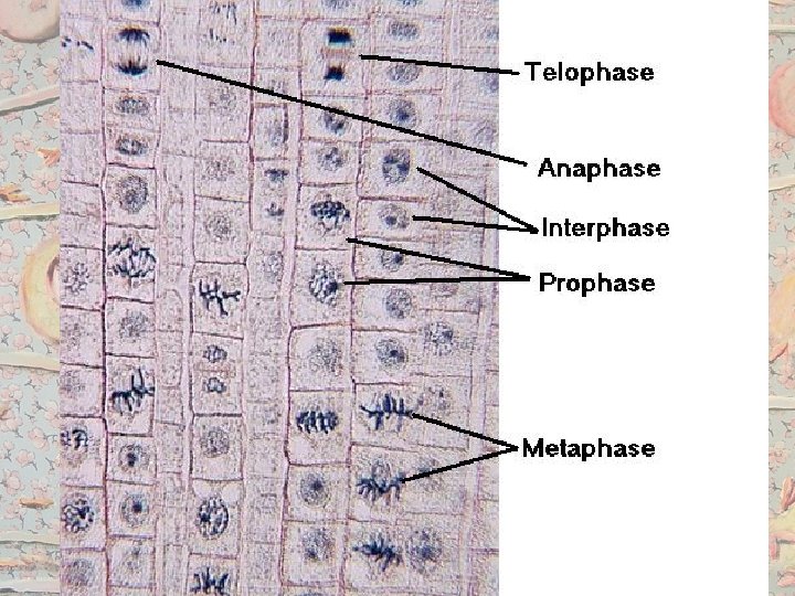
The Expectations Describe the events that occur in the four phases of mitosis -PMAT-
Prophase *chromatin condenses into chromosomes that contains 2 sister chromatids attached by a centromere * Centrioles move to poles *the nuclear envelope and nucleolus disintegrate *Mitotic spindle forms from microtubules *the pair of sister chromatids attach to the spindle at their * Sister chromatids centromeres. present *in animal cells, a pair of centrioles move to each end, called the poles * During late prophase the nuclear envelope breaks down and each chromosome is connected to a spindle fibre by its centromere
Metaphase *Each chromosome becomes completely condensed. The chromosomes line up at the centre of the cell. Line up at the metaphase plate *The mitotic spindle (made of tubes) is complete and extend from each pole (centrioles) to the middle of the cell.
Anaphase • The sister chromatids separate at the centromere. Each is now called a chromosome. The separated chromosome are pulled to opposite poles by the spindle fibres
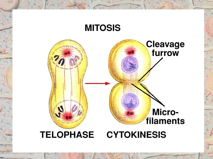
Telophase • • • Chromosomes have arrived at the poles Spindle disappears Centrioles replicate (in animals) Nuclear membrane reappears Nucleolus becomes visible Chromosomes become chromatin
Cytokinesis • The cell divides the cytoplasm and organelles into two portions (splitting known as cytokinesis) • The cell membrane cleaves inward and in plant cells a cell plate forms, nuclear membrane reforms and spindle disappears • 2 identical daughter cells result
Mitosis is an Animal cell Metaphase Anaphase Chromosomes line up. The centromeres divide and the resulting chromosomes, at the equatorial plate. Chromosomes continue move to opposite poles of the The nuclear membrane to condense. Early Prophase completely dissolves. cell. An identical set The centrioles assemble (homologues) of chromosomes and spindle moves to each pole. attach to the The chromosomesfibres condense, becoming shorter centromeres and thicker. of thetochromosomes. The centrioles move opposite nuclear membrane poles of the cell and spindle starts to dissolve. fibres start to form. Telophase DNA replicated and cell Chromosomes lengthen again, prepares for division. In the spindle fibres dissolve, and a Interphase humans, 46 chromosomes nuclear membrane forms around are duplicated (46 pairs). Telophase the chromosomes. In humans, Late Prophase Interphase each new nucleus contains 46 unique chromosomes http: //www. youtube. com/watch? v=Ahg. Rh. Xl
Mitosis Summary Two divisions occur during cell division • Nuclear division (mitosis) and cytoplasmic division (cytokinesis) During interphase genetic material is replicated 2 Identical daughter cells produced Embryonic growth is a result of repeated mitotic divisions • A zygote is one cell after fertilization. As divisions occur, specialization occurs.
Cancer • There is a strong correlation between smoking and cancer • Women smokers are 25. 7 times more likely than women who never smoked to develop lung cancer. For men smokers, it’s 25 times the risk of men who never smoked. (Source: US Surgeon General Report 2014) • Besides lung cancer, tobacco use also increases the risk for cancers of the mouth, lips, nose and sinuses, larynx (voice box), pharynx (throat), esophagus (swallowing tube), stomach, pancreas, kidney, bladder, uterus, cervix, colon/rectum, ovary (mucinous), and acute myeloid leukemia. (Source: Cancer Facts & Figures 2014)
Cancer • Mitosis and cell division are under strict control. Only producing cells when needed for growth and repair • Mutagens, oncogenes and metastasis are involved in the development of primary and secondary tumors • Tumour repressive genes- inhibit cell division • Prot-oncogenes- stimulate growth/division
Cancer • Cancer: Disorder in which some of the body's cells lose the ability to control their growth. • Tumour - repeated, uncontrolled cell division to form a mass of cells. This can happen in any organ. Some tumours grow large and spread to other parts of the body (metastasis). The diseases caused by the growth of tumours is known as cancer. – Tumours can be benign (harmless) or malignant (spread)
Causes • Carcinogens (increase chances of mutation leading to cancer) – Radiation (gamma, UV, x-ray etc. ) – Chemicals (textile dyes, paints and inks) – Viruses (hepititis B and C and HPV)
Removal • Surgical– physically excise the tumour cells • Radiation– using strong ionising or nuclear radiation beam which can be directed to a point and burn the cells • Chemotherapy– Uses chemicals to destroy all rapidly dividing cells by medication. Can destroy other rapidly dividing cells (hair, stomach/intestinal cells, sperm)
Cancer vs. Normal cells Cancer cells Normal Cells Make exact copies of themselves during mitosis Do not stop reproducing Behave independently Metstacize, no controlled death Reproduce 50 -60 cells Work dependently/stick together Self destruct “apoptosis” when old
Cell Death • Apoptosis: regulated or controlled cell death of cells that are no longer useful. This is also used to control cells that have stopped performing properly – E. g your body must produces cells to fight a viral infection. When the infection is gone the cells are removed by apoptosis
Any Questions?