Pleural diseases Case Studies Dr JM Nel Department


- Slides: 53

Pleural diseases: Case Studies Dr. JM Nel Department of Pulmonology

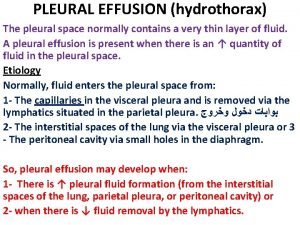
Pleural effusions n Case Presentation 1: – 68 year old lady – Known with hypertension – Presents with dyspnae – Pleural effusion clinically WHAT SPECIAL INVESTIGATION NEXT ?

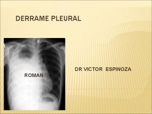
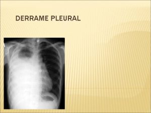
Pleural effusions n CXR § Curved shadow at lung base (meniscus) § Blunting of costophrenic angle

Pleural effusions WHAT NOW ? ? ? n Pleural tap – Transudate – Exudate

Pleural effusions n Pleural fluid features – A. Appearance of fluid – B. Biochemical analysis – C. Gram stain – D. Predominant cells in fluid – E. Other

Pleural effusion: Investigations LIGHT’S CRITERIA § Pleural fluid is an exudate if one or more of criteria is met: – – – Pleural fluid protein: Serum protein ratio > 0. 5 Pleural fluid LDH: Serum LDH ratio > 0. 6 Pleural fluid LDH > 2/3 upper limit of normal s- LDH

Pleural effusions n Pleural fluid biochemistry: – – – Protein: 20 Albumin: 10 LDH: 100 n Serum biochemistry: – – – Protein: 60 (60 -80 G/L) Albumin: 18 (35 -52 G/L) LDH: 200 (100 -190 U/L)

Pleural effusions TRANSUDATE

Pleural effusion: Causes n Transudate – Increased hydrostatic pressure § Congestive heart failure – Decreased plasma oncotic pressure § Nephrotic syndrome § Cirrhosis – Movement of transudative ascitic fluid through diaphragm § Cirrhosis

Pleural effusions n Case Presentation 2: – 32 year old man – Presents with fever, pleuritic chest pain and dyspnae – Pleural effusion clinically WHAT SPECIAL INVESTIGATION NEXT ?

Pleural effusions n CXR § Curved shadow at lung base (meniscus) § Blunting of costophrenic angle

Pleural effusions WHAT NOW ? ? ? n Pleural tap – Transudate – Exudate

Pleural effusion: Investigations LIGHT’S CRITERIA § Pleural fluid is an exudate if one or more of criteria is met: – – – Pleural fluid protein: Serum protein ratio > 0. 5 Pleural fluid LDH: Serum LDH ratio > 0. 6 Pleural fluid LDH > 2/3 upper limit of normal s- LDH

Pleural effusions n Pleural fluid biochemistry: – – – Protein: 60 Albumin: 20 LDH: 150 n Serum biochemistry: – – – Protein: 80 (60 -80 G/L) Albumin: 30 (35 -52 G/L) LDH: 180 (100 -190 U/L)

Pleural effusions EXUDATE

Pleural effusion: Causes n Exudate – Inflammatory § Infection – TB/ Pneumonia § Pulmonary embolus/ infarction § Connective tissue disease – RA/ SLE § Adjacent to subdiaphragmatic disease – Pancreatitis/ Subphrenic abscess – Malignancies

Pleural effusions n Pleural fluid biochemistry: – – – Protein: 60 Albumin: 20 LDH: 150 Glucose: 1. 8 p. H: 7. 0 n Serum biochemistry: – – – Protein: 80 (60 -80 G/L) Albumin: 30 (35 -52 G/L) LDH: 180 (100 -190 U/L)

Pleural effusions EMPYEMA

Empyema: Investigations n Aspiration of pus – Confirmation of empyema § 1. Appearance of fluid: pus § 2. Neutrophils § 3. Positive gram stain § 4. Low p. H < 7. 2 § 5. Low glucose < 3. 3

Pleural effusion: Investigations n E. Other – Low p. H § § § Infection/ Empyema RA/ SLE Malignancy TB Ruptured oesophagus – Low glucose § As low p. H – High ADA

Pulmonary Embolism: Case Studies Dr. JM Nel Department of Pulmonology

Pulmonary embolism n Case Presentation 1: – 64 year old male – Previous hip surgery 20 days ago – Sudden dyspnae – Pleuritic chest pain – Hypoxic – Clinically DVT

Pulmonary embolism DIFFERENTIAL DIAGNOSIS § Pulmonary embolism § Pneumonia § Pneumothorax § Musculoskeletal chest pain

Pulmonary embolism ASK 3 QUESTIONS – Is the presentation consistent with PE ? – Does the patient have risk factors for PE ? – Is there another diagnosis that can explain the patients presentation ?

Pulmonary embolism WHAT NOW ? ? ?

Pulmonary embolism n CXR – Exclude differential diagnoses § Heart failure § Pneumonia § Pneumothorax n High index of suspicion if normal CXR – Acute dyspnoeac and hypoxaemic patient

Pulmonary embolism n ECG – Exclude other differential diagnoses § Acute myocardial infarction § Pericarditis n Most common – Sinus tachycardia

Pulmonary embolism n Arterial n Low bloodgas Pa. O 2

Pulmonary embolism n D- dimer n POSITIVE n Other causes for elevation – Myocardial infarction – Pneumonia – Sepsis

Pulmonary embolism n Heartsonar n Massive PE – Acute dilatation of the right heart – Pulmonary hypertension – Thrombus can be seen n NORMAL n Alternative diagnoses – – – Left ventricular failure Aortic dissection Pericardial tamponade

Pulmonary embolism n Duplex n DVT doppler of legs in leg

Pulmonary embolism n V/Q scan n PULMONARY EMBOLISM

Pulmonary embolism: Management n General measures – Oxygen for all hyoxaemic patients § Keep arterial oxygen saturation > 90% n Anticoagulation – Clexane 80 mg bd sc § Give at least 5 days – Warfarin – Stop Clexane when INR is > 2

Pulmonary embolism: Management n n HOW LONG DO I TREAT THIS PATIENT WITH WARFARIN ? ? ? 3 Months n Duration of Warfarin therapy – If underlying prothrombotic risk or previous emboli § For life – If identifiable and reversible risk factor § 3 Months – If idiopathic § 6 Months

Pulmonary embolism n Case Presentation 2: – 28 year old lady – Oral contraceptives – 10 hour flight – Sudden dyspnae – BP 90/40 – Loud P 2/ Increased JVP – Hypoxic

Pulmonary embolism DIFFERENTIAL DIAGNOSIS § Massive pulmonary embolism § Myocardial infarction § Pericardial tamponade § Aortic dissection

Pulmonary embolism ASK 3 QUESTIONS – Is the presentation consistent with PE ? – Does the patient have risk factors for PE ? – Is there another diagnosis that can explain the patients presentation ?

Pulmonary embolism n CXR n NORMAL

Pulmonary embolism n ECG – S 1 Q 3 T 3 – RBBB n Arterial bloodgas – Low Pa. O 2 n D- dimer – POSITIVE

Pulmonary embolism n Heartsonar – Right ventricular dilatation – Increased pulmonary pressure

Pulmonary embolism n CT pulmonary angiography MASSIVE PULMONARY EMBOLISM

Pulmonary embolism: Management n General measures – Oxygen for all hypoxaemic patients § Keep arterial oxygen saturation > 90% – Treat hypotension with IVI fluids n. Thrombolytic – RV dilatation – Low BP therapy

Pulmonary embolism: Management n Complications of thrombolytic therapy – Intracranial haemorrhage – Haemorrhage at other sites – Anaphylaxis

Pulmonary embolism n Case Presentation 3: – 28 year old lady – Oral contraceptives – 10 hour flight – Sudden dyspnae – BP 130/80 – Loud P 2/ Increased JVP – Hypoxic

Pulmonary embolism n CXR n NORMAL

Pulmonary embolism n ECG – S 1 Q 3 T 3 – RBBB n Arterial bloodgas – Low Pa. O 2 n D- dimer – POSITIVE

Pulmonary embolism n Heartsonar – Right ventricular dilatation – Increased pulmonary pressure

Pulmonary embolism n CT pulmonary angiography PULMONARY EMBOLISM

Pulmonary embolism n Patient has normal BP n Patient has RV strain SUBMASSIVE PULMONARY EMBOLISM

Confirmed PE ECHO RV dysfunction NO YES Hemodynamically Stable ? Low risk Non-massive PE YES Anticoagulate UFH LMWH Submassive PE Anticoagulate NO Massive PE Thrombolysis if no contra-indication

Submassive PE n To thrombolise or not to thrombolise n THAT REMAINS THE QUESTION !!!

Thrombolytic therapy n Associated with rapid resolution of radiographic abnormality n No reduction in mortality !!!

Thrombolytic therapy n Indicated only in hemodynamically unstable patients !!! – SBP < 90 mm. Hg n All must be followed by therapeutic anticoagulation
 Best case worst case average case
 Paradigm shift from women studies to gender studies
 Advantages of case studies in psychology
 Times 100 business case studies
 The times 100 business case studies
 Holistic technology in human values
 Example of outbreak
 Case series
 Eu-gdpr-p
 Gcse geography manchester case study
 Euthanasia case studies
 E procurement case studies
 Types of case studies
 Definition of a case study
 Kellogg's crunchy nut bites market research
 Nasa case study
 Blood group discrepancy
 Organic chemistry case studies
 Nspe board of ethical review cases
 Organ donation case studies
 Jmp case studies
 Case studies of rtos
 Ferpa case studies
 Social engineering case studies
 Plagiarism case study
 Case studies financial management
 Digital marketing case studies 2012
 @the..porky..maggot:pixeldrain.com/u/vbkr37be
 The times 100 business case studies
 Ref impact case studies
 Tanya carmichael
 Food safety errors: case studies
 Growth hacking case studies
 Case study base
 Meniscus sign pleural effusion
 Vocal fermitus
 What causes tactile fremitus
 What is tactile fremitus
 émoussement sac pleural forum
 Pleural fluid
 Derrame pleural grados
 Pleural empyema
 Aspirimi i rrugeve te frymemarrjes
 Cupola pleurica
 Pleural effusion color chart
 بیمارستان مدرس
 Indications of intercostal drainage
 Effusion definition
 Light criteria
 Pleural aspiration site
 Serosangineous
 Mediastinum structures
 Lung pleura
 Exsudat transudat


