
Pleural Effusions Dr Sasan Tavana pulmonologist Modarres Hospital




- Slides: 62
Pleural Effusions Dr. Sasan Tavana pulmonologist Modarres Hospital Shahid Beheshti University
Summary Slide n
Normalcomposision composition pleural fluid Normal of of pleural fluid n n Volume Cells/mm 3 %mesothelial cells %monocyte %lymphocytes %granolocytes n Protein %albumin n Glucose LDH PH 0. 1 -0. 2 ml/kg 1000 -5000 3 -70% 30 -75% 2 -30% 1 -2 g/dl 50 -70% ≈ plasms level <50% plasma level ≥plasma
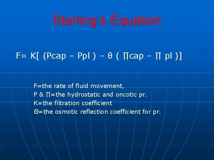
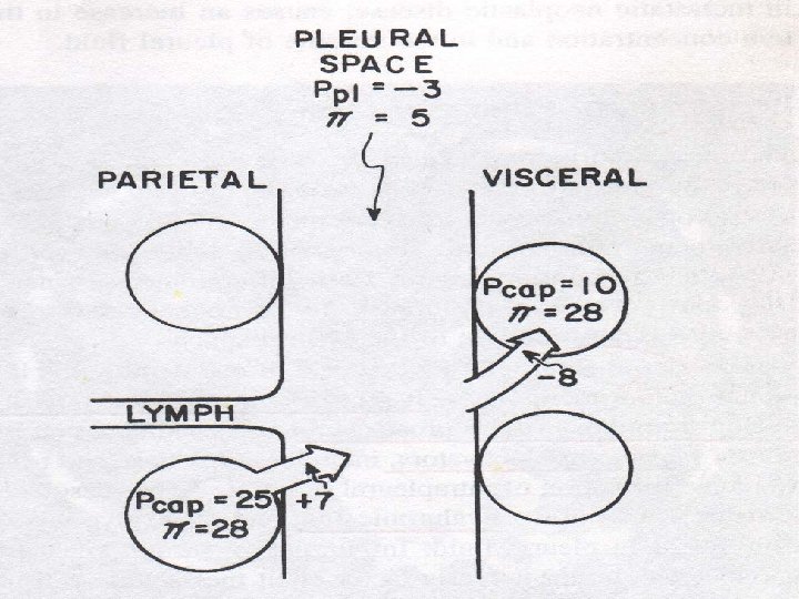
Starling’s Equation F= K[ (Pcap – Ppl ) – θ ( ∏cap – ∏ pl )] F=the rate of fluid movement, P & ∏=the hydrostatic and oncotic pr. K=the filtration coefficient Θ=the osmotic reflection coefficient for pr.
Summary Slide n
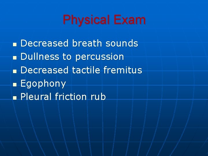
Physical Exam n n n Decreased breath sounds Dullness to percussion Decreased tactile fremitus Egophony Pleural friction rub
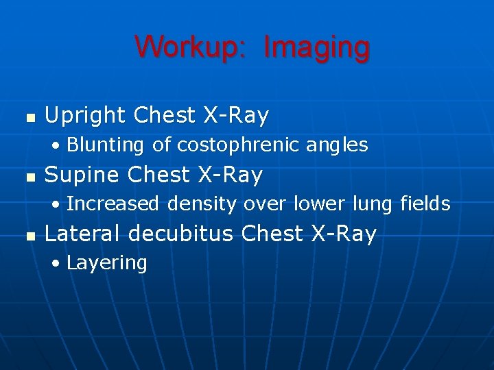
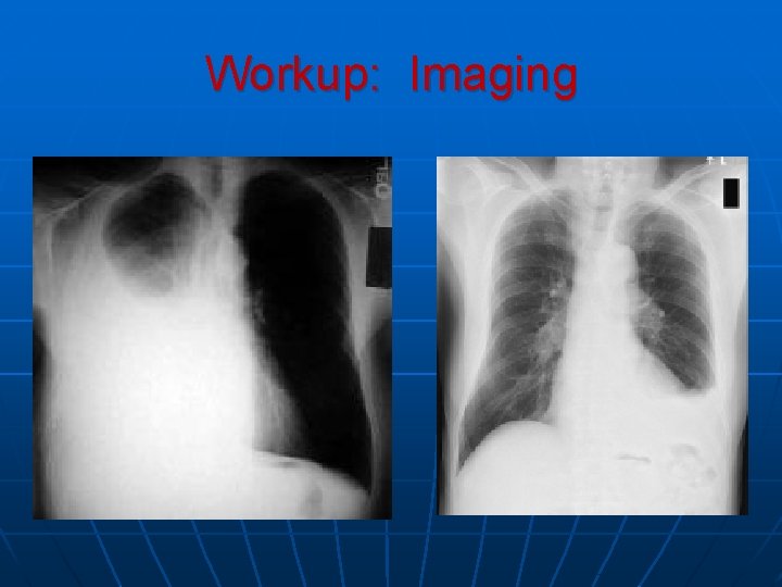
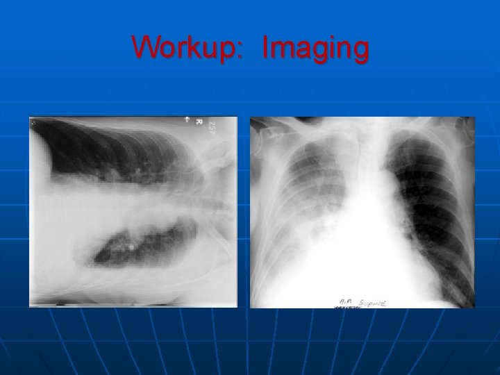
Workup: Imaging n Upright Chest X-Ray • Blunting of costophrenic angles n Supine Chest X-Ray • Increased density over lower lung fields n Lateral decubitus Chest X-Ray • Layering
Workup: Imaging
Workup: Imaging
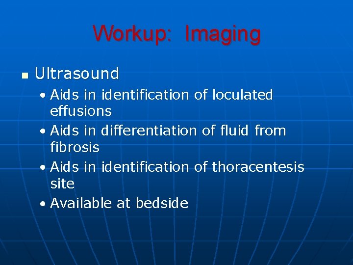
Workup: Imaging n Ultrasound • Aids in identification of loculated effusions • Aids in differentiation of fluid from fibrosis • Aids in identification of thoracentesis site • Available at bedside
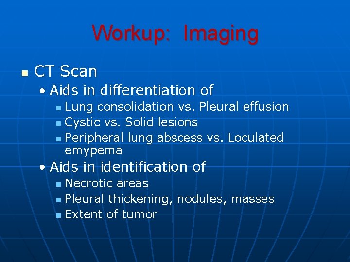
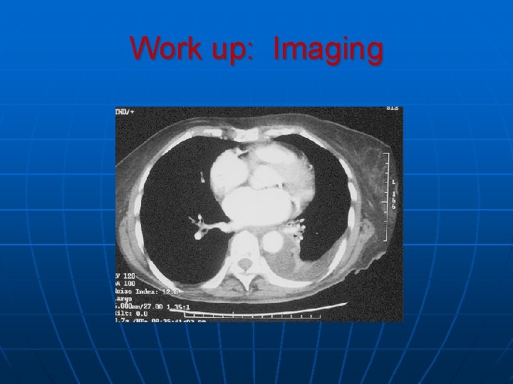
Workup: Imaging n CT Scan • Aids in differentiation of Lung consolidation vs. Pleural effusion n Cystic vs. Solid lesions n Peripheral lung abscess vs. Loculated emypema n • Aids in identification of Necrotic areas n Pleural thickening, nodules, masses n Extent of tumor n
Work up: Imaging
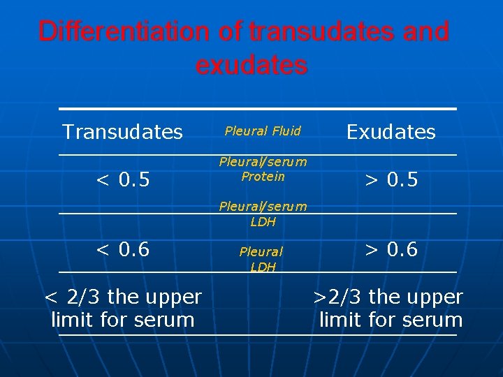
Differentiation of transudates and exudates Transudates < 0. 5 Pleural Fluid Pleural/serum Protein Exudates > 0. 5 Pleural/serum LDH < 0. 6 < 2/3 the upper limit for serum Pleural LDH > 0. 6 >2/3 the upper limit for serum
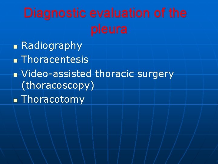
Diagnostic evaluation of the pleura n n Radiography Thoracentesis Video-assisted thoracic surgery (thoracoscopy) Thoracotomy
Causes of Transudative Effusions n n n n Congestive Heart failure Nephrotic Syndrome Cirrhosis Meig’s Syndrome Hydronephrosis Peritoneal dialysis Ex vacuo
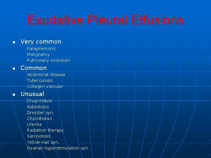
Exudative Pleural Effusions n Very common Parapnemonic Malignancy Pulmonary embolism n Common Abdominal disease Tuberculosis Collagen vascular n Unusual Drug-induce Asbestosis Dressler syn. Chylothorax Uremia Radiation therapy Sarcoidosis Yellow-nail syn. Ovarian hyperstimulation syn.
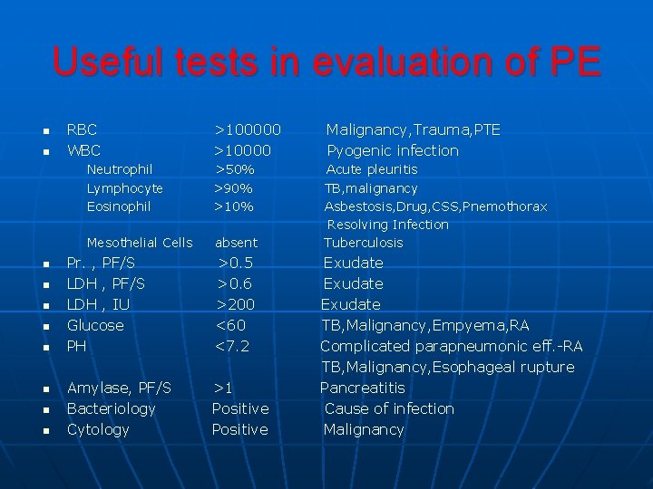
Useful tests in evaluation of PE n n n n n RBC WBC >100000 >10000 Malignancy, Trauma, PTE Pyogenic infection Neutrophil Lymphocyte Eosinophil >50% >90% >10% Mesothelial Cells absent Acute pleuritis TB, malignancy Asbestosis, Drug, CSS, Pnemothorax Resolving Infection Tuberculosis Pr. , PF/S LDH , IU Glucose PH >0. 5 >0. 6 >200 <60 <7. 2 Amylase, PF/S Bacteriology Cytology >1 Positive Exudate TB, Malignancy, Empyema, RA Complicated parapneumonic eff. -RA TB, Malignancy, Esophageal rupture Pancreatitis Cause of infection Malignancy
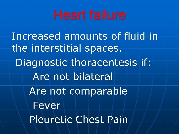
Heart failure Increased amounts of fluid in the interstitial spaces. Diagnostic thoracentesis if: Are not bilateral Are not comparable Fever Pleuretic Chest Pain
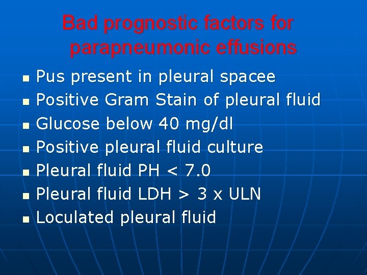
Bad prognostic factors for parapneumonic effusions n n n n Pus present in pleural spacee Positive Gram Stain of pleural fluid Glucose below 40 mg/dl Positive pleural fluid culture Pleural fluid PH < 7. 0 Pleural fluid LDH > 3 х ULN Loculated pleural fluid
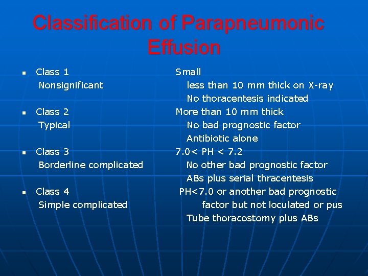
Classification of Parapneumonic Effusion n n Class 1 Nonsignificant Class 2 Typical Class 3 Borderline complicated Class 4 Simple complicated Small less than 10 mm thick on X-ray No thoracentesis indicated More than 10 mm thick No bad prognostic factor Antibiotic alone 7. 0< PH < 7. 2 No other bad prognostic factor ABs plus serial thracentesis PH<7. 0 or another bad prognostic factor but not loculated or pus Tube thoracostomy plus ABs
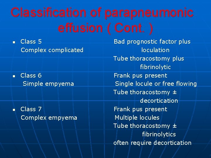
Classification of parapneumonic effusion ( Cont. ) n n n Class 5 Complex complicated Class 6 Simple empyema Class 7 Complex empyema Bad prognostic factor plus loculation Tube thoracostomy plus fibrinolytic Frank pus present Single locule or free flowing Tube thoracostomy ± decortication Frank pus present Multiple locules Tube thoracostomy ± fibrinolytics often require decortication
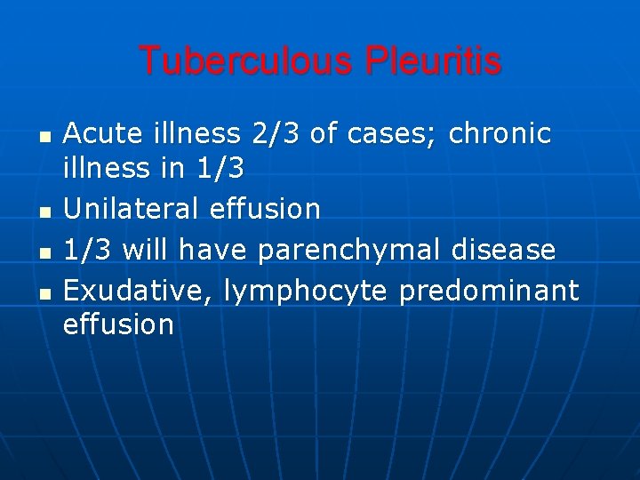
Tuberculous Pleuritis n n Acute illness 2/3 of cases; chronic illness in 1/3 Unilateral effusion 1/3 will have parenchymal disease Exudative, lymphocyte predominant effusion
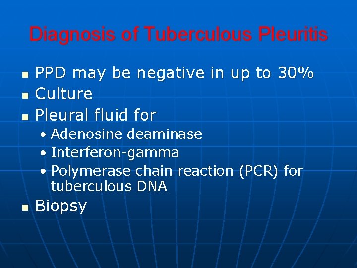
Diagnosis of Tuberculous Pleuritis n n n PPD may be negative in up to 30% Culture Pleural fluid for • Adenosine deaminase • Interferon-gamma • Polymerase chain reaction (PCR) for tuberculous DNA n Biopsy
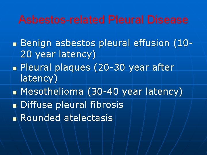
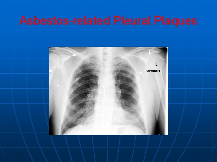
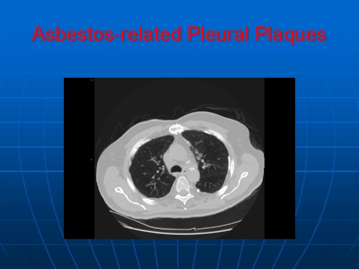
Asbestos-related Pleural Disease n n n Benign asbestos pleural effusion (1020 year latency) Pleural plaques (20 -30 year after latency) Mesothelioma (30 -40 year latency) Diffuse pleural fibrosis Rounded atelectasis
Asbestos-related Pleural Plaques
Asbestos-related Pleural Plaques
Mesotheolioma n Associated with asbestos exposure (even very modest exposures) • Latency of 35 -40 years n n n No association with smoking Difficult diagnosis by cytology. Therefore, usually a biopsy is recommended Three histological subtypes • Epithelial • Sarcomatous • Mixed
Treatment of Mesothelioma n Extrapleural pneumonectomy • 5% surgical mortality • Median survival of 21 months (best with epithelial histology) • 5 year survival 22% n There may be a role for multimodality therapy using chemotherapy and radiation therapy
Benign Mesothelioma n n Localized pleural tumors of mesenchymal origin Clinical manifestations • Asymptomatic in 50% • Cough, chest pain, dyspnea in 40% of symptomatic patients • 2 paraneoplastic syndromes n n Hypoglycemia – caused by secretion of insulin-like growth factor II Hypertrophic pulmonary osteoarthropathy Solitary mass Usually cured by surgical removal
Pulmonary Embolization Exudative Transudative
Hepatic Hydrothorax 5% of patient with cirrhosis Right side Large
Viral Infection Adenovirus Undiagnosed P. E
AIDS Kaposi Sarcoma Para pneumonic eff. TB Lymphoma
Collagen-Vascular Disease of the Pleura n n n Rheumatoid Arthritis Systemic Lupus Erythematosis Sarcoidosis Wegener’s Granulomatosis Sjogren’s syndrome
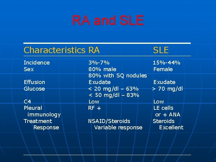
RA and SLE Characteristics RA Incidence Sex Effusion Glucose C 4 Pleural immunology Treatment Response SLE 3%-7% 15%-44% 80% male Female 80% with SQ nodules Exudate < 20 mg/dl – 63% > 70 mg/dl < 50 mg/dl – 83% Low RF + LE cells or + ANA NSAID/Steroids Variable response Excellent
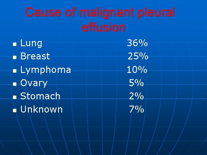
Cause of malignant pleural effusion n n n Lung Breast Lymphoma Ovary Stomach Unknown 36% 25% 10% 5% 2% 7%
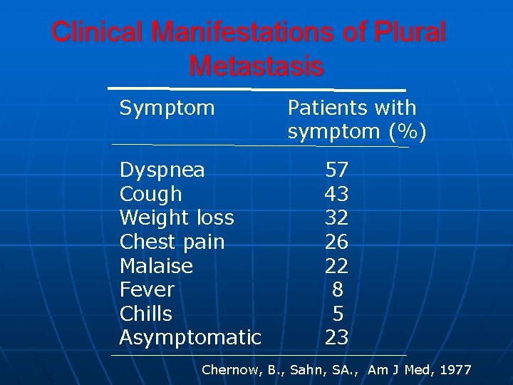
Clinical Manifestations of Plural Metastasis Symptom Dyspnea Cough Weight loss Chest pain Malaise Fever Chills Asymptomatic Patients with symptom (%) 57 43 32 26 22 8 5 23 Chernow, B. , Sahn, SA. , Am J Med, 1977
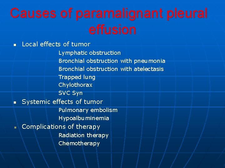
Causes of paramalignant pleural effusion n Local effects of tumor Lymphatic obstruction Bronchial obstruction with pneumonia Bronchial obstruction with atelectasis Trapped lung Chylothorax SVC Syn n Systemic effects of tumor Pulmonary embolism Hypoalbuminemia Complications of therapy Radiation therapy Chemotherapy
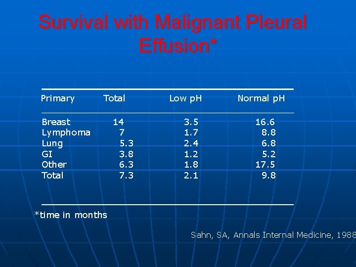
Survival with Malignant Pleural Effusion* Primary Total Breast Lymphoma Lung GI Other Total 14 7 5. 3 3. 8 6. 3 7. 3 Low p. H 3. 5 1. 7 2. 4 1. 2 1. 8 2. 1 Normal p. H 16. 6 8. 8 6. 8 5. 2 17. 5 9. 8 *time in months Sahn, SA, Annals Internal Medicine, 1988
Diagnosis of Malignant Pleural Effusion n Pleural fluid cytology Pleural biopsy Thoracoscopy
Treatment of Malignant and Paramalignent Pleural Effusions n n n Serial thoracentesis Chest tube with pleurodesis Thoracoscopy with talc poudrage Pleuroperitoneal shunt Pleurectomy
Pneumothorax n n n Primary spontaneous pneumothorax Secondary pneumothorax Iatrogenic pneumothorax Traumatic Catamenial
Summary Slide n
Summary Slide n
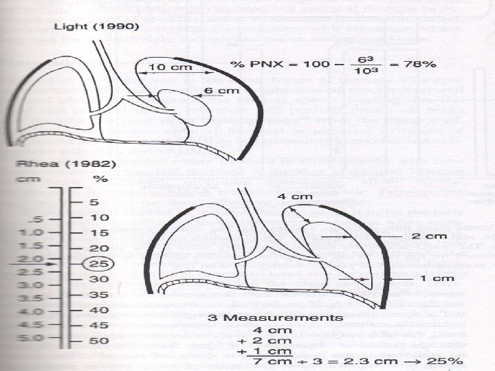
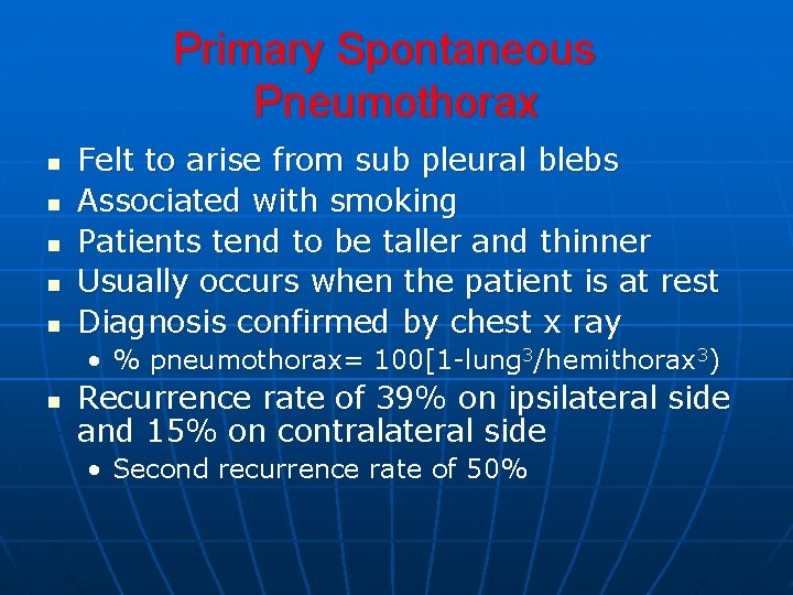
Primary Spontaneous Pneumothorax n n n Felt to arise from sub pleural blebs Associated with smoking Patients tend to be taller and thinner Usually occurs when the patient is at rest Diagnosis confirmed by chest x ray • % pneumothorax= 100[1 -lung 3/hemithorax 3) n Recurrence rate of 39% on ipsilateral side and 15% on contralateral side • Second recurrence rate of 50%
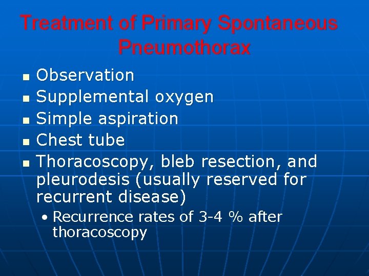
Treatment of Primary Spontaneous Pneumothorax n n n Observation Supplemental oxygen Simple aspiration Chest tube Thoracoscopy, bleb resection, and pleurodesis (usually reserved for recurrent disease) • Recurrence rates of 3 -4 % after thoracoscopy
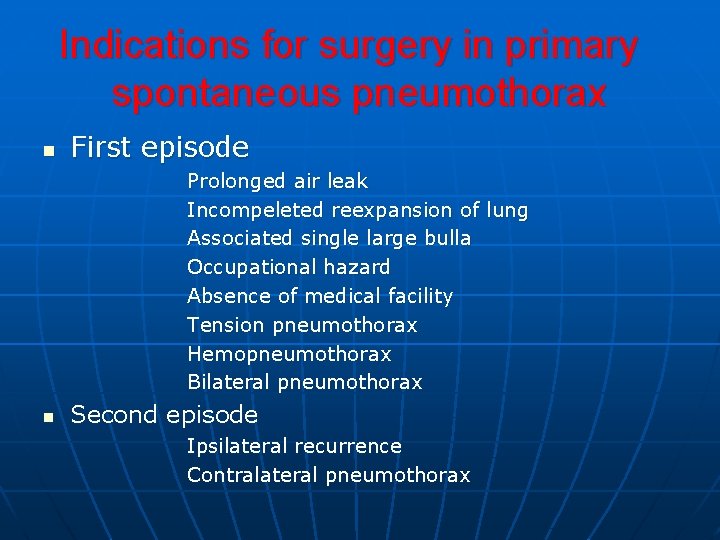
Indications for surgery in primary spontaneous pneumothorax n First episode Prolonged air leak Incompeleted reexpansion of lung Associated single large bulla Occupational hazard Absence of medical facility Tension pneumothorax Hemopneumothorax Bilateral pneumothorax n Second episode Ipsilateral recurrence Contralateral pneumothorax
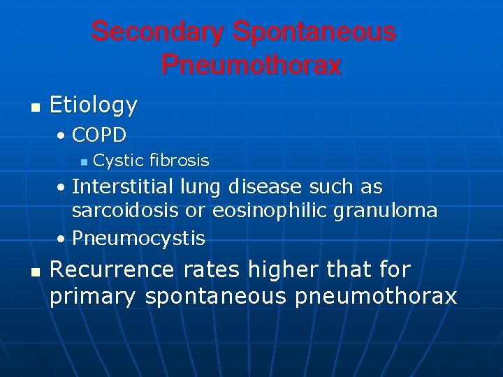
Secondary Spontaneous Pneumothorax n Etiology • COPD n Cystic fibrosis • Interstitial lung disease such as sarcoidosis or eosinophilic granuloma • Pneumocystis n Recurrence rates higher that for primary spontaneous pneumothorax
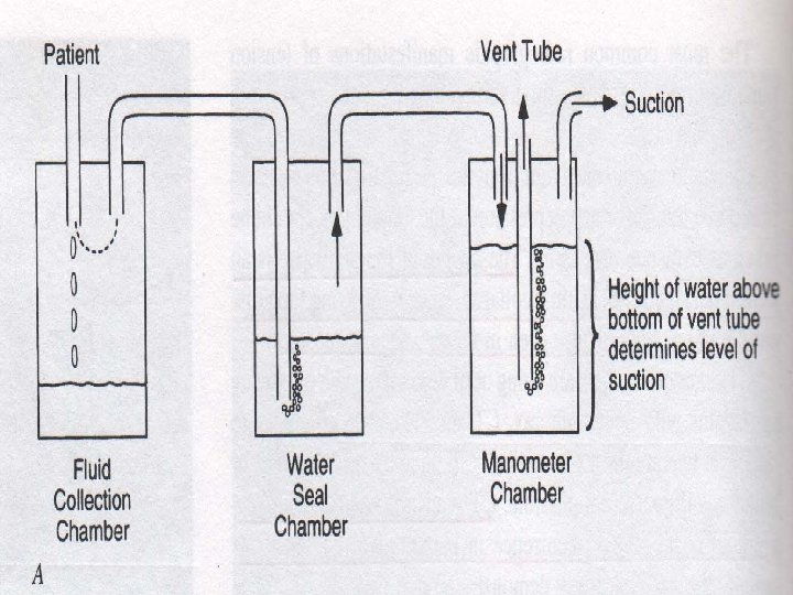
Treatment of Secondary Pneumothorax n n Chest tube Pleurodesis with first event with or without thoracoscopy
Summary Slide n
Iatrogenic Pneumothorax n n n Transthroacic needle aspiration – 20% Mechanical ventilation Thoracentesis Central line placement Transbronchial lung biopsy
Treatment of Iatrogenic Pneumothorax n n n Minimal symptoms and less that 15% pneumothorax: observe Symptomatic or > 15 % : aspiration or chest tube For patients on mechanical ventilation: chest tube
Traumatic Pneumothorax n n n Penetrating or non-penetrating trauma 40% are occult to plain chest film and are discovered with CT Consider rare but catastrophic diagnoses that require immediate thoracotomy • Rupture of the trachea • Rupture of the esophagus n Treatment is usually with a chest tube. If the pneumothorax is small and the patient is not in the ventilator, observation may be considered
Chylous Pleural Effusion n n Defined by the presence of chyle (lymph) in the pleural space. Diagnosis • Appearance often milky. Must differentiate chylous from chyliform effusion • Chemical confirmation n Triglyceride > 110 mg/d. L If triglyceride is between 50 -110 mg/d. L, send fluid for lipoprotein electrophoresis. Chylomicrons confirms a chylothorax If triglyceride is < 50, it is not chylous • Chyliform effusion has elevated cholesterol and occurs in long standing effusions.
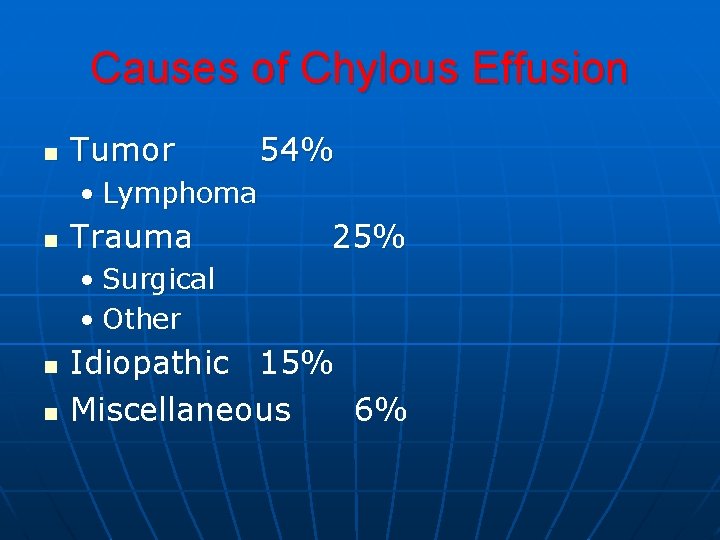
Causes of Chylous Effusion n Tumor 54% • Lymphoma n Trauma 25% • Surgical • Other n n Idiopathic 15% Miscellaneous 6%
Treatment of Traumatic Chylous Effusion • • Pleuroperitineal shunts Chest tube: Caution that the patient may become malnourished. Therefore, chyle flow is reduced by GI rest and the use of parenteral nutrition Chemical pleurodesis Thoracotomy or thoracoscopy and ligation of the thoracic duct.
Treatment of Non-traumatic Chylous Effusions • • Treat underlying lymphoma or carcinoma If ineffective, then insert a pleuroperitineal shunt
Lymphangioleiomyomatosis (LAM) n n n Rare disease of women of reproductive age A disease of proliferation of smooth muscle-like cells and affects the small airways, pulmonary microvasculature, and lymphatics Causes airway obstruction, cystic changes in the lung, and chylous effusions Typical patient is a young woman with interstitial disease on chest film, hyperinflated lungs (from air trapping) on pulmonary function, and a chylous effusion. Treatment with antiestrogen therapy (medroxyprogesterone, tamoxifen) or transplantation
Chyliform Effusions n n Milky pleural fluid due to elevated cholesterol of lecithin-globulin complexes Most commonly associated with tuberculosis, rheumatoid arthritis, therapeutic pneumothorax
Hemothorax n n Pleural fluid hematocrit greater that 50% that of peripheral blood Causes • • • Traumatic (penetrating or non-penetrating) Iatrogenic (thoracic surgery or line placement) Non traumatic (from metastatic pleural disease), spontaneous rupture of an intrathoracic vessel, bleeding disorders • Complication of anticoagulant therapy n Treatment is immediate chest tube (both to evacuate the fluid and monitor for additional bleeding)
Complications of Hemothorax n n Retention of clotted blood in the thorax (causing restriction) Infection Effusion (usually self limited) Fibrothorax (occurs in less that 1% of hemothoraces. Decortication is necessary)