Medical Genetics 14 chromosomal disorder Medical Genetics Any















- Slides: 116
Medical Genetics 14 畸变染色体引起的疾 病 chromosomal disorder
Medical Genetics Any syndrome characterized by malformations or malfunctions in any of the body's systems, and caused by abnormal chromosome number or constitution.
Medical Genetics
Medical Genetics 1. Down syndrome A. A Brief History The formal story began in 1866, when a physician named John Langdon Down published an essay in England in which he described a set of children with common features who were distinct from other children with mental retardation.
Medical Genetics Down was superintendent of an asylum for children with mental retardation in Surrey, England when he made the first distinction between children who were cretins (later to be found to have hypothyroidism) and what he referred to as "Mongoloids. "
Medical Genetics Down based this unfortunate name on his notion that these children looked like people from Mongolia, who were thought then to have an arrested development.
Medical Genetics This ethnic insult came under fire in the early 1960 s from Asian genetic researchers, and the term was dropped from scientific use. Instead, the condition became called "Down's syndrome. " In the 1970 s, an American revision of scientific terms changed it simply to "Down syndrome, " while it still is called "Down's" in the UK and some places in Europe.
Medical Genetics In the first part of the twentieth century, there was much speculation of the cause of Down syndrome. The first people to speculate that it might be due to chromosomal abnormalities were Waardenburg and Bleyer in the 1930 s.
Medical Genetics But it wasn't until 1959 that Jerome Lejeune and Patricia Jacobs, working independently, first determined the cause to be trisomy (triplication) of the 21 st chromosome. Cases of Down syndrome due to translocation and mosaicism were described over the next three years.
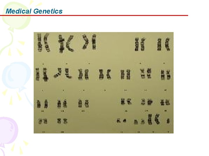
Medical Genetics B. The Chromosomes There are three different types of Down syndrome: Standard Trisomy 21, Translocation, and Mosaicism.
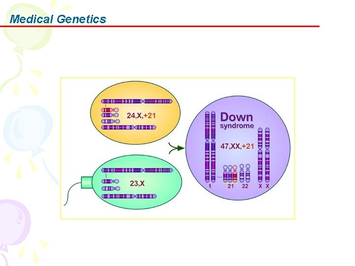
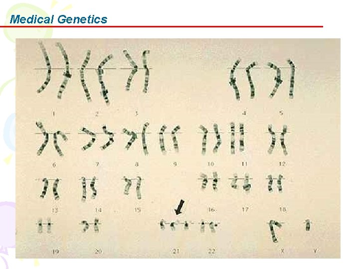
Medical Genetics In Down syndrome, 95% of all cases are caused by this event: one cell has two 21 st chromosomes instead of one, so the resulting fertilized egg has three 21 st chromosomes. Hence the scientific name, trisomy 21. Recent research has shown that in these cases, approximately 90% of the abnormal cells are the eggs.
Medical Genetics The cause of the nondisjunction error isn't known, but there is definitely connection with maternal age. Research is currently aimed at trying to determine the cause and timing of the nondisjunction event.
Medical Genetics
Medical Genetics
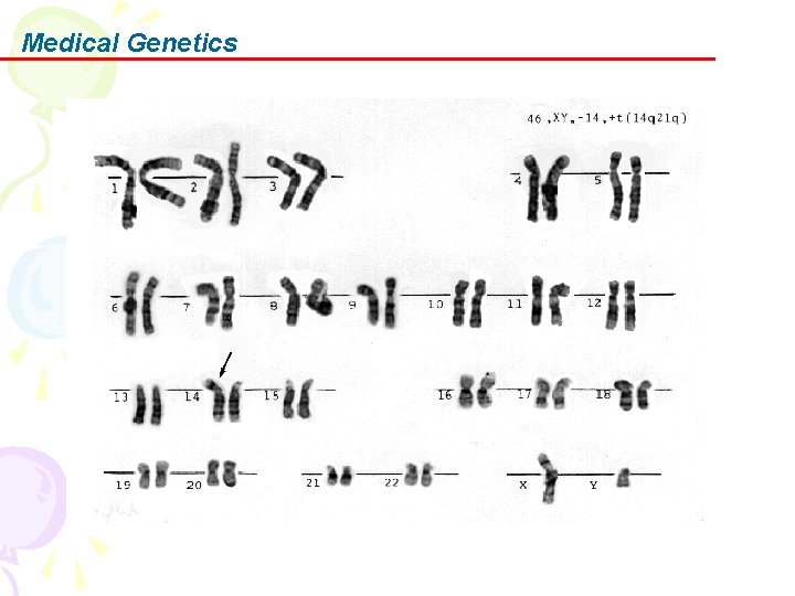
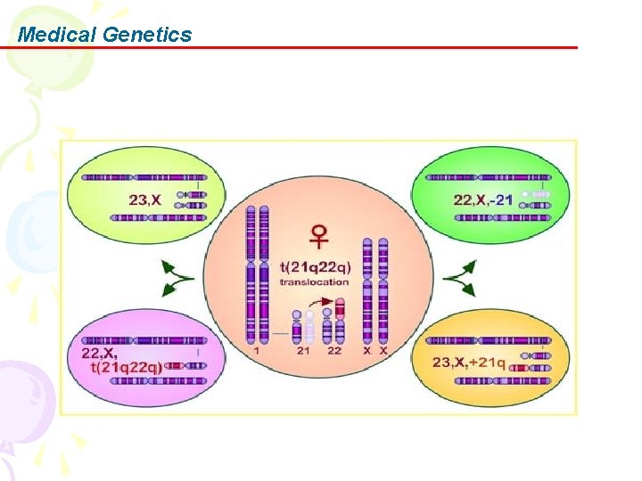
Medical Genetics Three to four percent of all cases of trisomy 21 are due to Robertsonian Translocation. In this case, two breaks occur in separate chromosomes, usually the 14 th and 21 st or 21 st and 22 st chromosomes. There is rearrangement of the genetic material so that some of the 14 th chromosome is replaced by extra 21 st chromosome.
Medical Genetics So while the number of chromosomes remain normal, there is a triplication of the 21 st chromosome material. Some of these children may only have triplication of part of the 21 st chromosome instead of the whole chromosome, which is called a partial trisomy 21.
Medical Genetics
Medical Genetics
Medical Genetics Translocations resulting in trisomy 21 may be inherited, so it's important to check the chromosomes of the parents in these cases to see if either may be a "carrier. "
Medical Genetics Mosaicism is when a person has a mix of cells, some containing 46 chromosomes and some containing 47 chromosomes.
Medical Genetics C. The 21 st Chromosome and Down Syndrome In trisomy 21, the presence of an extra set of genes leads to overexpression of the involved genes, leading to increased production of certain products.
Medical Genetics For most genes, their overexpression has little effect due to the body's regulating mechanisms of genes and their products. But the genes that cause Down syndrome appear to be exceptions.
Medical Genetics Which genes are involved? That's been the question researchers have asked ever since third 21 st chromosome was found. From years of research, one popular theory stated that only a small portion of the 21 st chromosome actually needed to be triplicated to get the effects seen in Down syndrome; this was called the Down Syndrome Critical Region(DSCR).
Medical Genetics However, this region is not one small isolated spot, but most likely several areas that are not necessarily side by side. The 21 st chromosome may actually hold 200 to 250 genes (being the smallest chromosome in the body in terms of total number of genes); but it's estimated that only a small percentage of those may eventually be involved in producing the features of Down syndrome.
Medical Genetics Genes that may have input into Down syndrome include: • Superoxide Dismutase (SOD 1)-- overexpression may cause premature aging and decreased function of the immune system; its role in Senile Dementia of the Alzheimer's type or decreased cognition is still speculative • COL 6 A 1 -- overexpression may be the cause of heart defects • ETS 2 -- overexpression may be the cause of skeletal abnormalities • CAF 1 A -- overexpression may be detrimental to DNA synthesis • Cystathione Beta Synthase (CBS) -- overexpression may disrupt metabolism and DNA repair • DYRK -- overexpression may be the cause of mental retardation • CRYA 1 -- overexpression may be the cause of cataracts • GART -- overexpression may disrupt DNA synthesis and repair • IFNAR -- the gene for expression of Interferon, overexpression may interfere with the immune system as well as other organ systems
Medical Genetics Other genes that are also suspects include APP, GLUR 5, S 100 B, TAM, PFKL, and a few others. Again, it is important to note that no gene has yet been fully linked to any feature associated with Down syndrome.
Medical Genetics One of the more notable aspects of Down syndrome is the wide variety of features and characteristics of people with trisomy 21: There is a wide range of mental retardation and developmental delay noted among children with Down syndrome.
Medical Genetics Some babies are born with heart defects and others aren't. Some children have associated illnesses such as epilepsy, hypothyroidism or celiac disease, and others don't. The first possible reason is the difference in the genes that are triplicated.
Medical Genetics The effect of overexpression of genes may depend on which allele is present in the person with trisomy 21. The second reason that might be involved is called "penetrance, " and that appears to be what happens with trisomy 21: the alleles don't do the same thing to every person who has it. Both reasons may be why there is such variation in children and adults with Down syndrome.
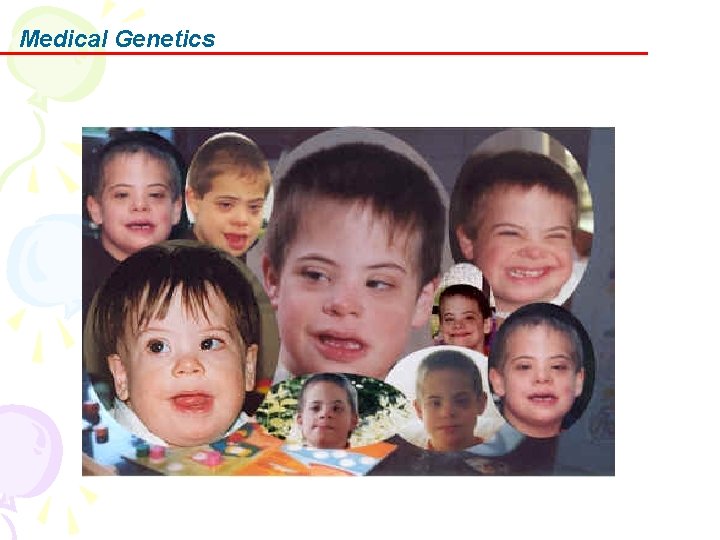
Medical Genetics D. Clinical features Growth: Short stature and obesity occurs during adolescence. CNS: Moderate-to-severe mental retardation occurs, with an intelligence quotient (IQ) of 20 -85. Congenital heart defects
Medical Genetics
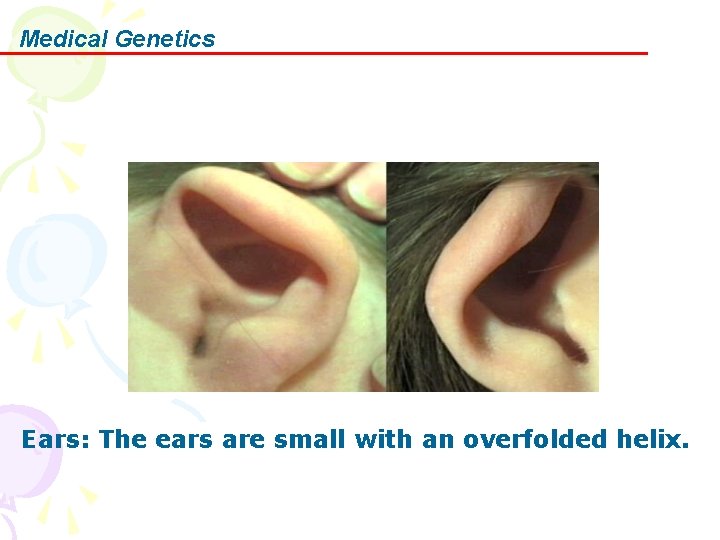
Medical Genetics Ears: The ears are small with an overfolded helix.
Medical Genetics Dermatoglyphic s: Distal axial triradius in the palms, transverse palmar creases, a single flexion crease in the fifth finger, ulnar loops (often 10), a pattern in hypothenar, and interdigital III regions are observed.
Medical Genetics E. Prenatal Testing Maternal serum alpha fetoprotein (MSAFP) is measured by a blood test in the pregnant woman at 15 -18 weeks of gestation.
Medical Genetics AFP is made in the fetal liver, and some escapes into the maternal circulation. It is widely used as a screening test for a DS fetus. MSAFP and at least 1 other screening test are recommended for all pregnancies not having amniocentesis by the American College of Obstetrics, and the American College of Medical Genetics.
Medical Genetics MSAFP testing is based on the fact that DS fetuses tend to be a little smaller on average, have smaller placentas, and thus secrete less MSAFP and other materials, which are determined in the serum of the pregnant woman. Factors that affect this test include gestational age, maternal weight, diabetes and ethnicity.
Medical Genetics MSAFP screening is not a definitive test. If the MSAFP test is low it suggests the risk of a DS fetus equal to the risk of a woman age 35, and prenatal testing/chromosome studies are suggested if the parents want this information.
Medical Genetics If MSAFP alone is tested, 20 per cent of DS fetuses will test low. If MSAFP and human chorionic gonadotrophin (HCG) are determined, 50 - 60 % of DS fetuses will be identified. If MSAFP, HCG and estradiol (E 2) are tested, 60 -70 % of DS fetuses will be identified. Some HCG testing employs a beta subunit but this is not widespread.
Medical Genetics A positive screening test suggests only that the risk of DS is increased, and the definitive testing of amniocentesis is indicated.
Medical Genetics Some ultrasonographers can determine suggestive findings of DS by changes in the neck, or heart. This is not yet confirmed or in wide spread use. Ultrasounds can be normal in a DS fetus.
Medical Genetics Amniocentesis, a sampling of the amniotic fluid surrounding the fetus, is routinely done at 14 -16 wks. Amniocentesis testing for chromosome disorders is 99. 8 per cent reliable for chromosome number, and there is a risk of miscarriage (usually 1/250 or less) after the procedure.
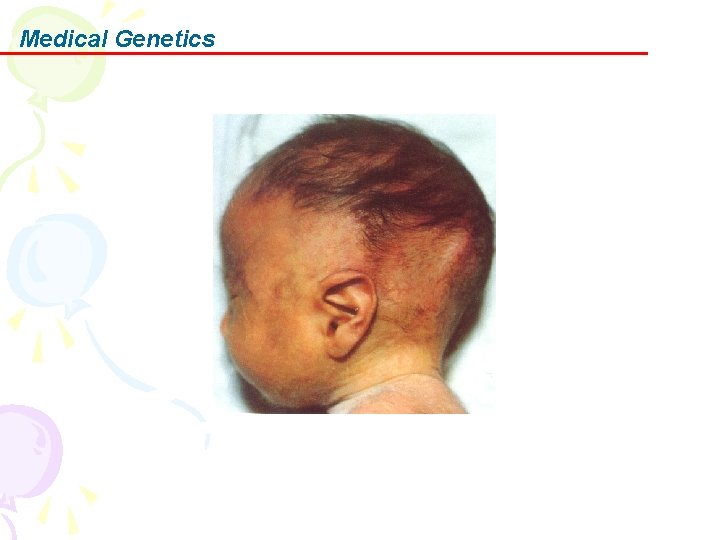
Medical Genetics 2. Trisomy 18 was independently described by Edwards et al and Smith et al in 1960. Among liveborn children, trisomy 18 is the second most common autosomal trisomy after trisomy 21. The disorder is characterized by severe psychomotor and growth retardation, microcephaly, microphthalmia, malformed ears, micrognathia or retrognathia, microstomia, distinctively clenched fingers, and other congenital malformations.
Medical Genetics Synonyms and related keywords: Edwards syndrome Edwards' syndrome trisomy 18 syndrome trisomy E syndrome
Medical Genetics A. Pathophysiology Trisomy 18 severely affects all organ systems. In translocations that result in partial trisomy or in cases of mosaic trisomy 18, clinical expression is less severe and longer survival is usual.
Medical Genetics B. Frequency In the US: Prevalence is approximately 1 in 6000 -8000 live births.
Medical Genetics C. Mortality/Morbidity • Approximately 95% of conceptuses with trisomy 18 die in embryonic or fetal life; 5 -10% of affected children survive beyond the first year of life. • The high mortality rate is usually due to the presence of cardiac and renal malformations, feeding difficulties, sepsis, and apnea caused by CNS defects. • Severe psychomotor and growth retardation are invariably present for those who survive beyond infancy.
Medical Genetics D. Sex Approximately 80% of cases occur in females. The preponderance of females with trisomy 18 in liveborn infants (sex ratio 0. 63) compared to fetuses with prenatal diagnoses (sex ratio 0. 90) indicates a prenatal selection against males with trisomy 18 after the time of amniocentesis.
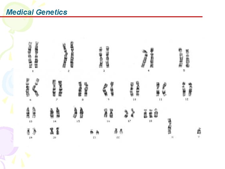
Medical Genetics E. Cause The cause of Edwards syndrome is full trisomy 18 in 95% of cases. Mosaicism and translocations cause few cases. An extra chromosome 18 is responsible for the phenotype.
Medical Genetics
Medical Genetics Incidence increases with advanced maternal age. In approximately 90% of cases, the extra chromosome is maternal in origin, with meiosis II errors occurring twice as frequently as meiosis I errors.
Medical Genetics This is in contrast to other human trisomies, which exhibit a higher frequency of nondisjunction in maternal meiosis I. Among cases resulting from paternal nondisjunction, most are the result of postzygotic mitotic errors.
Medical Genetics F. Clinical features Neurological Cranial Facial Skeletal Cardiac Pulmonary Gastrointestinal Genitourinary Endocrine Dermal
Medical Genetics
Medical Genetics
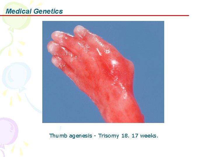
Medical Genetics Thumb agenesis - Trisomy 18. 17 weeks.
Medical Genetics
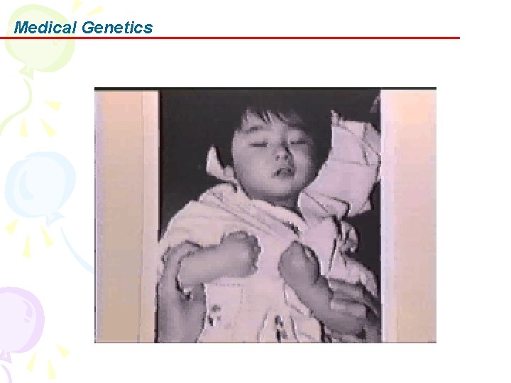
Medical Genetics 3. Patau syndrome Trisomy 13 syndrome is a disorder of human chromosomes which occurs in approximately 1 in 10, 000 live born infants.
Medical Genetics Synonyms: Trisomy 13 Patau Syndrome Trisomy 13 -15 D Trisomy Syndrome
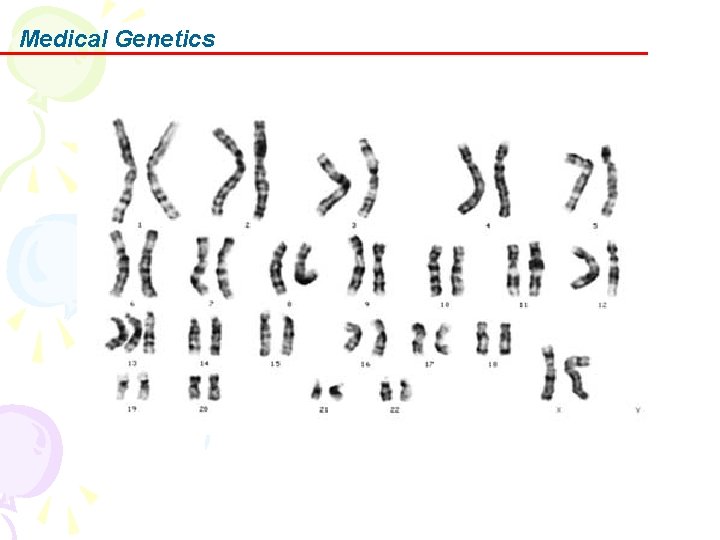
Medical Genetics Trisomy 13 is due to the presence of an extra 13 rd chromosome.
Medical Genetics Approximately 80% of infants with Trisomy 13 syndrome will have a full trisomy while the remainder will have a trisomy due to a rearrangement called a translocation or have mosaicism (two different cell lines).
Medical Genetics
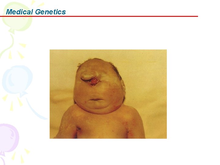
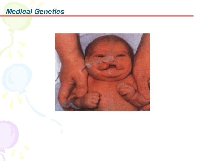
Medical Genetics Findings of significance include small head size (microcephaly); small eyes (microphthalmia) or sometimes absent eye or faulty development of the retina. Cleft lip or cleft palate or both occur in about 60% of children.
Medical Genetics In addition, there a number of less medically significant physical findings that are helpful in diagnosis. These include variations of ear shape, changes on the palm of the hand, and extra fingers and toes. Changes in foot development, including changes to the heel, the socalled rocker bottom foot, can occur.
Medical Genetics
Medical Genetics
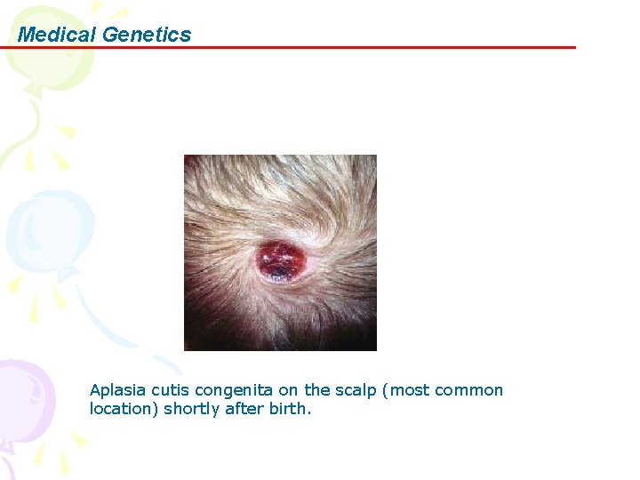
Medical Genetics Aplasia cutis congenita on the scalp (most common location) shortly after birth.
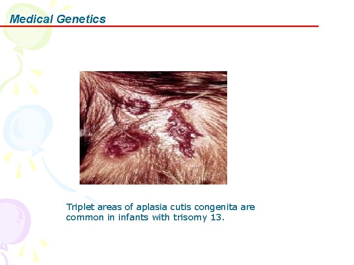
Medical Genetics Triplet areas of aplasia cutis congenita are common in infants with trisomy 13.
Medical Genetics 4. cri du cat syndrome In 1963, Lejeune et al described a syndrome of multiple congenital anomalies, mental retardation, microcephaly, abnormal face, and a mewing cry in infants with deletion of a B group chromosome (Bp-), later identified as 5 p-.
Medical Genetics Cri-du-chat syndrome is an autosomal deletion syndrome caused by a partial deletion of chromosome 5 p. It is characterized by a distinctive, high-pitched, catlike cry in infancy with growth failure, microcephaly, facial abnormalities, and mental retardation throughout life.
Medical Genetics Synonyms and related keywords cat cry syndrome chromosome deletion 5 p syndrome monosomy 5 p syndrome (Bp-) 5 p partial deletion of chromosome 5 p 5 p deletion 5 p monosomy
Medical Genetics A. Pathophysiology A partial deletion of the short arm of chromosome 5 is responsible for the characteristic phenotype. The characteristic cry is perceptually and acoustically similar to the mewing of kittens.
Medical Genetics This unusual cry is because of structural abnormalities of the larynx (such as laryngeal hypoplasia) and CNS dysfunction. The laryngeal appearance may be normal or may exhibit marked anatomical abnormalities such as floppy epiglottis, small larynx, and asymmetric vocal cords.
Medical Genetics However, the cause of the characteristic cry cannot be entirely ascribed to the larynx. A developmental field connecting the brain and the affected clivus region of the cranial base with the laryngeal region from which the characteristic cry derives may exist. This area of the brain is probably deformed in patients with cri-du-chat syndrome. The characteristic cry usually disappears with time.
Medical Genetics B. Frequency In the US: The estimated prevalence is about 1 in 50, 000 live births. The prevalence among individuals with mental retardation is about 1. 5 in 1000.
Medical Genetics C. Mortality/Morbidity With contemporary interventions, the chance of survival to adulthood is possible. Currently, death occurs in 6 -8% of the overall population affected with the syndrome. Pneumonia, aspiration pneumonia, congenital heart defects, and respiratory distress syndrome are the most common causes of death.
Medical Genetics D. Sex A significant female predominance exists in affected newborns, with a male-to-female ratio of 0. 72.
Medical Genetics E. Causes • Most cases (80 -85%) are due to sporadic de novo deletion of 5 p (15. 3 ->15. 2). • Approximately 10 -15% of cases result from the unequal segregation of a parental balanced translocation where the 5 p monosomy is often accompanied by a trisomic portion of the genome. The phenotypes in these individuals may be more severe than in those with isolated monosomy of 5 p because of this additional trisomic portion of the genome.
Medical Genetics
Medical Genetics
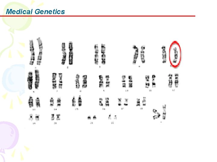
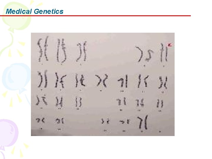
Medical Genetics • Most cases involve terminal deletions with 30 -60% loss of 5 p material. Fewer than 10% of cases have other rare cytogenetic aberrations. • The deleted chromosome 5 is paternal in origin in about 80% of the cases. • Loss of a small region in band 5 p 15. 2 (cridu-chat critical region) correlates with all the clinical features of the syndrome with the exception of the catlike cry, which maps to band 5 p 15. 3 (catlike critical region).
Medical Genetics • High-resolution mapping of genotype-phenotype relationships in cri-du-chat syndrome using array comparative genomic hybridization (CGH) – Localized the region associated with the cry to 1. 5 Mb in distal band 5 p 15. 31, between bacterial artificial chromosomes (BACs) containing markers D 5 S 2054 and D 5 S 676. – Localized the region associated with the speech delay to 3. 2 Mb in band 5 p 15. 32 -15. 33, between BACs containing markers D 5 S 417 and D 5 S 635. – Localized the region associated with the facial dysmorphology to 2. 4 Mb in band 5 p 15. 2 -15. 31, between BACs containing markers D 5 S 208 and D 5 S 2887.
Medical Genetics F. Clinical features History: Characteristic cry Developmental history: Early feeding problems are present because of swallowing difficulties; poor suck; failure to thrive; early ear infections; and severe cognitive, speech, and motor delays. Almost all patients have these problems. Behavioral history Physical: Neonatal period Childhood: Findings include severe mental retardation; developmental delay; microcephaly;
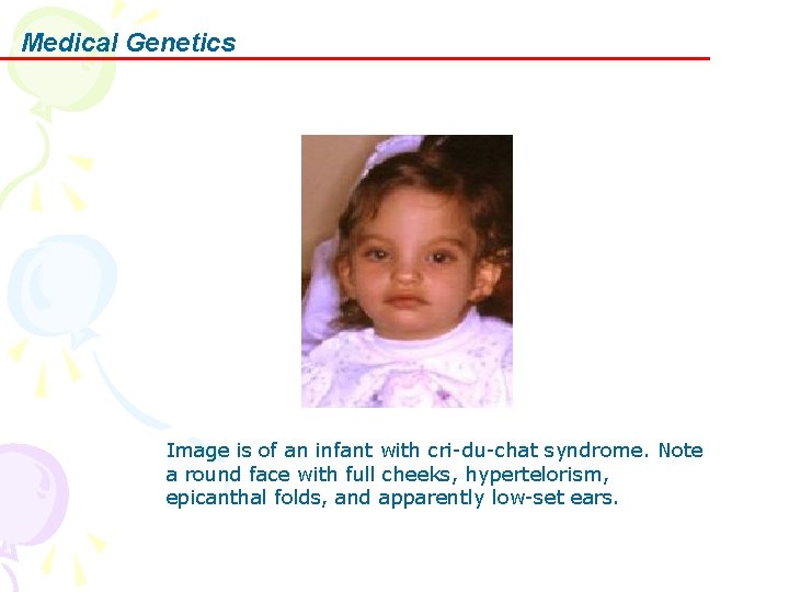
Medical Genetics Image is of an infant with cri-du-chat syndrome. Note a round face with full cheeks, hypertelorism, epicanthal folds, and apparently low-set ears.
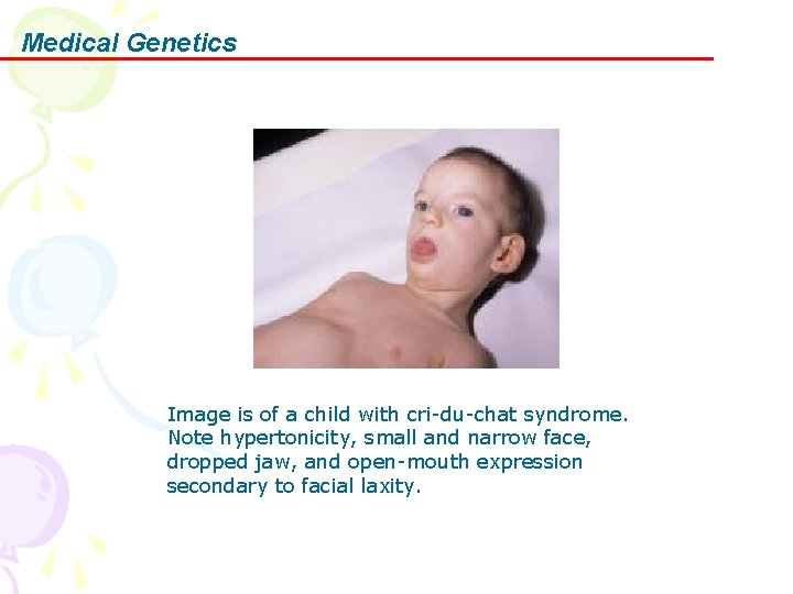
Medical Genetics Image is of a child with cri-du-chat syndrome. Note hypertonicity, small and narrow face, dropped jaw, and open-mouth expression secondary to facial laxity.
Medical Genetics 5. Klinefelter syndrome In 1942, Klinefelter et al published a report on 9 men who had enlarged breasts, sparse facial and body hair, small testes, and inability to produce sperm.
Medical Genetics In 1959, these men with Klinefelter syndrome were discovered to have an extra sex chromosome (genotype XXY) instead of the usual male sex complement (genotype XY).
Medical Genetics Klinefelter syndrome is the most common chromosomal disorder associated with male hypogonadism and infertility. It is defined classically by a 47, XXY karyotype with variants demonstrating additional X and Y chromosomes.
Medical Genetics The syndrome is characterized by hypogonadism (small testes, azoospermia/oligospermia), gynecomastia at late puberty, psychosocial problems, hyalinization and fibrosis of the seminiferous tubules, and elevated urinary gonadotropins.
Medical Genetics A. Pathophysiology The addition of more than 1 extra X or Y chromosome to a male karyotype results in variable physical and cognitive abnormalities. In general, the extent of phenotypic abnormalities, including mental retardation, is related directly to the number of supernumerary X chromosomes.
Medical Genetics As the number of X chromosomes increases, somatic and cognitive development are more likely to be affected. Skeletal and cardiovascular abnormalities can become increasingly severe. Gonadal development is particularly susceptible to each additional X chromosome, resulting in seminiferous tubule dysgenesis and infertility as well as hypoplastic and malformed genitalia in polysomy X males.
Medical Genetics Moreover, mental capacity diminishes with additional X chromosomes. The IQ is reduced by approximately 15 points for each supernumerary X chromosome, but conclusions about reduced mental capacity must be drawn cautiously. All major areas of development, including expressive and receptive language and coordination, are affected by extra X chromosome material.
Medical Genetics B. Frequency In the US: Approximately 1 in 5001, 000 males is born with an extra sex chromosome; over 3, 000 affected males are born yearly. The prevalence is 5 -20 times higher in the mentally retarded than in the general newborn population.
Medical Genetics C. Mortality/Morbidity • About 40% of concepti with Klinefelter syndrome survive the fetal period. • In general, severity of somatic malformations in Klinefelter syndrome is proportional to the number of additional X chromosomes; mental retardation and hypogonadism are more severe in 49, XXXXY than in 48, XXXY. • Mortality rate is not significantly higher than in healthy individuals.
Medical Genetics D. Causes • Klinefelter syndrome is caused by the presence of an additional X chromosome in a male. • About 50 -60% of cases are due to maternal nondisjunction (75% meiosis I errors). In cases in which these maternal meiosis I errors are identified, maternal age is increased. The remaining cases are due to paternal nondisjunction. • The most common karyotype is 47, XXY (about 80 -90% of all cases). Mosaicism (46, XY/47, XXY) is observed in about 10% of cases. Other variant karyotypes, including 48, XXYY, 48, XXXY, 49, XXXYY, and 49, XXXXY, are rare.
Medical Genetics
Medical Genetics • The mosaic forms of Klinefelter syndrome – The mosaic forms of Klinefelter syndrome are due to mitotic nondisjunction after fertilization of the zygote. – These forms can arise from a 46, XY zygote or a 47, XXY zygote. • The variant forms are as follows: – 48, XXXY – 49, XXXXY – 48, XXYY – 49, XXXYY
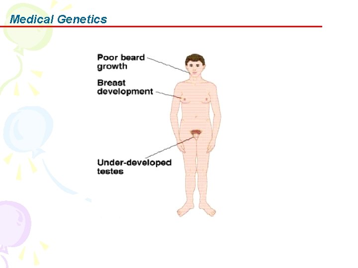
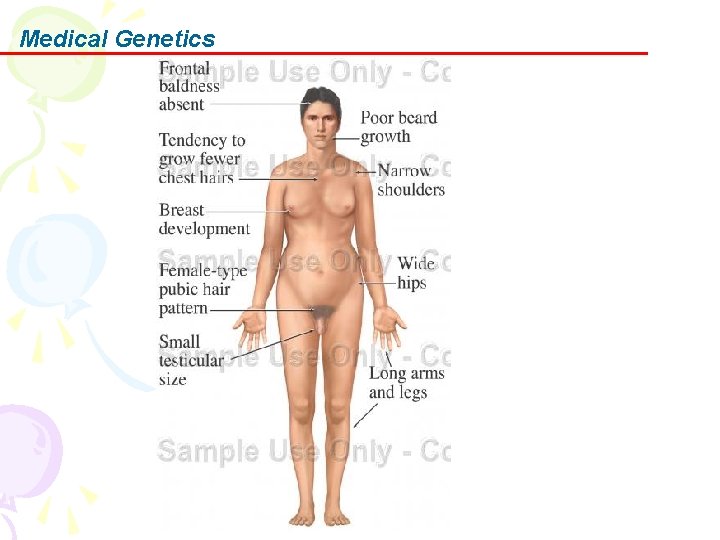
Medical Genetics E. Clinical features Growth Central nervous system Dental Sexual characteristics Cardiac and circulatory problems
Medical Genetics
Medical Genetics
Medical Genetics
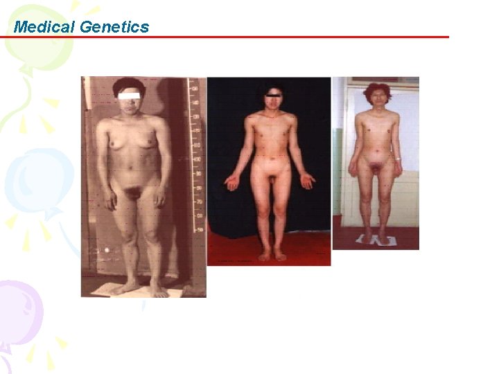
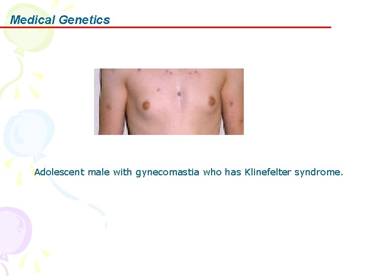
Medical Genetics Adolescent male with gynecomastia who has Klinefelter syndrome.
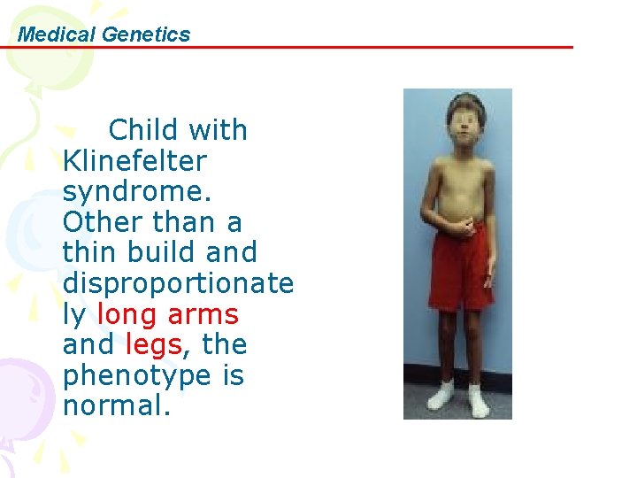
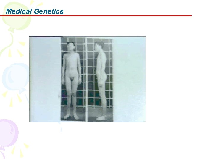
Medical Genetics Child with Klinefelter syndrome. Other than a thin build and disproportionate ly long arms and legs, the phenotype is normal.
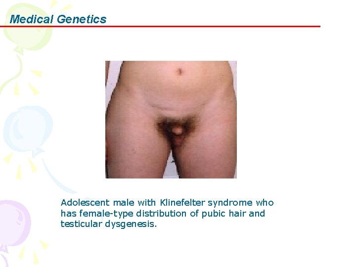
Medical Genetics Adolescent male with Klinefelter syndrome who has female-type distribution of pubic hair and testicular dysgenesis.
Medical Genetics
Medical Genetics 6. Turner syndrome In 1938, Henry Turner first described Turner syndrome, which is one of the most common chromosomal abnormalities. More than 95% of adult women with Turner syndrome exhibit short stature and infertility.
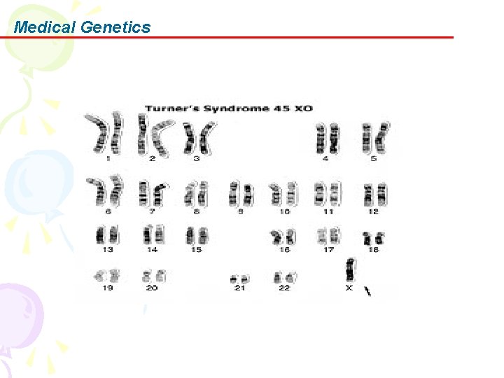
Medical Genetics A. Pathophysiology Turner syndrome is caused by the absence of one set of genes from the short arm of one X chromosome. In patients with 45 X karyotype, about two thirds are missing the paternal X chromosome.
Medical Genetics In addition to monosomy X, a similar clinical picture is found with a 46 XXiq karyotype and in some individuals with mosaic karyotypes. A deletion of the SHOX gene can cause a similar skeletal phenotype known as Leri-Weill dyschondrosteosis.
Medical Genetics B. Frequency • In the US: Frequency is approximately 1 in 2000 live-born female infants. As many as 15% of spontaneous abortions have a 45 X karyotype. • Internationally: Incidence is the same as for the United States. No known ethnic or racial factors influence frequency.
Medical Genetics C. Mortality/Morbidity • Mortality may be increased in the neonatal period because of coarctation of the aorta and in adulthood because of cardiovascular disease. • Renal anomalies found in some individuals may cause a predisposition to urinary tract infections or hypertension. Even in the absence of cardiac or renal anomalies, patients are prone to develop hypertension.
Medical Genetics D. Causes • Advanced maternal age is not associated with an increased incidence. • In patients with a single X chromosome, the chromosome is of maternal origin in two thirds of cases. • Many of the features of Turner syndrome, including the short stature, are due to the lack of a second SHOX gene, which is on the X chromosome.
Medical Genetics
Medical Genetics E. Clinical features Approximately 95% of individuals with Turner syndrome have both short stature and signs of ovarian failure on physical examination.
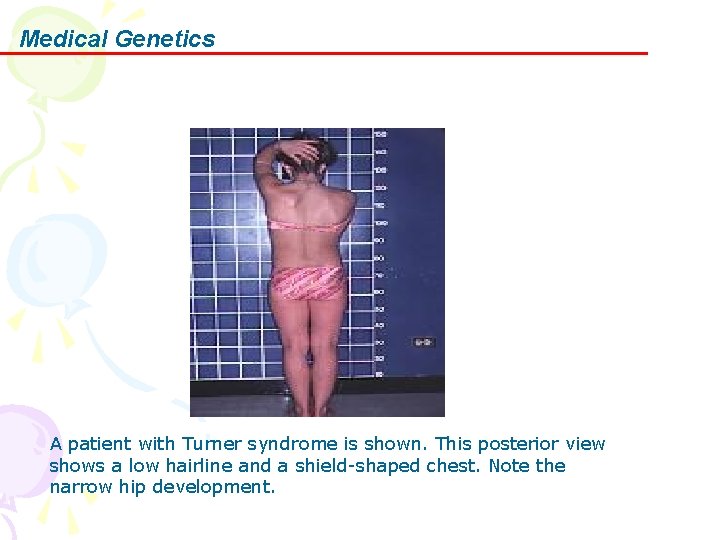
Medical Genetics A patient with Turner syndrome is shown. This posterior view shows a low hairline and a shield-shaped chest. Note the narrow hip development.
Medical Genetics Women and girls with TS tend to have a wide carrying angle of the arms (Figure A). This means that when they hold their arms at their sides, with their elbows touching their sides, their hands stand away from their bodies. Girls with TS can have low hairlines at the back of their heads (Figure B).
Medical Genetics Their ears may be slightly lower than those of other girls and their jawbones may be slightly less prominent (Figure A). The nails on their hands and feet tend to turn up slightly (Figure B).
Medical Genetics