
Recurrent Pregnancy loss Genetic Recurrent miscarriage RM is




- Slides: 37
Recurrent Pregnancy loss & Genetic
Recurrent miscarriage (RM) is defined as the presence of two or more spontaneous abortions, which imposes emotional, physical, and financial complications for the couples. It has been estimated that 1% to 3% of couples experience RM. The disease could be considered as a multifactorial disease, affected by factors such as maternal nutrition, hormonal and endocrine disorders, anatomical abnormalities, infections, immune system and metabolic pathway malfunctions, endometriosis, sperm quality, and genetic abnormalities.
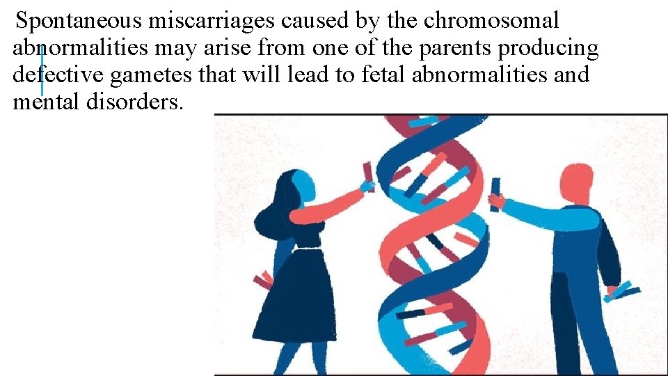
Spontaneous miscarriages caused by the chromosomal abnormalities may arise from one of the parents producing defective gametes that will lead to fetal abnormalities and mental disorders.
v. Most spontaneous miscarriages result from chromosomal abnormalities in the embryo or fetus. v. Approximately 50% of all first-trimester pregnancy losses, 30% of second-trimester abortuses, and 3% of stillbirths are chromosomally abnormal.
However, these have very likely underestimated the prevalence of chromosomal abnormalities among abortuses because the data are biased by unrecognized maternal cell contamination and because normal euploid cells (from the mother or abortus) are less likely to fail culture than abnormal cell lines.
ØAnalyses using newer techniques not dependent on cell culture (comparative genomic hybridization or microarray) and more recent careful cytogenetic studies of early missed abortions suggest that the true incidence of chromosomal abnormalities in miscarried early pregnancies is closer to 75%.
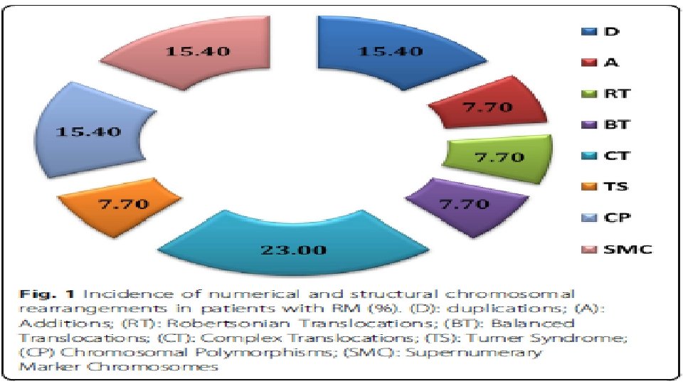
q. Over 90% of the chromosomal abnormalities observed among abortuses are numerical (aneuploidy, polyploidy); The remainder are split between structural abnormalities (translocations, inversions) and mosaicism.
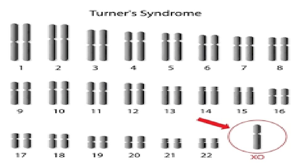
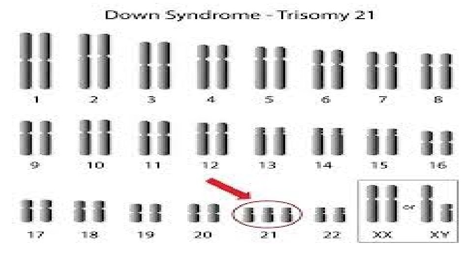
Overall, autosomal trisomies are the most common abnormality (usually involving chromosomes 13– 16, 21, or 22), followed by monosomy X (45, X) and polyploidies.
Fetal aneuploidy is present at a frequency of up to 90% in specimens obtained from losses aged 0– 6 wk of gestation, ∼ 50% in sporadic losses occurring at 8– 11 wk gestation, and 30% in tissues from losses at 16– 19 wk gestation. Six to 12% of miscarriage specimens obtained from demises that occur after 20 wk of gestational display chromosomal abnormalities
The frequency of parental karyotypic abnormalities, including balanced translocations, is higher among couples with a history of RPL (2%– 5%) than in the general population (0. 2%). The prevalence of RPL among first degree relatives of women with RPL is increased approximately sixfold compared with controls.
Preimplantation genetic screening (PGS) in age matched populations shows that embryos from women with RPL have a higher incidence of aneuploidy than those from women undergoing screening for reasons not related to pregnancy loss.
o. Among women with history of recurrent pregnancy loss, chromosomally normal (euploid) abortuses are more common, particularly in those age 35 and under.
There is a very high frequency of sporadic karyotypic abnormalities in products of conception, while the actual incidence of karyotypic abnormalities in the parents is low. Majority of chromosomally abnormal conceptions result from the chance union of one normal and one aneuploid gamete or from nondisjunction during early embryonic development.
q. Balanced translocations (reciprocal, Robertsonian) are the most common abnormalities; sex chromosome mosaicism, chromosome inversions, and other structural abnormalities can also be observed.
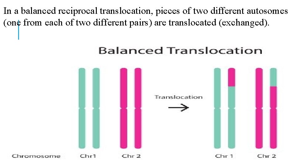
In a balanced reciprocal translocation, pieces of two different autosomes (one from each of two different pairs) are translocated (exchanged).
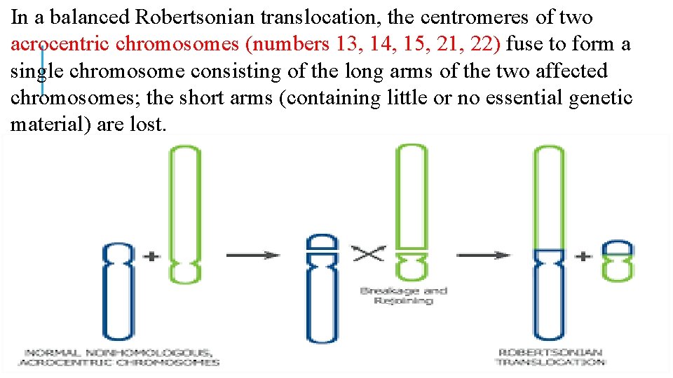
In a balanced Robertsonian translocation, the centromeres of two acrocentric chromosomes (numbers 13, 14, 15, 21, 22) fuse to form a single chromosome consisting of the long arms of the two affected chromosomes; the short arms (containing little or no essential genetic material) are lost.
In both cases, the translocation carrier is genetically balanced and phenotypically normal. Unfortunately, when their oogonia or spermatogonia undergo meiosis to yield haploid oocytes or sperm, a large proportion of the gametes end up genetically unbalanced and abnormal, having either a deficiency or an excess of genetic material.
Depending on how the chromosomes segregate during meiosis, the gametes may be chromosomally normal (containing only the normal copy of each of the two affected chromosome pairs), abnormal but balanced (containing the translocated member of each of the two affected chromosome pairs), or abnormal and unbalanced (containing two copies or no copies of an affected chromosome or chromosome segment).
When such chromosomally unbalanced gametes combine with a normal gamete from an unaffected partner, the conceptus will have a trisomy and/or a monosomy and will almost always abort; an unbalanced conceptus may occasionally survive, but those that do are at high risk for malformations and mental retardation.
The probability of a subsequent genetically normal conceptus and live birth depends on the chromosome(s) involved and the type of rearrangement. In theory, one-fourth of the gametes produced by reciprocal translocation carriers should be normal, one-fourth should be abnormal but balanced, and one-half should be abnormal and unbalanced, yielding a 50% probability of a normal pregnancy (normal or balanced conceptus) and a 50% probability of an abnormal pregnancy (abortion or a viable but anomalous fetus), assuming union with a chromosomally normal gamete from the unaffected partner.
The probability of a successful pregnancy and the risk of a chromosomally abnormal but viable fetus vary with the specific chromosomes involved and the size and location of the translocated segments!!!
Abnormalities of some chromosomes (chromosome 21) are better tolerated than others and risk of an unbalanced but viable conceptus is higher when the exchanged chromosomal segments are small.
Chromosomal inversions occur less frequently than translocations and may or may not have reproductive implications, depending on their size and location. Pericentric inversions (those that involve the centromere) often have no clinical consequences; a pericentric inversion of chromosome 9, inv(9)(p 11 q 13), is so common (1– 1. 5% in the general population) that some consider it a normal variant with no importance.
NOTICE…………. . The most common reproductive history in translocation carrier couples includes: both a normal child and early pregnancy losses (6– 7%); other histories involving only spontaneous miscarriages or combinations of malformed children, stillbirths, and abortions are slightly less common (4– 5%).
The probability of identifying a balanced chromosomal translocation in a couple with three or more previous pregnancy losses is not significantly higher than in those having had only two. In some couples, family history (recurrent pregnancy loss, stillbirths, or birth defects) suggests the possibility of an occult chromosomal abnormality after only one spontaneous miscarriage.
Couples with miscarriages interspersed with normal pregnancies and outcomes should be evaluated in the same fashion as couples with consecutive miscarriages!!!!
AGING…… Prevalence studies indicate the aneuploidy in oocytes is relatively low before age 35 (<10%) but increases abruptly thereafter, reaching nearly 100% after age. These observations offer a logical explanation for the overall age-related increase in the incidence of miscarriage and the higher prevalence of aneuploidy among the abortuses of aging women. Indeed, most trisomies observed among abortuses can be traced to maternal meiotic errors and oocyte aneuploidy.
The prevalence of abnormal ovarian reserve tests in women with unexplained recurrent pregnancy loss is higher than in women with other defined causes of recurrent pregnancy loss and comparable to that observed in the general population of infertile women.
Women who have had a trisomic abortus reach menopause at an earlier average age. Other women may have their ovarian follicular pool depleted by disease that destroys ovarian tissue or requires its removal. Either way, the end result is the same accelerated follicular depletion, declining fertility, and increasing risk for miscarriage begin at an earlier than normal age.
Besides offering information that may help to explain recurrent pregnancy loss, ovarian reserve testing may identify young women at increased risk for fetal aneuploidy in subsequent pregnancies who would otherwise not be considered candidates for prenatal diagnostic studies.
CONCLUSION Any pregnancy in an affected couple becomes a candidate for prenatal diagnostic studies, regardless of the mother’s age or previous reproductive history.
CONCLUSION…. . The recent advent of genome-wide high-resolution technologies, such as chromosome microarray analysis and next-generation sequencing (NGS), including whole exome sequencing (WES), has opened new possibilities for discovery of genetic changes that are too small to be detected by traditional chromosome analysis.