CHROMOSOMAL ABNORMALITIES By Dr Samina Anjum CAUSES OF

CHROMOSOMAL ABNORMALITIES By Dr Samina Anjum

CAUSES OF BIRTH DEFECTS & SPONTANEOUS ABORTIONS ARE • Chromosomal abnormalities • Genetic factors

INCIDENCE FOR MAJOR CHROMOSOMAL ABNORMALITIES • 50% of conceptions end in spontaneous abortions and 50% of these abortions have major chromosomal abnormalities • Thus approx. 25% of conceptuses have major chromosomal defects • Chromosomal abnormalities account for 7% of major birth defects; Commonest is Turner’s syndrome • Gene mutations account for an additional 8% cases

• A Karyotype refers to a full set of chromosomes from an individual which can be compared to a "normal" Karyotype for the species via genetic testing. • Ploidy Is the number of sets of chromosomes in a biological cell.
 • Diploid=2 n (in Normal somatic")
• Haploid = n (in normal gametes) • Diploid=2 n (in Normal somatic cell) • Euploid = An exact or multiple of n or of the monoploid number. A human with abnormal, but integral multiple of the monoploid number, (69 chromosomes) would also be considered as euploid e. g. ( 2 n, 3 n, 4 n etc)

POLYPLOID • Many organisms have more than two sets of homologous chromosomes and are called polyploid. • A chromosome number that is a multiple of haploid number of 23 other than the diploid number eg. 69 • True polyploidy rarely occurs in humans, although it occurs in some tissues (especially in the liver).

ANEUPLOID • Is any chromosome number that is not euploid. • Aneuploidy is an abnormal number of chromosomes such as having a single extra chromosome (47), or a missing chromosome (45). • Aneuploid (not good) karyotypes are given names with the suffix somy (rather than -ploidy, used for euploid karyotypes), such as trisomy and monosomy.

Therefore the distinction between aneuploidy and polyploidy is: Aneuploidy refers to a numerical change in part of the chromosome set, whereas polyploidy refers to a numerical change in the whole set of chromosomes.

CHROMOSOMAL ABNORMALITIES Can occur during meiotic or mitotic divisions Two types: • Numerical • Structural

NUMERICAL CHROMOSOMAL ABNORMALITIES • Meiotic Non disjunction • Mitotic Non disjunction • Chromosomal translocations

MEIOTIC NON DISJUNCTION • May involve autosomes or sex chromosomes • In females incidence increases with age 35 yrs or more. • Meiosis I: Two members of homologous chromosomes fails to separate and both members of a pair move into one cell. • Meiosis II: When sister chromatids fail to separate.
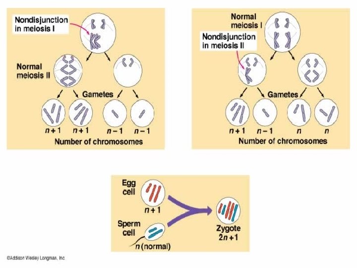

MITOTIC NONDISJUNCTION Mosaicism: • Some cells have abnormal chromosomal number and others have normal • Occurs in the earliest cell divisions • Affected individuals exhibit characteristics of a particular syndrome for e. g. down syndrome in 1% cases

CHROMOSOMAL TRANSLOCATIONS • When a portion of one chromosome is transferred to another non homologous chromosome and a fusion gene is created. There are two main types of translocations: • Balanced: An even exchange of material with no genetic information is extra or missing, and individual is normal. • Unbalanced: Where the exchange of genetic material is unequal and part of one chromosome is lost & altered phenotype is produced ( Down’s syndrome – 4% cases)

BALANCED TRANSLOCATION If no genetic material is lost during the exchange, the translocation is considered to be a balanced translocation.

UNBALANCED TRANSLOCATIONS • An entire chromosome has attached to another at the Centromere • long q arms of two chromosomes (14 & 21) become joined at a single centromere. • 4% cases of down syndrome, unbalanced translocation can occur during meiosis I or meiosis II.
 Unbalanced translocation 4% b/w 21 and")
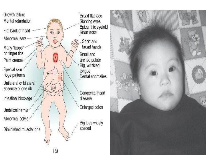
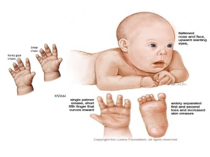
DOWN’S SYNDROME Causes: Meiotic nondisjunction 95% (trisomy 21) Unbalanced translocation 4% b/w 21 and 13, 14, 15 Mosaicism due to mitotic non dysjunction-1% Incidence: Female under 25 --- 1: 2000 At 35 --- 1: 300 At 40 --- 1: 40

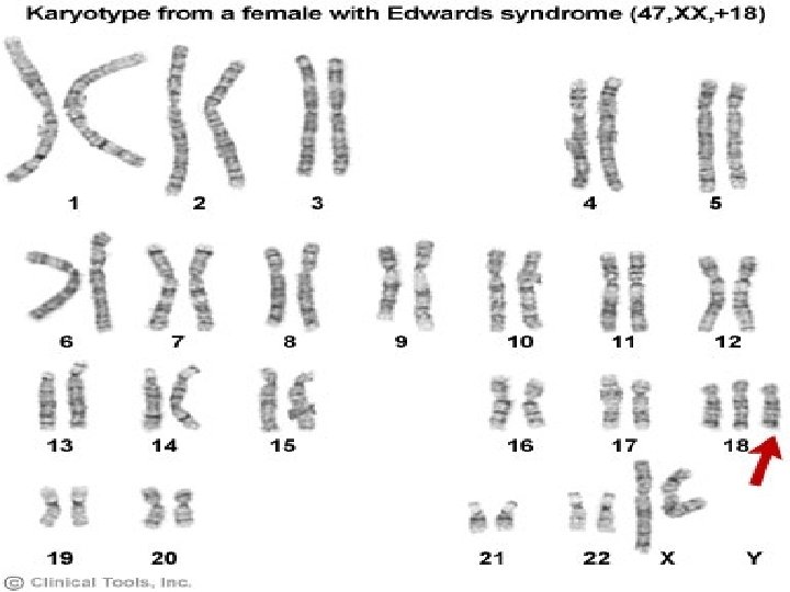
TRISOMY 18 1: 5000; Infants usually die by age of 2 months S/S: Mental retardation, congenital heart defects, low set ears, flexion of fingers

TRISOMY 13 1: 5000 ; most of the infants die by age 3 months S/S: mental retardation, holoprosencephly, congenital heart defects
 & a sex chromatin Barr body or 48(XXXY);")
KLINEFELTER’S SYNDROME Have 47 chromosomes (XXY) & a sex chromatin Barr body or 48(XXXY); more the number of X more the chances of mental impairment Cause: Nondisjunction of XX homologue Found only in males, detected at puberty Incidence ---1 in 500 males S/S Sterility, testicular atrophy, hyalinization of seminiferous tubules, gynecomastia.

TURNER SYNDROME 45 X karyotype Only monosomy compatible with life Cause Nondisjunction in male gamete Structural abnormalities of X chromosome One X chromosome is missing Mitotic nondisjunction

STRUCTURAL ABNORMALITIES • Occur when the chromosome's structure is altered, this can take several forms: Translocation, deletion or duplication of chromosomes • Chromosome breaks occur either as a result of damage to DNA (by radiation or chemicals) or as part of the mechanism of recombination. • However, the total number of chromosomes is usually normal.

CHROMOSOMAL DELETION • A part of a chromosome is missing or "deleted. " • Breaks are caused by environmental factors • A very small piece of a chromosome can contain many different genes. • When genes are missing, "instructions" are missing resulting in errors in the development of a fetus.

CRI-DU-CHAT SYNDROME Partial deletion of chromosome 5 S/S • High pitched cat like cry, a small head size , low birth weight, mental retardation and congenital heart disease.
 on long arm of chromosome 15. Inherited")
ANGELMAN’S SYNDROME Microdeletion (span few contiguous genes) on long arm of chromosome 15. Inherited on maternal chromosome S/S Mentally retarded, Cannot speak Prolonged periods of laughter

PRADER-WILLI SYNDROME Microdeletion occurs on long arm of chromosome 15 Inherited on paternal chromosome S/S Obesity Mental retardation Hypogonadism Cryptorchidism

FRAGILE X SYNDROME • Fragile X is a genetic disorder that is caused by a break or weakness on the long arm of the X chromosome. • Syndrome occurs in 1: 5000 individuals with males affected more than females. • Is the 2 nd most common inherited cause of mental retardation due to chromosomal abnormalities S/S Mental retardation, large ears, prominent jaw and pale blue irises

Genomic imprinting • These syndromes depend on whether the affected genetic material is inherited from the mother or father they are also an example of imprinting.
- Slides: 32