Lecture Power Point to accompany Inquiry into Life


")


")


- Slides: 57
Lecture Power. Point to accompany Inquiry into Life Twelfth Edition Sylvia S. Mader Chapter 22 Copyright © The Mc. Graw-Hill Companies, Inc. Permission required for reproduction or display.
22. 1 Principles of Animal Development • Fertilization • Sperm squeezes through follicle cells • Sperm releases acrosomal enzymes so it can penetrate the zona pellucida • Sperm cell membrane fuses with egg cell membrane • Sperm enters egg, nucleus is released • Egg nucleus and sperm nucleus fuse forming a zygote
Fertilization
22. 1 Principles of Animal Development • Early Stages of Animal Development – Cleavage: first mitotic divisions resulting in a multicellular embryo • Cell divisions without growth in size • Increases number of cells but not the total volume of cytoplasm – Cleavage Divisions • Divisions of zygote are equal • Forms a multicellular stage called a morula (ball of cells) • The next stage is the Blastula (a hollow ball of cells) – The fluid-filled cavity is called a blastocoel
Lancelet Early Development
22. 1 Principles of Animal Development • Early Stages of Animal Development – All vertebrates have a blastula stage, but the appearance may be different – Human blastulas resemble chick blastulas even though we have little yolk
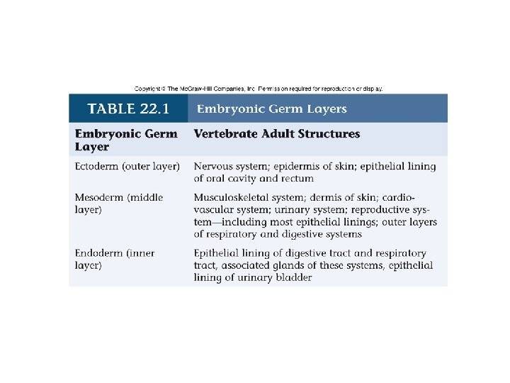
22. 1 Principles of Animal Development • Tissue stages of Development – Gastrulation: The invagination of cells into the blastocoel • Cells migrate to specific destinations • Form distinct cell layers- “germ” layers • Pore created by invagination is the blastopore – Early Gastrula has two layers of cells • Ectoderm- outer layer of cells • Endoderm- inner layer of cells – Lines the archenteron – Late gastrula- has three layers of cells • A middle mesoderm layer is formed
Comparative Development of Mesoderm
22. 1 Principles of Animal Development • Organ Stages of Development – Mesodermal cells alongitudinal axis of the embryo form the notochord • Notochord persists in the lancelet but is replaced by the vertebral column in vertebrates – The nervous system develops from the ectoderm • Neural plate stage-thickening of ectoderm above notochord forms neural plate • Neural folds form moving upward and joining to form a neural tube – Anterior end becomes the brain, the rest becomes spinal cord • Neural crest cells form where tube pinches off from ectoderm– Cells migrate to contribute to skin, muscles, adrenal medulla, ganglia
Development of Neural Tube and Coelom in a Frog Embryo
Vertebrate Embryo (Cross Section)
22. 1 Principles of Animal Development • Processes of Animal Development – Cellular Differentiation • Cells become specialized in structure and function – Morphogenesis • Produces the shape and form of the body – Pattern Formation • How tissues and organs are arranged in the body
22. 1 Principles of Animal Development • Processes of Animal Development – Cellular Differentiation • Due to differential gene expression • Frogs: egg cytoplasm is different in different regions – Gray crescent is visible after fertilization – Egg has polarity- both a dorsal/ventral and anterior/posterior axis – A classic experiment » If egg is divided so both halves get gray crescent, then two complete tadpoles develop » If egg is divided so only half gets gray crescent, then that half develops into a tadpole and the other half stops developing
Experimental Determination of Cytoplasmic Influence on Development
22. 1 Principles of Animal Development • Processes of Animal Development – Cellular Differentiation Continued • Cytoplasmic Segregation – Frog experiment shows egg contains substances called maternal determinants- influence development – Parceling out of maternal determinants (like the gray crescent) during mitosis – Determines how cells of morula will develop – Specialization of cells is influenced by maternal determinants and signals from surrounding cells • Induction – Ability of one embryonic tissue to influence another by chemical signals
22. 1 Principles of Animal Development • Processes of Animal Development – Cellular Differentiation Continued • Dorsal blastopore lip in the frog is the primary organizer – Contains gray crescent – Cells closest to dorsal lip become endoderm, those farthest become ectoderm, those in the middle become mesoderm – Location of the gray crescent indicates the dorsal surface » Mesoderm here forms the notochord » Notochord induces ectoderm to become the neural plate • Another Example of Induction – Vertebrate eye- optic vesicle induces overlying ectoderm to become the lens – Lens then induces the optic vesicle to form retina
22. 1 Principles of Animal Development • Processes of Animal Development – Morphogenesis • Processes by which specific body plan develops • Morphogen genes- determine relationship of individual parts – Some genes control which end becomes the head and which the tail » These genes code for proteins in a morphogen gradient » Cells at one end have high levels, at the other end they have low levels – Other morphogen genes determine how many segments the body will have – Determines shape of organism
22. 1 Principles of Animal Development • Processes of Animal Development – Morphogenesis Continued • Sequential sets of master genes code for morphogen gradients that activate the next set of master genes, etc. • Homeotic genes-control the organization of differentiated cells into 3 dimensional structures like wings, legs, etc. – All share a sequence of nucleotides called a homeobox » Codes for a sequence of 60 amino acids called a homeodomain » Homeodomain protein binds to DNA and determines which genes are turned on – Homeodomain protein from one homeotic gene binds to the next homeotic gene and turns it on, etc.
Morphogen Gradients in the Fruit Fly
22. 1 Principles of Animal Development • Morphogenesis Continued – Homeotic genes of many different organisms contain the same homeodomain- indicates this sequence originated early in evolutionary history – Apoptosis-plays role in morphogenesis • Hands and feet of humans are shaped by apoptosis • Cell receives a death signal – An inhibiting protein becomes inactivated – Cell-death cascade proceeds – Enzymes destroy cell
Pattern Formation in Drosophila
22. 2 Human Embryonic and Fetal Development • Human Development – From conception to birth is approximately nine months – Embryonic Development- Months 1 and 2 • Development of all organ systems – Fetal Development- Months 3 -9 • Refinement of organ systems
Extraembryonic Membranes
22. 2 Human Embryonic and Fetal Development • Extraembryonic Membranes – In the Chick • • Chorion: lies next to the shell and functions in gas exchange Amnion: contains amniotic fluid which bathes embryo Allantois: collects nitrogenous wastes Yolk sac: surrounds yolk which provides nourishment – Humans • • Chorion: develops into the fetal side of the placenta Yolk sac: the earliest site of blood cell formation Allantoic vessels: become the umbilical vessels Amnion: contains the amniotic fluid
22. 2 Human Embryonic and Fetal Development • Embryonic Development - The First Week – Fertilization occurs within the first 1/3 of the oviduct – Zygote undergoes the first cleavage divisions as it migrates through the oviduct toward the uterus – After about three days it is in the morula stage • Morula enters the uterus • At day five it has become a blastocyst – Outer layer of trophoblast cells becomes the chorion – The inner cell mass becomes the embryo
Human Development Before Implantation
22. 2 Human Embryonic and Fetal Development • Embryonic Development - The Second Week – Implantation- trophoblast cells secrete digestive enzymes to burrow into the endometrium – Trophoblast cells begin to secrete human chorionic gonadotropin – Inner cell mass detaches itself from the trophoblast – Amnion and yolk sac develop – Gastrulation occurs- inner cell mass now becomes the flattened embryonic disk • At this stage ectoderm and endoderm differentiate – Embryonic disk elongates to form primitive streak and mesoderm develops
Human Embryonic Development
22. 2 Human Embryonic and Fetal Development • Embryonic Development • The Third Week – The nervous system becomes visually evident – Development of the heart begins • The Fourth and Fifth Weeks – The body stalk (mesoderm) connects the tail end of the embryo with the chorion • Allantois is contained within this stalk – Limb buds appear – Sense organs are distinguishable
Human Embryonic Development
Human Embryo at the Beginning of the Fifth Week
22. 2 Human Embryonic and Fetal Development • Embryonic Development • The Sixth Through Eighth Weeks • The embryo becomes recognizable as human • Nervous system continues to develop – Reflexes are present • All organ systems are now established
22. 2 Human Embryonic and Fetal Development • Fetal Development – The Third and Fourth Months • The head is large, nose is flat, eyes are far apart • Epidermal structures develop – Eyelashes, hair on head, eyebrows, fingernails, and nipples • Cartilage begins to be replaced by bones • Sex of the individual may be determined • In the fourth month, the heartbeat can be heard
Three to Four Month Old Fetus
22. 2 Human Embryonic and Fetal Development • Fetal Development – The Fifth Through Seventh Months • The mother begins to feel movement • Fetal skin is covered by fine hair called lanugo • The skin is also covered with a thick, cheesy coating called the vernix caseosa • Eyelids are open • Survival is now possible if birth occurs prematurely
22. 2 Human Embryonic and Fetal Development • Fetal Development – Features of Fetal Circulation • A fetus does not use its lungs for gas exchange • Blood entering the right atrium is shunted through the left atrium through the oval opening (foramen ovale) • Any blood that enters the right ventricle ends up being shunted into the aorta by way of the arterial duct (ductus arteriosus) • Blood in the aorta is distributed to iliac arteries leading to the placenta
22. 2 Human Embryonic and Fetal Development • Fetal Development – Features of Fetal Circulation continued • Exchange of gases and nutrients between the fetus and mother occur in the placenta • Blood leaves the placenta via the umbilical vein • Blood enters the fetal liver and joins the venous duct which merges with the inferior vena cava • The inferior vena cava returns blood to the heart
Fetal Circulation and the Placenta
22. 2 Human Embryonic and Fetal Development • Fetal Development – Features of Fetal Circulation continued • At birth blood in the left atrium should cause a flap to close off oval opening • Arterial duct closes as endothelial cells proliferate. Remains of the arterial duct and umbilical vessels are converted to connective tissue • The most common cardiac defects in newborns is that the oval opening fails to close. – Failure to close can usually be corrected by surgery
22. 2 Human Embryonic and Fetal Development • Fetal Development – Structure and Function of the Placenta • The placenta is attached to the uterine wall by the allantois and chorionic villi • Placenta is fully formed by the tenth week – Produces progesterone and estrogen – Functions in gas, nutrient, and waste exchange between the fetal and maternal circulatory systems – Fetal and maternal blood do not mix
Anatomy of the Placenta in a Fetus at Six to Seven Months
22. 2 Human Embryonic and Fetal Development • Fetal Development – Structure and Function of the Placenta • The umbilical cord functions to take fetal blood to and from the placenta. • Harmful substances (ex: alcohol, some medications) can cross the placenta and cause irreversible birth defects
22. 2 Human Embryonic and Fetal Development • Birth – Contractions occur throughout the third trimester and become stronger and more frequent toward the end of pregnancy. – A positive feedback mechanism is involved. • Stretching of the uterus causes oxytocin release – Oxytocin causes further contractions which causes the uterus to be stretched further. – More oxytocin is then released
22. 2 Human Embryonic and Fetal Development • Birth – Events That Occur Shortly Before Birth • Strong uterine contractions occur about every five minutes • The “water breaks”, meaning the amnion has ruptured and the amniotic fluid is released • A mucus plug (from the cervix) is expelled – The plug prevents bacteria and sperm from entering the vagina during pregnancy
Three Stages of Parturition (birth)
22. 2 Human Embryonic and Fetal Development • Female Breast and Lactation – Each contains 15 -25 lobules • Lobule has a milk duct which branches from the nipple into numerous smaller ducts that terminate in alveoli • During pregnancy the number of ducts and alveoli increase • Prolactin stimulates milk synthesis – Inhibited by estrogen and progesterone during pregnancy – When placenta is delivered, the anterior pituitary produces prolactin • First secretions are colostrum which is rich in protein and antibodies – Suckling stimulus-causes oxytocin release • Oxytocin causes milk let-down into the ducts
Female Breast Anatomy
22. 2 Human Embryonic and Fetal Development • Benefits of Breast Feeding – Breast milk contains antibodies – Breast-fed babies are less likely to develop stomach and intestinal illnesses in the first 13 weeks of life – Suckling by the baby helps return the uterus to normal size – Breast-feeding burns calories helping the mother to return to her normal weight
22. 3 Human Development After Birth • Development continues throughout the stages of life. • • Infancy Childhood Adolescence Adulthood • Gerontology is the study of aging.
22. 3 Human Development After Birth • Hypotheses About Aging – Genetic in Origin • Cells divide a specific number of times – This is species specific • Some cell lines become nonfunctional before the maximum number of divisions occurs – Mutations may accumulate and affect function • Children of long-lived parents tend to live longer than children of shorter-lived parents
22. 3 Human Development After Birth • Hypotheses About Aging – Whole-Body Process • Decline in hormonal systems affect many different organs • Could also be due to a change in a specific tissue type that affects many organs – Cross-linkages in collagen lead to stiffness, decreased elasticity – Extrinsic Factors • Could be that we view as aging is actually the result of poor life choices in nutrition, habits • Sensible diet and exercise may prevent many signs of aging
22. 3 Human Development After Birth • Effect of Age on Body Systems – Skin • • Thinner, less elastic, less subcutaneous fat Causes wrinkles, decreased insulation Number of oil glands reduced, skin is dry Pigmented blotches may appear
22. 3 Human Development After Birth • Effect of Age on Body Systems – Processing and Transport • Cardiovascular disorders are the leading cause of death – Heart shrinks because of reduction in cardiac muscle cell size – Arteries become more rigid; plaque buildup may narrow lumen of vessels – Blood pressure increases with age
22. 3 Human Development After Birth • Effect of Age on Body Systems – Processing and Transport • Cardiovascular disorders are the leading cause of death – Blood flow to the liver is reduced, and the liver does not metabolize drugs as efficiently. » Smaller doses of medications are needed – Blood supply to the kidneys is also reduced » Salt and water balance are difficult to maintain » The elderly dehydrate easier than young people
22. 3 Human Development After Birth • Effect of Age on Body Systems – Integration and Coordination • • • Brain and muscle cells do not replace lost cells Few neurons in cerebral cortex are lost. Cognitive functions are not affected in normal aging Short-term memory skills may decrease If neurons do die, may be due to decreased oxygen availability and not from aging • Loss of skeletal muscle mass may prevented by exercise • Osteoporosis may be prevented by calcium intake and exercise
22. 3 Human Development After Birth • Effect of Age on Body Systems – The Reproductive System • Females undergo menopause • Males produce less androgens – Still produce sperm until death • Females tend to live longer than males