INTRODUCTION TO NUCLEIC ACIDS Friedrich Miescher in 1869


- Slides: 50

INTRODUCTION TO NUCLEIC ACIDS

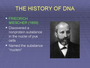
Friedrich Miescher in 1869 • isolated what he called nuclein from the nuclei of pus cells • Nuclein was shown to have acidic properties, hence it became called nucleic acid

Two types of nucleic acid are found • Deoxyribonucleic acid (DNA) • Ribonucleic acid (RNA)

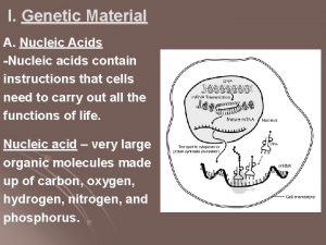
The distribution of nucleic acids in the eukaryotic cell • DNA is found in the nucleus with small amounts in mitochondria • RNA is found throughout the cell

DNA as genetic material 1. 2. 3. Present in all cells and virtually restricted to the nucleus The amount of DNA in somatic cells (body cells) of any given species is constant (like the number of chromosomes) The DNA content of gametes (sex cells) is half that of somatic cells. In cases of polyploidy (multiple sets of chromosomes) the DNA content increases by a proportional factor

Deoxyribose Nucleic Acid. DNA contains all the information necessary for all living organisms to grow and live. (i. e. a blueprint for life).

NUCLEIC ACID STRUCTURE • Nucleic acids are polynucleotides • Their building blocks are nucleotides

NUCLEOTIDE STRUCTURE PHOSPATE SUGAR Ribose or Deoxyribose BASE PURINES PYRIMIDINES Adenine (A) Cytocine (C) Guanine(G) Thymine (T) Uracil (U) NUCLEOTIDE

Ribose is a pentose C 5 O C 1 C 4 C 3 C 2

Spot the difference DEOXYRIBOSE CH 2 OH O C H H H C OH OH CH 2 OH C C H H OH O C H H C C C OH OH H H

5 The bases The most common organic bases are Adenine (A) Thymine (T) Cytosine (C) Guanine (G)

6 Nucleotides The deoxyribose, the phosphate and one of the bases Combine to form a nucleotide PO 4 adenine deoxyribose

Nitrogenous bases Pyrimidines cytosine (C) thymine (T) uracil (U) Have One ring; Purines Have two rings adenine (A) guanine (G) Complementarity of bases T (U) and A, C and G.

Pyrimidines and Purines • Pyrimidine and purine are the names of the parent compounds of two types of nitrogencontaining heterocyclic aromatic compounds. Pyrimidine Purine

• The nitrogen containing bases are the only difference in the four nucleotides.

Nucleotide & Nucleoside Basic building block of nucleic acid It consist of A sugar (ribose or deoxyribose) A nitrogenous base (purine & pyrimidine rings) A phosphate Nucleosides= Base + Sugar They glycosylamines consisting of a nucleobase bound to a ribose or deoxyribose sugar via a beta-glycosidic linkage.

Structures of Nucleosides

Structures of Nucleotides

When phosphate group is added to the above nucleosides then the nucleotide is formed. When a single phosphate group is added to the nucleoside then it is called as mononucleotide. The bond between pentose and base is b-N-glycosidic bond. N 9 of purine ring binds with C 1 of pentose sugar. In pyrimidines the bond is in between N 1 of pyrimidine and C 1 of pentose. When a phosphate group is attached at 5' position of a sugar then it is written as Adenosine 5'-monophosphate (AMP). The nucleotides are abbreviated by three capital letters, preceded by d- in case of deoxy series i. e. , d. AMP = deoxyadenosine monophosphate ATP = adenosine triphosphate UDP = Uridine diphosphate

Ribose Sugar • The double-bonded oxygen on the first carbon in the linear form becomes the beta OH that is used to bond to a base unit. • The OH in the second position serves to distinguish between the ribose in RNA and the deoxyribose in DNA. • The OH on the third carbon will bond to the phosphate group of other nucleotides. • The OH group on the fourth carbon is involved in the closure of the ring. • The OH group on the fifth carbon is what bonds to the phosphate unit of this particular nucleotide. The carbon atoms in the ribose portion of a nucleotide are numbered with primes (1', 2', 3', 4', 5') to distinguish them from the carbon and nitrogen atoms in the base which are numbered without primes.

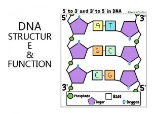
Phosphate Group DNA is a double helical structure. Its backbone is made up of a repeated pattern of a sugar group and a phosphate group. The deoxyribose sugars are joined at both the 3'-hydroxyl and 5'-hydroxyl groups to phosphate groups in ester links, also known as "phosphodiester" bonds.

Watson & Crick Base pairing

Nucleotides • Nucleotides are phosphoric acid esters of nucleosides.

Adenosine 5'Monophosphate (AMP) • Adenosine 5'-monophosphate (AMP) is also called 5'-adenylic acid. NH 2 N O HO P HO 5' N 4' 1' OCH 2 O 3' HO 2' OH N N

Adenosine Diphosphate (ADP) NH 2 N HO O O P OCH 2 O HO N HO HO OH N N

Adenosine Triphosphate (ATP) • ATP is an important molecule in several biochemical processes including: energy storage (Sections 28. 4 -28. 5) phosphorylation NH 2 N O HO O P O HO HO O P OCH 2 O N HO HO OH N N

ATP and Phosphorylation HOCH 2 ATP + HO HO This is the first step in the metabolism of glucose. hexokinase OH O ADP + (HO)2 POCH 2 HO HO OH

Joined nucleotides 7 PO 4 sugar-phosphate + bases backbone A molecule of DNA is formed by millions of nucleotides joined together in a long chain

8 In fact, the DNA usually consists of a double strand of nucleotides The sugar-phosphate chains are on the outside and the strands are held together by chemical bonds between the bases

2 -stranded DNA PO 4 PO 4 PO 4 PO 4 9

Bonding 1 10 The bases always pair up in the same way Adenine forms a bond with Thymine Adenine Thymine and Cytosine bonds with Guanine Cytosine Guanine

The Double Helix (1953)

8 In fact, the DNA usually consists of a double strand of nucleotides The sugar-phosphate chains are on the outside and the strands are held together by chemical bonds between the bases

A History of Discovery DNA • Watson and Crick’s discovery built on the work of Rosalind Franklin and Erwin Chargaff. – Franklin’s x-ray images suggested that DNA was a double helix of even width. – Chargaff’s rules stated that A=T and C=G.

A History of Discovery DNA • Chargaff’s Rules A + G = T + C (Chargaff’s rule) • Rosalind Franklin’s data provide clues about DNA’s 3 -D shape • • Helix Width = 2 nm; probably two strands (DOUBLE HELIX) Nitrogenous bases = 0. 34 n. M apart One turn every 3. 4 n. M (10 base pairs per turn) • Nobel prize won by Francis Crick, James Watson &Maurice Wilkins 1962.

13 The paired strands are coiled into a spiral called A DOUBLE HELIX

14 THE DOUBLE HELIX bases sugar-phosphate chain

Watson JD and Crick FC • Watson J. D. and Crick F. H. C. "A Structure for Deoxyribose Nucleic Acid" Nature 171, 737738 (1953). • Wilkins M. H. F. , Stokes A. R. & Wilson, H. R. "Molecular Structure of Deoxypentose Nucleic Acids" Nature 171, 738 -740 (1953). • Franklin R. and Gosling R. G. "Molecular Configuration in Sodium Thymonucleate" Nature 171, 740 -741 (1953)

Structure of DNA Sequence of nucleotides is the primary structure. They bond together in a double-strand which twists into a double-helix (secondary structure). This double-helix then wraps around some proteins (tertiary structure) Then these coils wrap and fold around each other and this wrapping and folding continues until the chromosomes is formed

Polynucleotide chain Primary Structure of Single Stranded DNA

DNA Polynucleotides DNA is a normally double stranded directional (antiparallel) macromolecule. Two polynucleotide chains, held together by weak thermodynamic forces, form a DNA molecule. Two chains run in opposite directions, Chain has a direction (known as polarity), 5'- to 3'- from top to bottom.

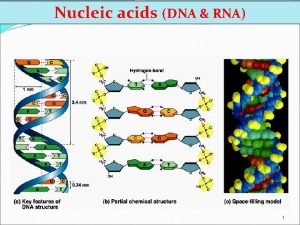
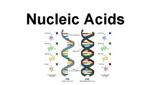
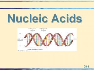
Features of DNA double helix (secondary structure) § § § § DNA is double stranded: each molecule of DNA is composed of two polynucleotide chain that are joined together by formation of hydrogen bonds between the bases. DNA strands are twisted to form a double helix. The backbone of each consists of alternating deoxyribose and phosphate groups. The phosphate group bonded to the 5' carbon atom of one deoxyribose is covalently bonded to the 3' carbon of the next. The two strands are "antiparallel"; that is, one strand runs 5′ to 3′ while the other runs 3′ to 5′ The DNA strands are assembled in the 5′ to 3′ direction The purine or pyrimidine attached to each deoxyribose projects in toward the axis of the helix. Each base forms hydrogen bonds with the one directly opposite it, forming base pairs.

§ 3. 4 Å separate the planes in which adjacent base pairs are located. § The double helix makes a complete turn in just over 10 nucleotide pairs, so each turn takes a little more (35. 7 Å to be exact) than the 34 Å. § There is an average of 25 hydrogen bonds within each complete turn of the double helix providing a stability of binding about as strong as what a covalent bond would provide. § The diameter of the helix is 20 Å. § The path taken by the two backbones forms a major (wider) groove and a minor (narrower) groove.

Pitch = 3. 4 nm

Homologous pairs contain identical genes, but the chromosomes may have different forms of the genes (alleles).

What is Genome? Genome of an organism is a sequence of nucleic acid that provides information needed to construct the organism. It contains the complete set of hereditary information for any organism. What is Gene? Gene is the functional unit of genome. Gene is a sequence of nucleic acid that produces another nucleic acid. Gene and Chromosome? DNA is organized into chromosomes which are found within the nuclei of cells. Genetic material of living organisms is either DNA or RNA. DNA – Deoxyribonucleic acid RNA – Ribonucleic acid

RNA • Most cellular RNA molecules are single stranded. §many self-complementary regions , RNA commonly exhibits an intricate secondary structure (relatively short, double helical segments alternated with single stranded regions) • complex tertiary interactions fold the RNA in its final three dimensional form. • Double standed RNA can also exists and is generally similar to A-DNA (present is few viruses)

RNA structure Primary structure A) single stranded regions formed by unpaired nucleotides Secondary structure B) duplex double helical RNA (A-form with 11 bp per turn) C C) hairpin duplex bridged by a loop of unpaired nucleotides D) internal loop D nucleotides not forming Watson-Crick base pairs E F G E) bulge loop unpaired nucleotides in one strand, other strand has contiguous base pairing F) junction B A three or more duplexes separated by single stranded regions G) pseudoknot tertiary interaction between bases of hairpin loop and outside bases

Nucleic acids are DNA and RNA DNA RNA • The principal location of • The different types of DNA is the nucleus of RNA are located in eukaryotic cells. nucleus, cytoplasm, mitochondria, ribosomes. • Secondary locations of DNA are mitochondria.

Three classes of RNA Messenger RNA (m. RNA) Transfer RNA (t. RNA) Ribosomal RNA (r. RNA) Getting from DNA to protein: Two parts: 1. Transcription in which a short portion of chromosomal DNA is used to make a RNA molecule small enough to leave the nucleus. 2. Translation in which the RNA code is used to assemble the protein at the ribosome The “central dogma” of modern biology: DNA RNA protein
 Cuadro comparativo entre adn y arn
 Johan friedrich miescher
 Where is dna replicated in the cell cycle
 Friedrich miescher
 Alloburinol
 Hydoxyle
 What is an anticodon
 Nucleic acid building block
 Nucleic acid monomer
 Nucleic acid function
 Ribonucleotide vs deoxyribonucleotide
 Nucleic acids
 Nucleoide funcion
 Store and transmit hereditary information
 Nucleic acid
 Food that rich in nucleic acid
 Nucleic acids
 Features of nucleic acid
 Miescher nuclein
 Mendeleev lab answer key
 Mahatma gandhi was born on 2nd october 1869
 Matisse nationality
 Scoperta dna 1869
 In 1869, dmitri ivanovich mendeléev
 October 2 1869
 Ernst haeckel 1869
 Henri matisse portrait of lydia
 Rīgas latviešu biedrība dibināta
 Introduction to acids and bases webquest
 Acid definition
 Dna structure labeled diagram
 Nucleic acid made up of
 Dideoxyribonucleic
 Fructose
 Principle of hybridization
 Nucleic acid chart
 Quinolones mode of action
 Nucleic acid monomer
 Nucleic
 Nucleic acid test
 Infectious nucleic acid
 Kalju kahn
 Dna fragment length
 Types of rna and dna
 Nucleic acid
 Types of nucleic acid
 We love dna song
 Nucleic acid
 Nucleic acid dna structure
 Nucleic acid
 Biological molecules you are what you eat
