In The Name Of GOD Mild asymptomatic hyperglycemia







- Slides: 51
In The Name Of GOD
Mild asymptomatic hyperglycemia Soheila Sadeghi
1) What is the diagnosis ? 2) What are the differences between MODY and type 1 , 2 DM ? 3) MODYs subtypes? 4) Indications for genetic testing ? 5) What are the Benefits of Diagnosing MODY? 6) Treatment ? 7) Complication?
What is the diagnosis ?
Diabetes, Maturity Onset in the Young (MODY)
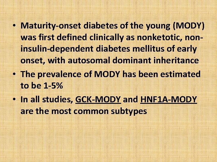
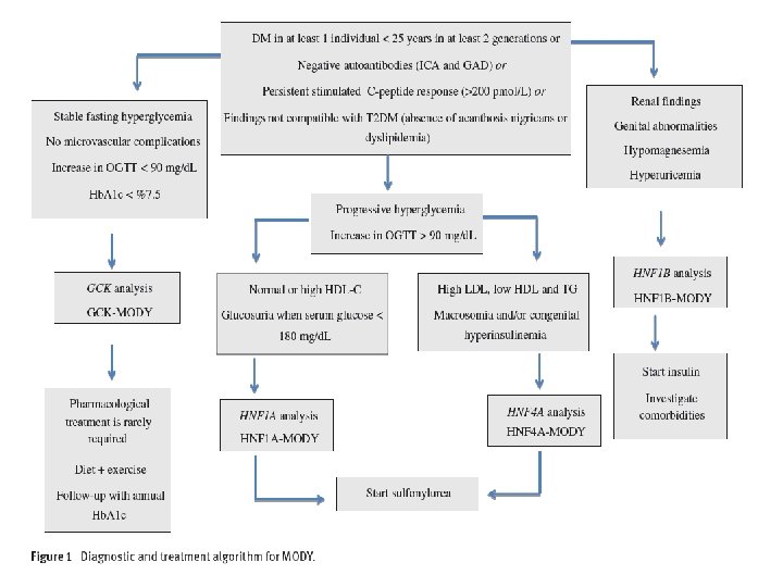
• Maturity-onset diabetes of the young (MODY) was first defined clinically as nonketotic, noninsulin-dependent diabetes mellitus of early onset, with autosomal dominant inheritance • The prevalence of MODY has been estimated to be 1 -5% • In all studies, GCK-MODY and HNF 1 A-MODY are the most common subtypes
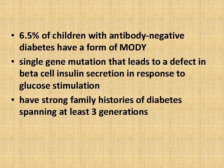
• 6. 5% of children with antibody-negative diabetes have a form of MODY • single gene mutation that leads to a defect in beta cell insulin secretion in response to glucose stimulation • have strong family histories of diabetes spanning at least 3 generations
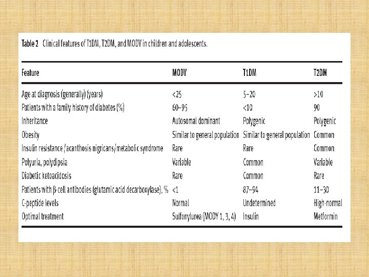
What are the differences between MODY and type 1 , 2 DM ?
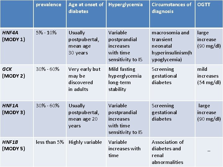
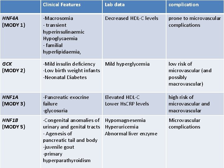
MODYs subtypes?
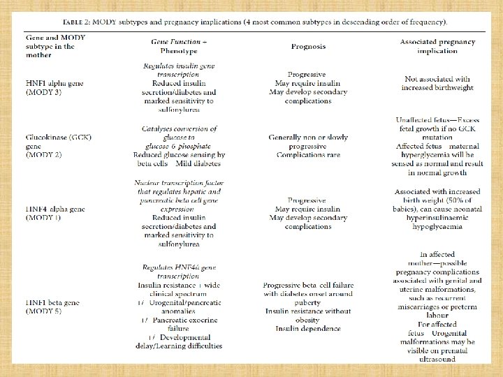
prevalence Age at onset of Hyperglycemia diabetes Circumstances of OGTT diagnosis HNF 4 A (MODY 1) 5% - 10% Usually postpubertal, mean age 30 years Variable postprandial increases with time sensitivity to IS macrosomia and large transient increase neonatal (90 mg/dl) hyperinsulinism(h ypoglycemia) GCK (MODY 2) 30% - 60% Very early but may be discovered in adults Mild fasting hyperglycemia long-term stability Screening gestational diabetes mild increases (54 mg/dl) HNF 1 A (MODY 3) 30% - 60% Usually postpubertal, mean age 20 years Variable postprandial increases with time sensitivity to IS Screening gestational diabetes large increase (90 mg/dl) HNF 1 B (MODY 5) less than 5% Highly variable Variable increases with time Association of diabetes and renal abnormalities _
Clinical Features Lab data complication HNF 4 A (MODY 1) -Macrosomia - transient hyperinsulinaemic Hypoglycaemia - familial hyperlipidaemia, Decreased HDL-C levels prone to microvascular complications GCK (MODY 2) -Mild insulin deficiency Mild hyperglycemia -Low birth weight infants -Neonatal Diabetes low risk of microvascular (and possibly macrovascular) HNF 1 A (MODY 3) -Pancreatic exocrine failure -glycosuria high risk of microvascular and macrovascular HNF 1 B (MODY 5) -Congenital anomalies of Hypomagnesemia urinary and genital tracts Hyperuricemia - Agenesis of Abnormal liver enzyme pancreatic tail and body -juvenile gout -primary hyperparathyroidism Elevated HDL-C Lower Hs. CRP levels Microvascular complications
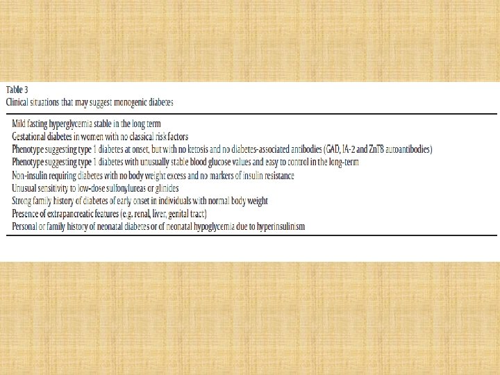
Indications for genetic testing ?
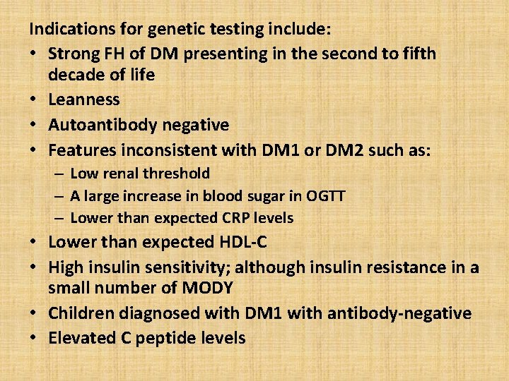
Indications for genetic testing include: • Strong FH of DM presenting in the second to fifth decade of life • Leanness • Autoantibody negative • Features inconsistent with DM 1 or DM 2 such as: – Low renal threshold – A large increase in blood sugar in OGTT – Lower than expected CRP levels • Lower than expected HDL-C • High insulin sensitivity; although insulin resistance in a small number of MODY • Children diagnosed with DM 1 with antibody-negative • Elevated C peptide levels
What are the Benefits of Diagnosing MODY? • The diagnosis of monogenic diabetes has many consequences in terms of prognosis, therapeutics and family screening
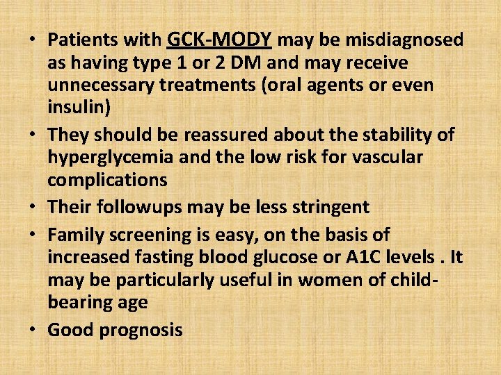
• Patients with GCK-MODY may be misdiagnosed as having type 1 or 2 DM and may receive unnecessary treatments (oral agents or even insulin) • They should be reassured about the stability of hyperglycemia and the low risk for vascular complications • Their followups may be less stringent • Family screening is easy, on the basis of increased fasting blood glucose or A 1 C levels. It may be particularly useful in women of childbearing age • Good prognosis
• In patients with HNF 1 A-MODY or HNF 4 A-MODY : - Switching from an unnecessary insulin therapy to oral treatment with sulfonylureas or glinides, even long after the onset of diabetes - Careful followup is necessary because insulin secretion decreases with time - Encourage to maintain normal body weights because obesity may accelerate the onset of diabetes - Identifying siblings who carry the mutation is mandatory to prevent the insidious onset of vascular complications and, in women, to avoid unrecognized hyperglycemia in the early stages of pregnancy
• Patients with HNF 1 B-MODY should be systematically evaluated for damage to other organs • because of the progression of insulin secretion defects and of renal disease, insulin therapy is needed
Treatment • Treatment of the hyperglycemia in GCK is generally unnecessary except during pregnancy ( ins to prevent excess fetal growth, 50%, if the fetus does not carry the MODY gene ) • Sulfonylureas have been shown to be effective in treating HNF 1 A • Similar response to sulfonylureas has been reported in HNF 4 A • HNF 1 B do not generally respond to sulfonylureas and typically require insulin early
Complication
• Some patients with HNF-related MODY are at increased risk for developing tumours. • Patients with HNF 1 A-MODY have a 5% to 10% risk for liver adenomatosis • This risk deserves screening by liver ultrasound avoidance of estrogencontaining contraceptives because they may favour the development of liver adenomas.
• In patients with HNF 1 B-MODY, a biallelic inactivation of HNF 1 B has been reported in chromophobe renal carcinomas • HNF 1 B may also be a suppressor gene for some ovarian cancers.
• A 35 -year-old woman, G 3 P 1 • Her first pregnancy in 2005 resulted in an early miscarriage. • Her second pregnancy in 2014 was complicated by GDM diagnosed at 13 weeks’ gestation and managed with a multiple daily insulin regimen from 20 weeks. Her pre-pregnancy weight was 52 kg (BMI 21. 4 kg/m 2) and gestational weight gain was 13 kg. Following induction of labour at 38+2 weeks’ gestation she had a vaginal delivery of a healthy girl weighing 3200 g. • She did not attend a postnatal 75 -g oral glucose tolerance test (OGTT).
• The couple were non-consanguineous. • There was a family history of impaired glucose tolerance affecting her sister and brother. Another sister had GDM in both her pregnancies and her father was treated for Type 2 diabetes mellitus. There was no family history of significant congenital malformations.
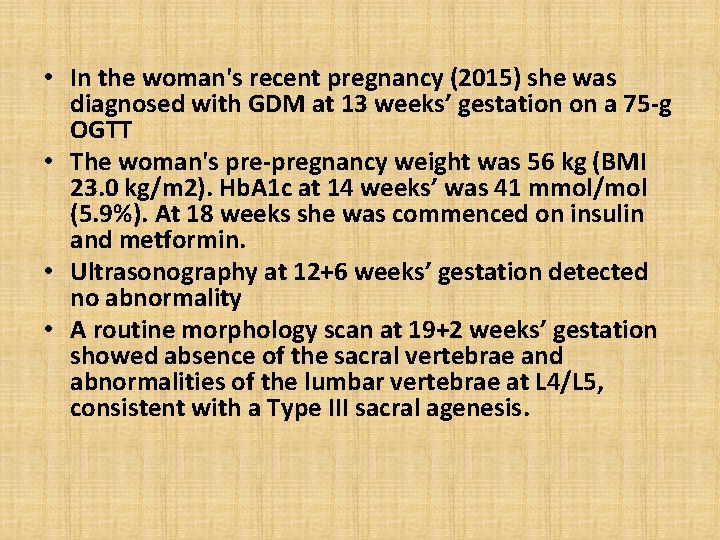
• In the woman's recent pregnancy (2015) she was diagnosed with GDM at 13 weeks’ gestation on a 75 -g OGTT • The woman's pre-pregnancy weight was 56 kg (BMI 23. 0 kg/m 2). Hb. A 1 c at 14 weeks’ was 41 mmol/mol (5. 9%). At 18 weeks she was commenced on insulin and metformin. • Ultrasonography at 12+6 weeks’ gestation detected no abnormality • A routine morphology scan at 19+2 weeks’ gestation showed absence of the sacral vertebrae and abnormalities of the lumbar vertebrae at L 4/L 5, consistent with a Type III sacral agenesis.
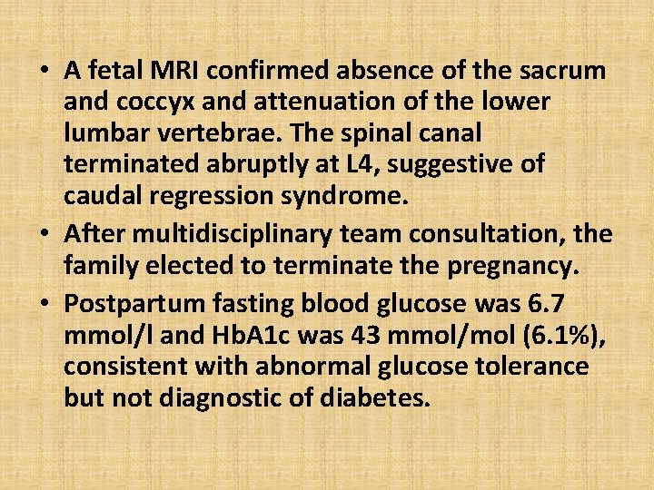
• A fetal MRI confirmed absence of the sacrum and coccyx and attenuation of the lower lumbar vertebrae. The spinal canal terminated abruptly at L 4, suggestive of caudal regression syndrome. • After multidisciplinary team consultation, the family elected to terminate the pregnancy. • Postpartum fasting blood glucose was 6. 7 mmol/l and Hb. A 1 c was 43 mmol/mol (6. 1%), consistent with abnormal glucose tolerance but not diagnostic of diabetes.
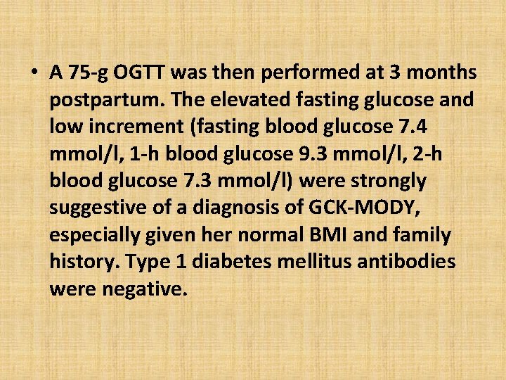
• A 75 -g OGTT was then performed at 3 months postpartum. The elevated fasting glucose and low increment (fasting blood glucose 7. 4 mmol/l, 1 -h blood glucose 9. 3 mmol/l, 2 -h blood glucose 7. 3 mmol/l) were strongly suggestive of a diagnosis of GCK-MODY, especially given her normal BMI and family history. Type 1 diabetes mellitus antibodies were negative.
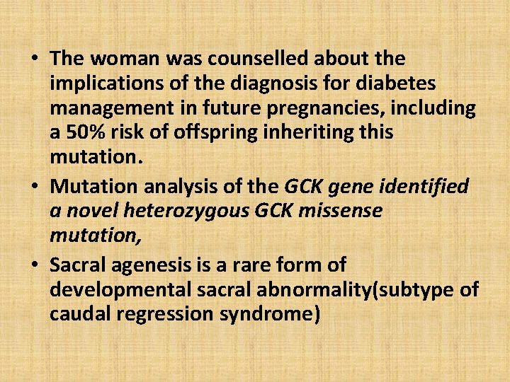
• The woman was counselled about the implications of the diagnosis for diabetes management in future pregnancies, including a 50% risk of offspring inheriting this mutation. • Mutation analysis of the GCK gene identified a novel heterozygous GCK missense mutation, • Sacral agenesis is a rare form of developmental sacral abnormality(subtype of caudal regression syndrome)
• Caudal regression syndrome is a cardinal feature of diabetic embryopathy. The relative risk of caudal regression syndrome in infants born to mothers with pre-GDM is 170– 200 times higher than in the general population • Sacral agenesis is a rare form of sacral abnormality affecting 0. 005% to 0. 1% of pregnancies. It is a subtype of the caudal regression sequence, a cardinal feature of diabetic embryopathy. • It is not thought to be strongly associated with major abnormalities, but with insufficient data and relatively low numbers of pregnancies in women with GCK-MODY
• In contrast to the well established link between glycaemic control in pre-GDM and congenital abnormalities, it is unclear whether a similar association exists with the mild hyperglycaemia seen in GCK-MODY • However, the risk increase was minimal at the Hb. A 1 c levels typically seen in GCK-MODY of 38– 56 mmol/mol (5. 6– 7. 3%),
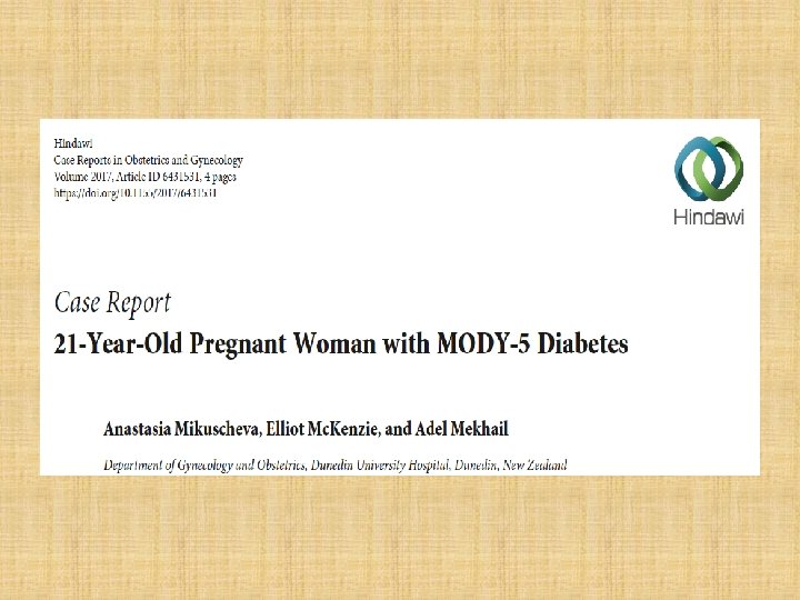
• Miss A. was a 21 -year-old fit and well woman without a prior documented significant medical or surgical history, who was referred to our antenatal clinic by her midwife because a bicornuate uterus was discovered • An Hb. A 1 C, routinely done in New Zealand for diabetes screening, was mildly elevated at 41 mmol/mol(5. 9 %). • She had persistent hyperglycemia and investigations to exclude type I diabetes were performed. The anti-GAD antibodies and Islet Cell Cytoplasmic autoantibodies came back negative.
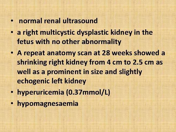
• normal renal ultrasound • a right multicystic dysplastic kidney in the fetus with no other abnormality • A repeat anatomy scan at 28 weeks showed a shrinking right kidney from 4 cm to 2. 5 cm as well as a prominent in size and slightly echogenic left kidney • hyperuricemia (0. 37 mmol/L) • hypomagnesaemia
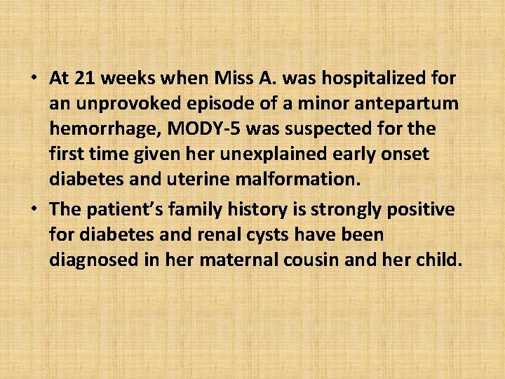
• At 21 weeks when Miss A. was hospitalized for an unprovoked episode of a minor antepartum hemorrhage, MODY-5 was suspected for the first time given her unexplained early onset diabetes and uterine malformation. • The patient’s family history is strongly positive for diabetes and renal cysts have been diagnosed in her maternal cousin and her child.
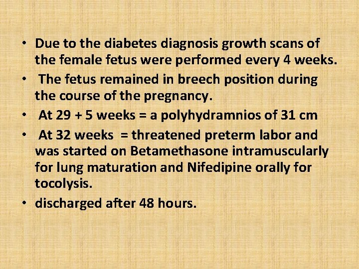
• Due to the diabetes diagnosis growth scans of the female fetus were performed every 4 weeks. • The fetus remained in breech position during the course of the pregnancy. • At 29 + 5 weeks = a polyhydramnios of 31 cm • At 32 weeks = threatened preterm labor and was started on Betamethasone intramuscularly for lung maturation and Nifedipine orally for tocolysis. • discharged after 48 hours.
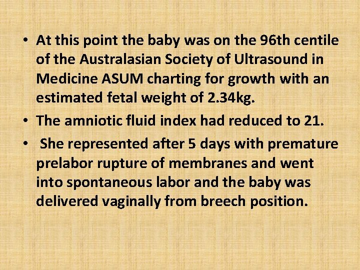
• At this point the baby was on the 96 th centile of the Australasian Society of Ultrasound in Medicine ASUM charting for growth with an estimated fetal weight of 2. 34 kg. • The amniotic fluid index had reduced to 21. • She represented after 5 days with premature prelabor rupture of membranes and went into spontaneous labor and the baby was delivered vaginally from breech position.
• The baby girl was born alive, and a birth weight of 2105 g. The physical examination was normal. • However, at one week postpartum a neonatal ultrasound scan confirmed an absent right kidney and the presence of a bicornuate uterus • Genetic testing was arranged and showed a heterozygous deletion of the HNF 1 B gene.
• We suggest that genetic testing should be offered to young female patients with uterine abnormalities and otherwise unexplained diabetes even in the absence of kidney abnormalities on imaging if kidney impairment is present on laboratory functions.
• The optimal care for pregnant patients with MODY-5 due to HNF 1 B mutations is multidisciplinary and involves obstetricians, endocrinologists, geneticists, nephrologists, and pediatricians. • Despite the more common association of MODY-5 with renal malformation we think that genetic testing is also warranted in cases where genital malformations are diagnosed and renal impairment is only present on laboratory testing.
• Miss S, a 19 -year-old woman, presented to antenatal clinic at 19 weeks gestation for a first consultation because of a preexisting hepatocyte nuclear factor �� (HNF-1�� ) mutation causing MODY 3 diabetes • Since age of 11 years when her condition first became apparent through recurrent mucosal candidiasis and mild postprandial hyperglycaemia. Due to a strong family history of diabetes and negative testing for type I diabetes, an HNF 1�� gene mutation was suspected and subsequently confirmed on molecular genetic testing.
• The patient was initially successfully treated with the sulfonylurea (SU) gliclazide, which more recently was switched to insulin due to increasing hyperglycaemia. • The father of the fetus is a 21 -year-old man also well • known to endocrinology and diabetes teams from age of 9 years, due to persistent mild hyperglycaemia and very significant family history of diabetes (Figure 2). Genetic testing for a Glucokinase (GCK) mutation was performed • asymptomatic and did not require any further treatment
• She was managed with insulin glargine (Lantus) daily and insulin aspart (Novorapid) with meals, and insulin requirements gently increased over the pregnancy, • From 28 weeks, doses were further reduced, until she presented to the emergency department at 33+3 weeks of gestation for frequent hypoglycaemia. • The patient never developed hypertension, and laboratory screening for preeclampsia was performed multiple times and was always within normal limits.
• Fetal growth until this presentation had been measured by fortnightly ultrasound and was on the 50 th centile • Given the unknown significance of the falling insulin requirements, biweekly monitoring of fetal wellbeing via Doppler measurements was commenced, which was satisfactory at all times. • At 34+3 weeks of gestation the patient went into spontaneous labour and delivered a healthy baby girl via forceps, weight 2. 22 kg
• Postnatal genetic testing in the baby showed a heterozygous mutation for the maternal familial likely nonpathogenic HNF 1 A gene variant
• In a pregnancy where the mother is affected by MODY 2 the fetus will not inherit the GCK mutation in 50% of cases, and will respond to maternal hyperglycaemia by excess insulin production and therefore excess growth (by approximately 550– 700 g) [2]. Alternatively, if the fetus does inherit the GCK abnormality it will sense the maternal hyperglycaemia as normal, produce normal amounts of insulin, and have normal growth
• Where mother and fetus are both affected by MODY 2, a fetus with MODY 2 to a mother with MODY 3 would be able to tolerate maternal hyperglycaemia better than a fetus with no MODY.