
PTH Related Hypercalcemia in an Adolescent case report


- Slides: 52
“PTH Related Hypercalcemia in an Adolescent” case report B. Rezvankhah. MD January. 2017
• What is the diagnosis? • What is the treatment?
Hypercalcemia & Elevated or Inappropriately NL PTH Levels • Primary Hyperparathyroidism • Familial Hypocalcuric Hypercalcemia(FHH) • Multiple Endocrine Neoplasia Type 1 (MEN 1) ! Considering young age of Pt, PTH level, no other clinical manifestation, negative imaging investigations & no side effects of Hypercalcemia, the diagnosis of FHH would rather be ruled out.
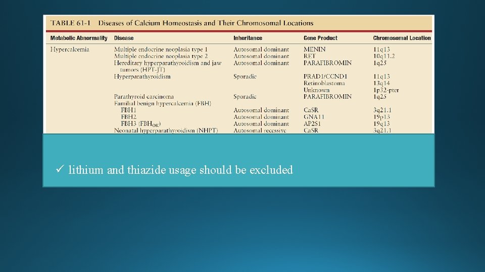
ü lithium and thiazide usage should be excluded
üThe upper normal range for plasma PTH depends on several variables including plasma 25 OHD, age, body weight and smoking. üPTH levels normally rise with age & the broad nl range(10 -65 pg/ml) reflects values for entire population; A narrower lower nl range (10 -45 pg/ml) in younger than 45 individual De Groot 2016
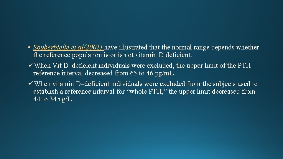
• Souberbielle et al(2001) have illustrated that the normal range depends whether the reference population is or is not vitamin D deficient. üWhen Vit D–deficient individuals were excluded, the upper limit of the PTH reference interval decreased from 65 to 46 pg/m. L. üWhen vitamin D–deficient individuals were excluded from the subjects used to establish a reference interval for “whole PTH, ” the upper limit decreased from 44 to 34 ng/L.
Primary Hyperparathyroidism (PHPT) • PHPT currently is recognized as the third most common endocrine disorder in adults, • PHPT is most common in patients who are between the ages of 50 and 60 yrs, • It has an annual incidence of 30/100, 000; with a lifetime prevalence of approximately 1/1000 persons, • Females outnumber males with PHPT by an approximate 3: 1 ratio
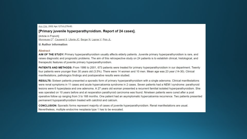
Primary Hyperparathyroidism in Adolescent • Prior to 1984, fewer than 150 children with PHPT had been described in the literature Primary studies have quoted MEN and familial non-MEN HPT to constitute as much as 30% to 50% of pediatric PHPT • Estimated incidence of 2 -5/100, 000 • Most often sporadic and caused by a parathyroid adenoma
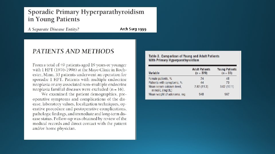
Arch Surg 1999
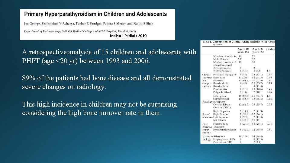
Indian J Pediatr 2010 A retrospective analysis of 15 children and adolescents with PHPT (age <20 yr) between 1993 and 2006. 89% of the patients had bone disease and all demonstrated severe changes on radiology. This high incidence in children may not be surprising considering the high bone turnover rate in them.
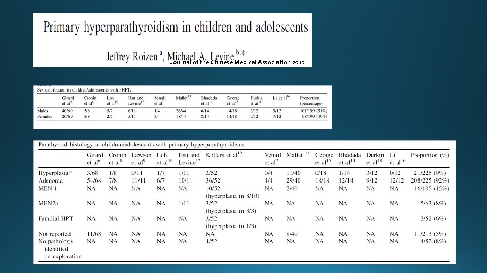
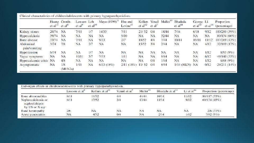
Journal of the Chinese Medical Association 2012
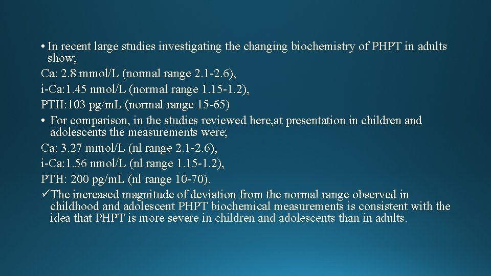
• In recent large studies investigating the changing biochemistry of PHPT in adults show; Ca: 2. 8 mmol/L (normal range 2. 1 -2. 6), i-Ca: 1. 45 nmol/L (normal range 1. 15 -1. 2), PTH: 103 pg/m. L (normal range 15 -65) • For comparison, in the studies reviewed here, at presentation in children and adolescents the measurements were; Ca: 3. 27 mmol/L (nl range 2. 1 -2. 6), i-Ca: 1. 56 nmol/L (nl range 1. 15 -1. 2), PTH: 200 pg/m. L (nl range 10 -70). üThe increased magnitude of deviation from the normal range observed in childhood and adolescent PHPT biochemical measurements is consistent with the idea that PHPT is more severe in children and adolescents than in adults.
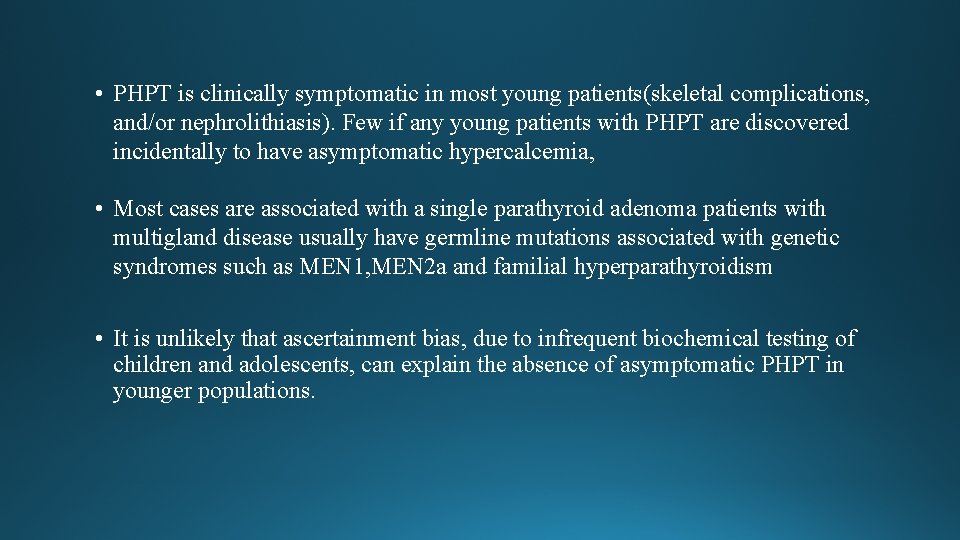
• PHPT is clinically symptomatic in most young patients(skeletal complications, and/or nephrolithiasis). Few if any young patients with PHPT are discovered incidentally to have asymptomatic hypercalcemia, • Most cases are associated with a single parathyroid adenoma patients with multigland disease usually have germline mutations associated with genetic syndromes such as MEN 1, MEN 2 a and familial hyperparathyroidism • It is unlikely that ascertainment bias, due to infrequent biochemical testing of children and adolescents, can explain the absence of asymptomatic PHPT in younger populations.
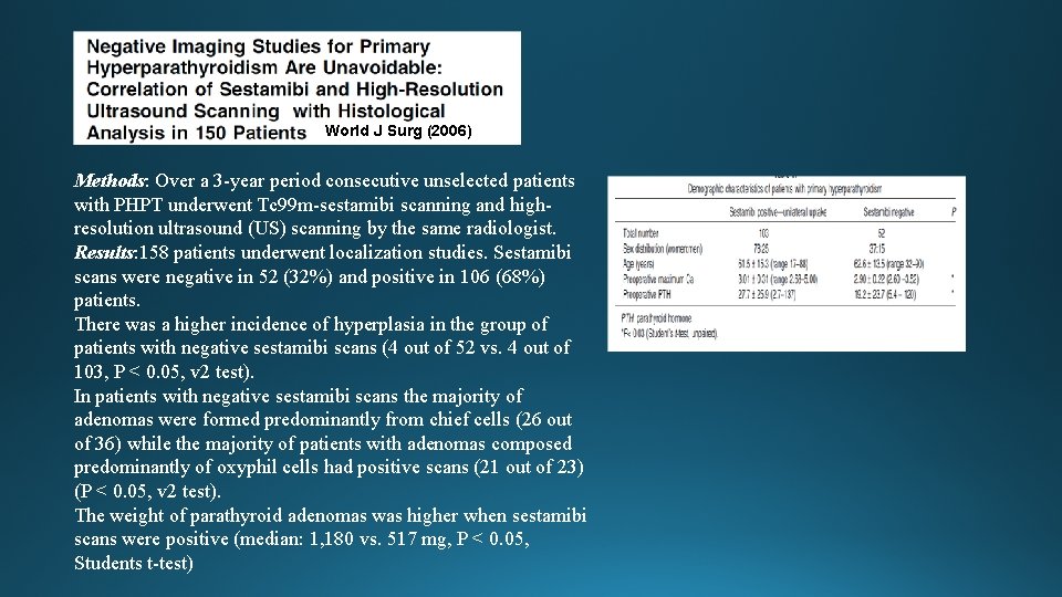
World J Surg (2006) Methods: Over a 3 -year period consecutive unselected patients with PHPT underwent Tc 99 m-sestamibi scanning and highresolution ultrasound (US) scanning by the same radiologist. Results: 158 patients underwent localization studies. Sestamibi scans were negative in 52 (32%) and positive in 106 (68%) patients. There was a higher incidence of hyperplasia in the group of patients with negative sestamibi scans (4 out of 52 vs. 4 out of 103, P < 0. 05, v 2 test). In patients with negative sestamibi scans the majority of adenomas were formed predominantly from chief cells (26 out of 36) while the majority of patients with adenomas composed predominantly of oxyphil cells had positive scans (21 out of 23) (P < 0. 05, v 2 test). The weight of parathyroid adenomas was higher when sestamibi scans were positive (median: 1, 180 vs. 517 mg, P < 0. 05, Students t-test)
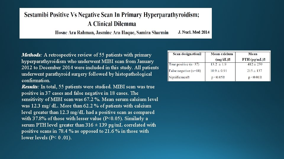
J. Nucl. Med 2014 Methods: A retrospective review of 55 patients with primary hyperparathyroidism who underwent MIBI scan from January 2012 to December 2014 were included in this study. All patients underwent parathyroid surgery followed by histopathological confirmation. Results: In total, 55 patients were studied. MIBI scan was true positive in 37 cases and false negative in 18 cases. The sensitivity of MIBI scan was 67. 2 %. Mean serum calcium level was 12. 3 mg/ d. L. More than 62. 2 % of patients with calcium level greater than 12. 3 mg/d. L had a positive scan as compared with 37. 8% of those with lesser value (P<0. 05). Similarly a serum PTH level greater than 316 ± 139 pg/m. L correlated with positive scans in 78. 4 % as opposed to 21. 6 % in those with lower levels (P< 0. 01).
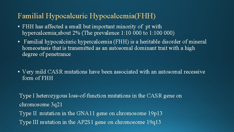
Familial Hypocalcuric Hypocalcemia(FHH) • FHH has affected a small but important minority of pt with hypercalcemia; about 2% (The prevalence 1: 10 000 to 1: 100 000) • Familial hypocalciuric hypercalcemia (FHH) is a heritable disorder of mineral homeostasis that is transmitted as an autosomal dominant trait with a high degree of penetrance • Very mild CASR mutations have been associated with an autosomal recessive form of FHH Type I heterozygous loss-of-function mutations in the CASR gene on chromosome 3 q 21 Type II mutation in the GNA 11 gene on chromosome 19 p 13 Type III mutation in the AP 2 S 1 gene on chromosome 19 q 13
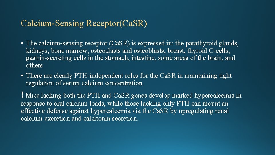
Calcium-Sensing Receptor(Ca. SR) • The calcium-sensing receptor (Ca. SR) is expressed in: the parathyroid glands, kidneys, bone marrow, osteoclasts and osteoblasts, breast, thyroid C-cells, gastrin-secreting cells in the stomach, intestine, some areas of the brain, and others • There are clearly PTH-independent roles for the Ca. SR in maintaining tight regulation of serum calcium concentration. ! Mice lacking both the PTH and Ca. SR genes develop marked hypercalcemia in response to oral calcium loads, while those lacking only PTH can mount an effective defense against hypercalcemia via the Ca. SR by upregulating renal calcium excretion and calcitonin secretion.
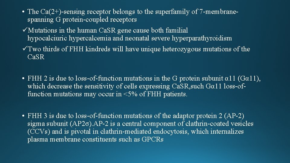
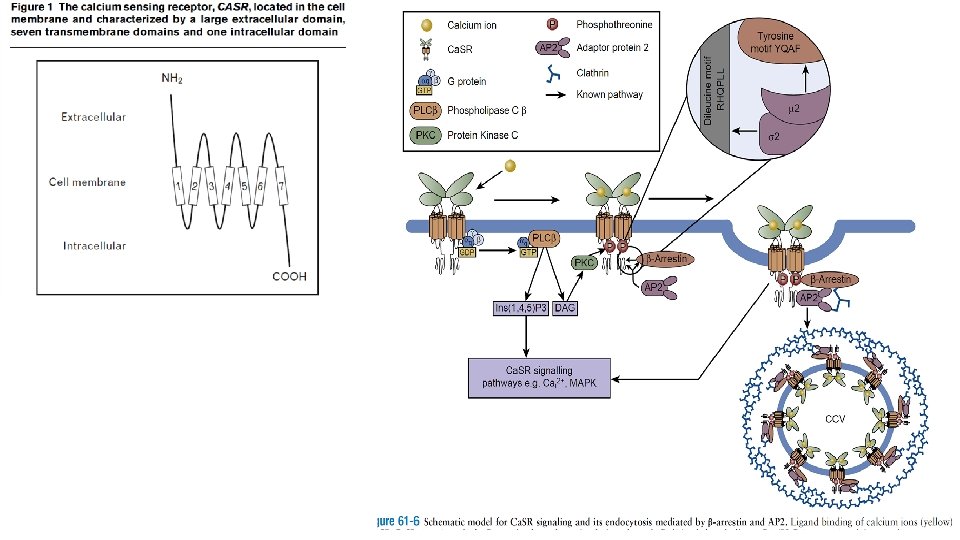
• The Ca(2+)-sensing receptor belongs to the superfamily of 7 -membranespanning G protein-coupled receptors üMutations in the human Ca. SR gene cause both familial hypocalciuric hypercalcemia and neonatal severe hyperparathyroidism üTwo thirds of FHH kindreds will have unique heterozygous mutations of the Ca. SR • FHH 2 is due to loss-of-function mutations in the G protein subunit α 11 (Gα 11), which decrease the sensitivity of cells expressing Ca. SR, such Gα 11 loss-offunction mutations may occur in <5% of FHH patients. • FHH 3 is due to loss-of-function mutations of the adaptor protein 2 (AP-2) sigma subunit (AP 2σ). AP-2 is a central component of clathrin-coated vesicles (CCVs) and is pivotal in clathrin-mediated endocytosis, which internalizes plasma membrane constituents such as GPCRs
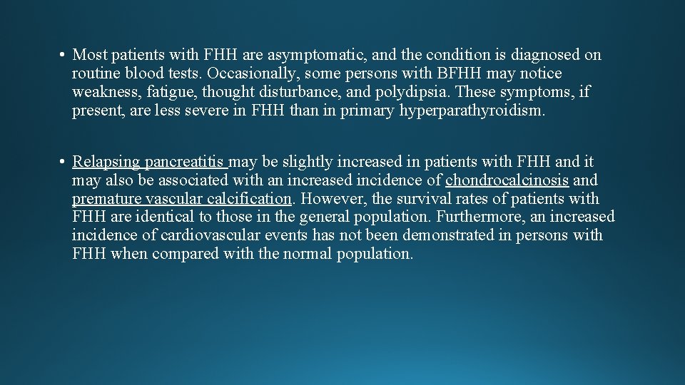
• Most patients with FHH are asymptomatic, and the condition is diagnosed on routine blood tests. Occasionally, some persons with BFHH may notice weakness, fatigue, thought disturbance, and polydipsia. These symptoms, if present, are less severe in FHH than in primary hyperparathyroidism. • Relapsing pancreatitis may be slightly increased in patients with FHH and it may also be associated with an increased incidence of chondrocalcinosis and premature vascular calcification. However, the survival rates of patients with FHH are identical to those in the general population. Furthermore, an increased incidence of cardiovascular events has not been demonstrated in persons with FHH when compared with the normal population.
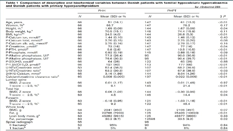
Eur J Endocrinol 2008
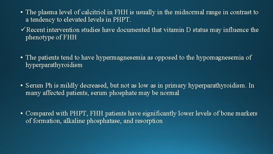
• The plasma level of calcitriol in FHH is usually in the midnormal range in contrast to a tendency to elevated levels in PHPT. üRecent intervention studies have documented that vitamin D status may influence the phenotype of FHH • The patients tend to have hypermagnesemia as opposed to the hypomagnesemia of hyperparathyroidism • Serum Ph is mildly decreased, but not as low as in primary hyperparathyroidism. In many affected patients, serum phosphate may be normal • Compared with PHPT, FHH patients have significantly lower levels of bone markers of formation, alkaline phosphatase, and resorption
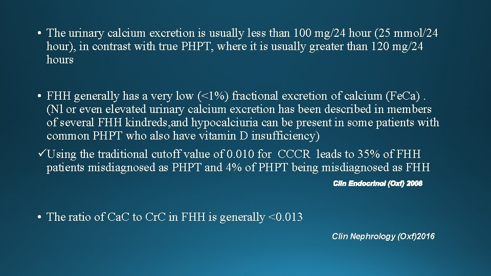
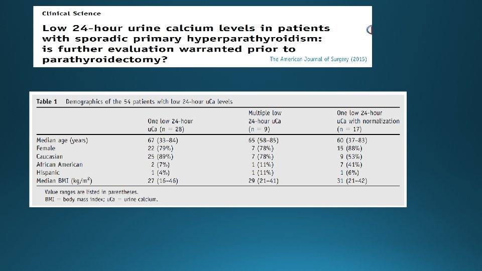
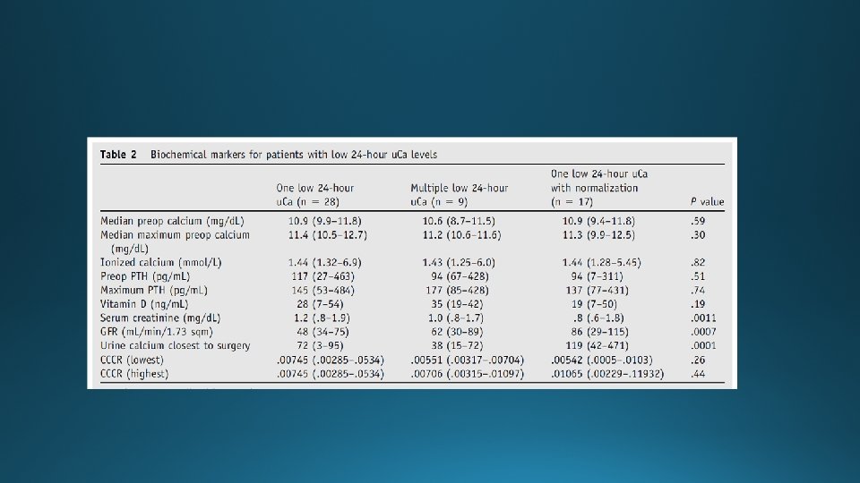
• The urinary calcium excretion is usually less than 100 mg/24 hour (25 mmol/24 hour), in contrast with true PHPT, where it is usually greater than 120 mg/24 hours • FHH generally has a very low (<1%) fractional excretion of calcium (Fe. Ca). (Nl or even elevated urinary calcium excretion has been described in members of several FHH kindreds, and hypocalciuria can be present in some patients with common PHPT who also have vitamin D insufficiency) üUsing the traditional cutoff value of 0. 010 for CCCR leads to 35% of FHH patients misdiagnosed as PHPT and 4% of PHPT being misdiagnosed as FHH • The ratio of Ca. C to Cr. C in FHH is generally <0. 013 Clin Nephrology (Oxf)2016
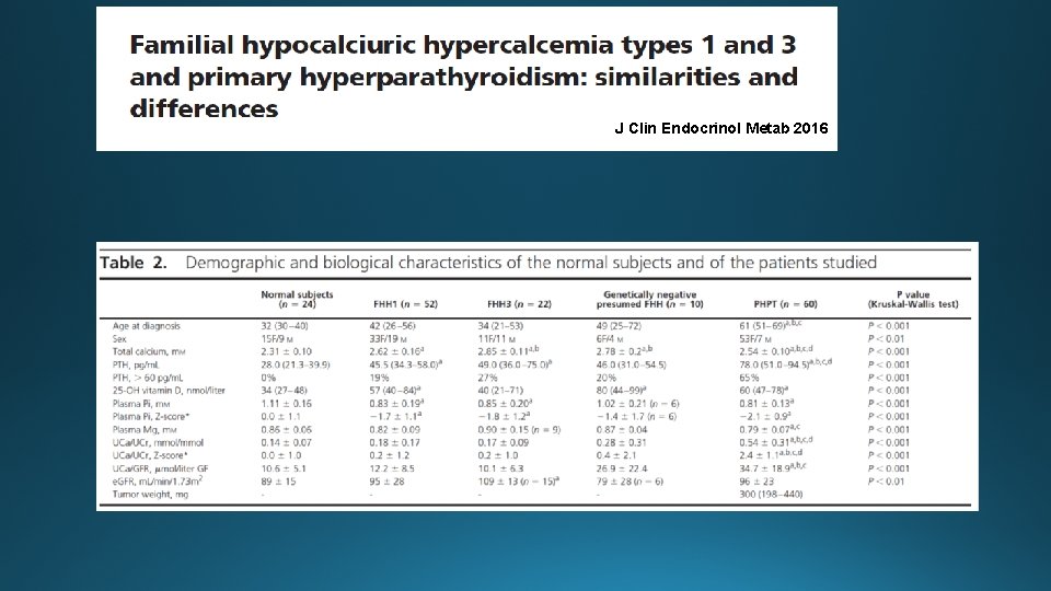
J Clin Endocrinol Metab 2016
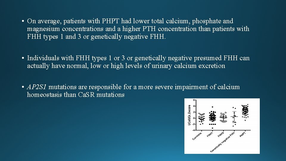
• On average, patients with PHPT had lower total calcium, phosphate and magnesium concentrations and a higher PTH concentration than patients with FHH types 1 and 3 or genetically negative FHH. • Individuals with FHH types 1 or 3 or genetically negative presumed FHH can actually have normal, low or high levels of urinary calcium excretion • AP 2 S 1 mutations are responsible for a more severe impairment of calcium homeostasis than Ca. SR mutations
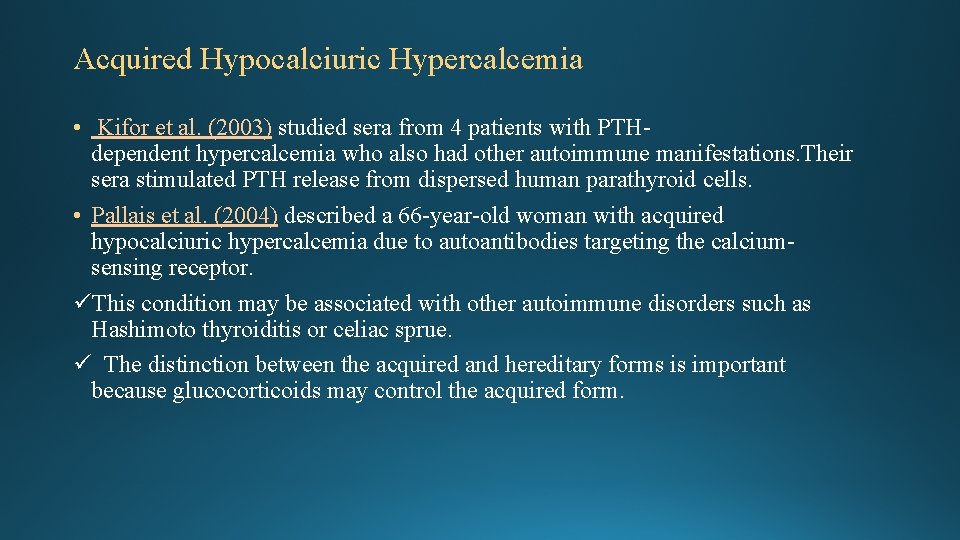
Acquired Hypocalciuric Hypercalcemia • Kifor et al. (2003) studied sera from 4 patients with PTHdependent hypercalcemia who also had other autoimmune manifestations. Their sera stimulated PTH release from dispersed human parathyroid cells. • Pallais et al. (2004) described a 66 -year-old woman with acquired hypocalciuric hypercalcemia due to autoantibodies targeting the calciumsensing receptor. üThis condition may be associated with other autoimmune disorders such as Hashimoto thyroiditis or celiac sprue. ü The distinction between the acquired and hereditary forms is important because glucocorticoids may control the acquired form.
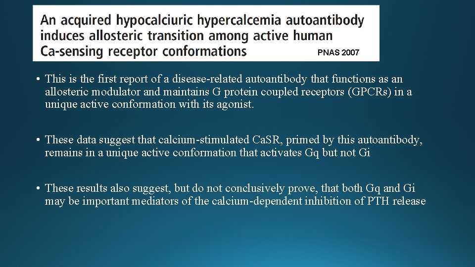
PNAS 2007 • This is the first report of a disease-related autoantibody that functions as an allosteric modulator and maintains G protein coupled receptors (GPCRs) in a unique active conformation with its agonist. • These data suggest that calcium-stimulated Ca. SR, primed by this autoantibody, remains in a unique active conformation that activates Gq but not Gi • These results also suggest, but do not conclusively prove, that both Gq and Gi may be important mediators of the calcium-dependent inhibition of PTH release
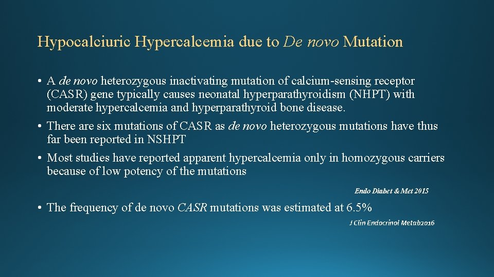
Hypocalciuric Hypercalcemia due to De novo Mutation • A de novo heterozygous inactivating mutation of calcium-sensing receptor (CASR) gene typically causes neonatal hyperparathyroidism (NHPT) with moderate hypercalcemia and hyperparathyroid bone disease. • There are six mutations of CASR as de novo heterozygous mutations have thus far been reported in NSHPT • Most studies have reported apparent hypercalcemia only in homozygous carriers because of low potency of the mutations Endo Diabet & Met 2015 • The frequency of de novo CASR mutations was estimated at 6. 5% J Clin Endocrinol Metab 2016
! Some reports have described infants with manifestation of NSHPT who possess only a single defective CASR allele. In these cases the mutant CASR was inherited from an affected father or arose as a de novo mutation. Presumably, exposure of the affected fetus to normal maternal calcium concentrations intensifies development of hyperparathyroidism in utero and leads to severe hypercalcemia after birth
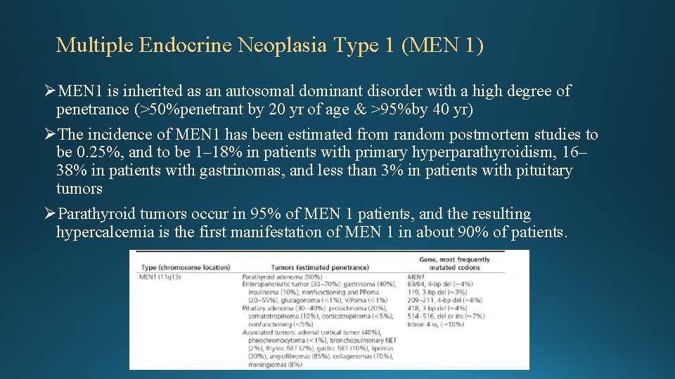
Multiple Endocrine Neoplasia Type 1 (MEN 1) ØMEN 1 is inherited as an autosomal dominant disorder with a high degree of penetrance (>50%penetrant by 20 yr of age & >95%by 40 yr) ØThe incidence of MEN 1 has been estimated from random postmortem studies to be 0. 25%, and to be 1– 18% in patients with primary hyperparathyroidism, 16– 38% in patients with gastrinomas, and less than 3% in patients with pituitary tumors ØParathyroid tumors occur in 95% of MEN 1 patients, and the resulting hypercalcemia is the first manifestation of MEN 1 in about 90% of patients.
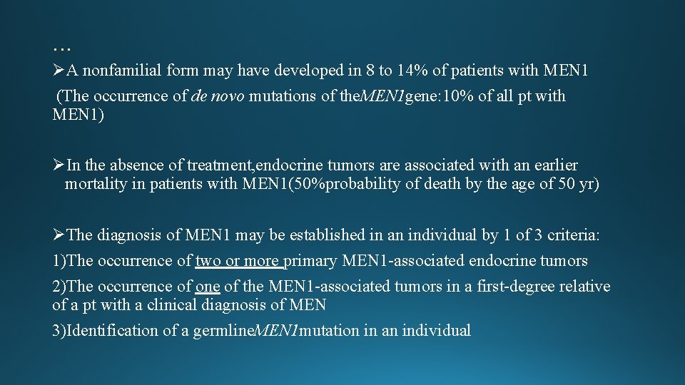
… ØA nonfamilial form may have developed in 8 to 14% of patients with MEN 1 (The occurrence of de novo mutations of the. MEN 1 gene: 10% of all pt with MEN 1) ØIn the absence of treatment, endocrine tumors are associated with an earlier mortality in patients with MEN 1(50%probability of death by the age of 50 yr) ØThe diagnosis of MEN 1 may be established in an individual by 1 of 3 criteria: 1)The occurrence of two or more primary MEN 1 -associated endocrine tumors 2)The occurrence of one of the MEN 1 -associated tumors in a first-degree relative of a pt with a clinical diagnosis of MEN 3)Identification of a germline. MEN 1 mutation in an individual
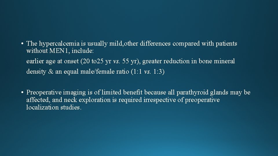
• The hypercalcemia is usually mild, other differences compared with patients without MEN 1, include: earlier age at onset (20 to 25 yr vs. 55 yr), greater reduction in bone mineral density & an equal male/female ratio (1: 1 vs. 1: 3) • Preoperative imaging is of limited benefit because all parathyroid glands may be affected, and neck exploration is required irrespective of preoperative localization studies.
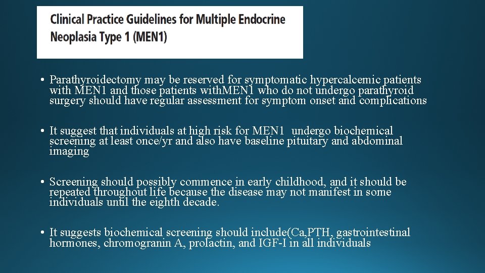
• Parathyroidectomy may be reserved for symptomatic hypercalcemic patients with MEN 1 and those patients with. MEN 1 who do not undergo parathyroid surgery should have regular assessment for symptom onset and complications • It suggest that individuals at high risk for MEN 1 undergo biochemical screening at least once/yr and also have baseline pituitary and abdominal imaging • Screening should possibly commence in early childhood, and it should be repeated throughout life because the disease may not manifest in some individuals until the eighth decade. • It suggests biochemical screening should include(Ca, PTH, gastrointestinal hormones, chromogranin A, prolactin, and IGF-I in all individuals
Coexisting AIT and parathyroid disease; are they related? • The association between thyroid diseases and PHPT was first described in 1947 (AIT association with PHPT was find in 3. 8 -17. 6%) • The inflammatory process found in AIT may interfere the uptake mechanism of 99 m. Tc-MIBI, giving a false negative image • Earlier experimental studies in rats have shown that chronic high TSH induces parathyroid hyperplasia and high PTH values. • Recent studies have shown that vitamin D & Selenium deficiency may be associated with both thyroid and parathyroid diseases Arch Clin Cases 2015
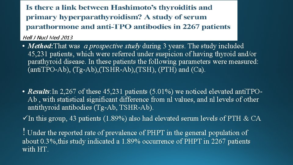
Hell J Nucl Med 2013 • Method: That was a prospective study during 3 years. The study included 45, 231 patients, which were referred under suspicion of having thyroid and/or parathyroid disease. In these patients the following parameters were measured: (anti. TPO-Ab), (Tg-Ab), (TSHR-Ab), (TSH), (PTH) and (Ca). • Results: In 2, 267 of these 45, 231 patients (5. 01%) we noticed elevated anti. TPOAb , with statistical significant difference from nl values, and nl levels of other antithyroid antibodies (Tg-Ab, TSHR-Ab). üIn this group, 43 patients (1. 89%) also had elevated serum levels of PTH & CA ! Under the reported rate of prevalence of PHPT in the general population of about 0. 3%, this study indicated a 1. 89% occurrence of PHPT in 2267 patients with HT.
NOTE: Althouth overall findings are more consistent with FHH because of the lack of genetic studies, diagnosis of the pt chould’t be confirmed, hence considering the fact that no clinical findings showed hypercalcemia side effects, we can follow “watch & wait” approach!
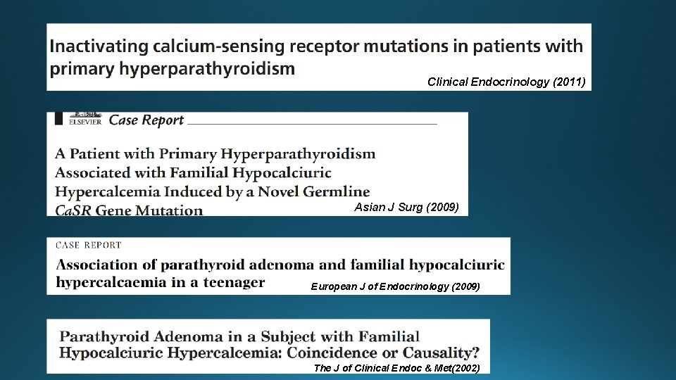
• Parathyroid hyperplasia is found in some cases of FHH • Single or multiple parathyroid adenomas have recently been described in several family members in a setting of apparent FHH based on a mutation in the cytoplasmic tail of the Ca. SR • It remains unclear whether the parathyroid hyperplasia seen in some patients may predispose to PHPT AACE/AAES Task Force on Primary Hyperparathyroidism 2005
Clinical Endocrinology (2011) Asian J Surg (2009) European J of Endocrinology (2009)) The J of Clinical Endoc & Met(2002)
!The urinary calcium excretion remaines low in coexistence of PHPT & FHH, and bone turnover markers remains normal in the patient despite PHPT !Symptoms may be obscured by Ca. SR mutation in PHPT
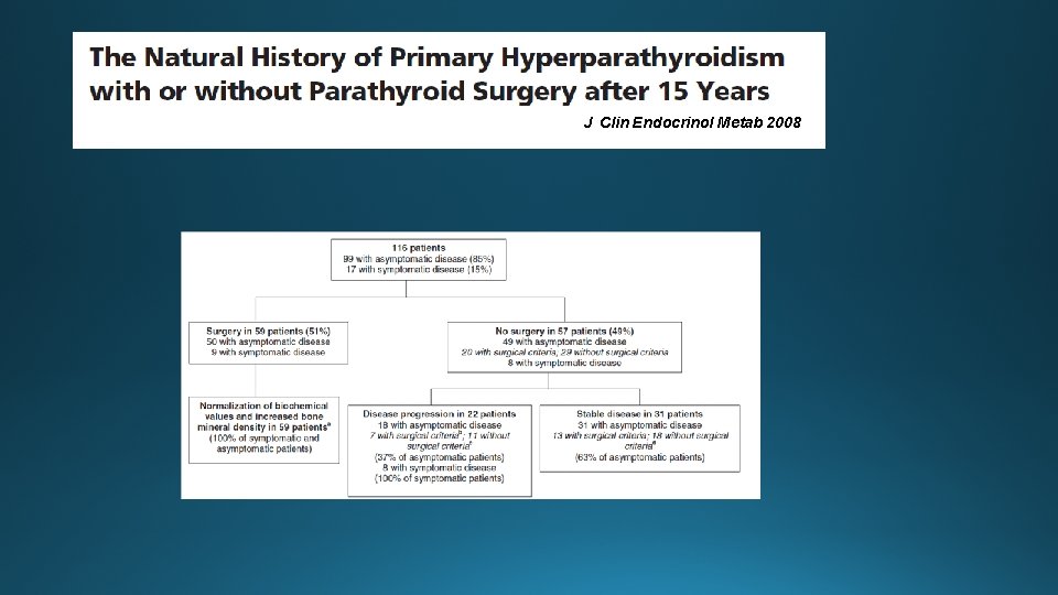
J Clin Endocrinol Metab 2008
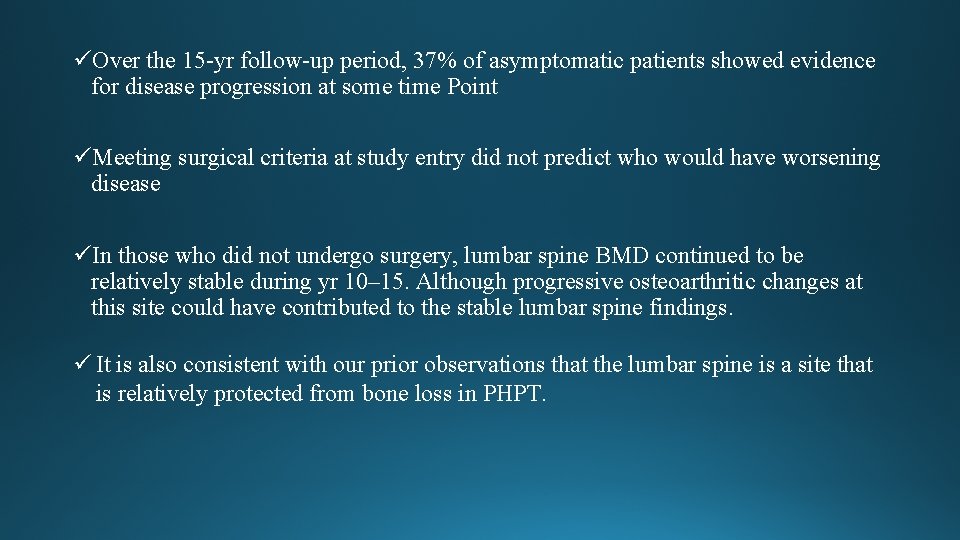
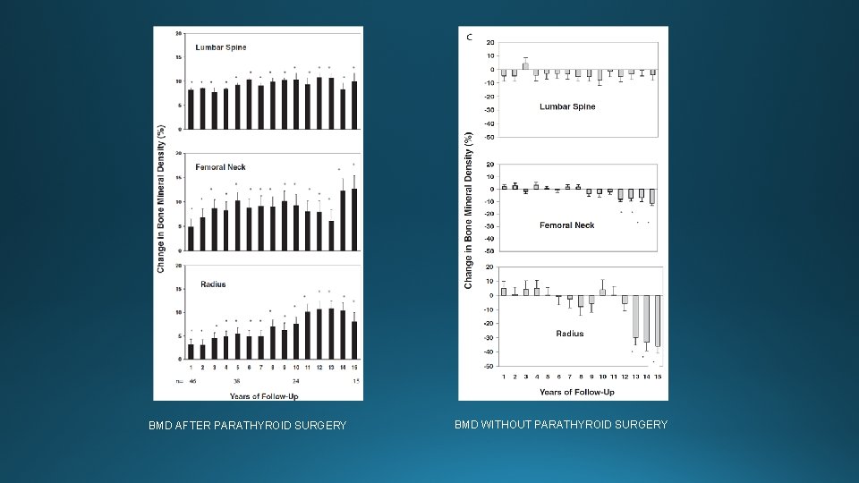
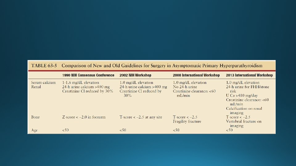
üOver the 15 -yr follow-up period, 37% of asymptomatic patients showed evidence for disease progression at some time Point üMeeting surgical criteria at study entry did not predict who would have worsening disease üIn those who did not undergo surgery, lumbar spine BMD continued to be relatively stable during yr 10– 15. Although progressive osteoarthritic changes at this site could have contributed to the stable lumbar spine findings. ü It is also consistent with our prior observations that the lumbar spine is a site that is relatively protected from bone loss in PHPT.
BMD AFTER PARATHYROID SURGERY BMD WITHOUT PARATHYROID SURGERY
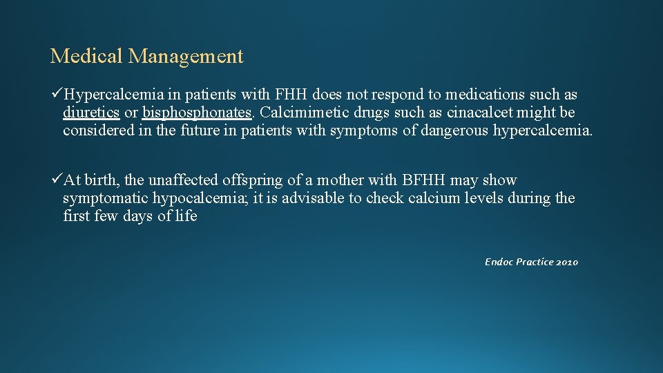
Medical Management üHypercalcemia in patients with FHH does not respond to medications such as diuretics or bisphonates. Calcimimetic drugs such as cinacalcet might be considered in the future in patients with symptoms of dangerous hypercalcemia. üAt birth, the unaffected offspring of a mother with BFHH may show symptomatic hypocalcemia; it is advisable to check calcium levels during the first few days of life Endoc Practice 2010
Diet • Dietary management of PHPT has long been an area of controversy. Many patients are advised to limit their dietary calcium intake because of the hypercalcemia. • Locker et al noted no difference in urinary calcium excretion between those on high (1000 mg/day) and low (500 mg/day) calcium intake diets in patients with nl levels of 1, 25(OH)D. On the other hand, in those with elevated levels of 1, 25(OH)2 D 3, high calcium diets were associated with worsening hypercalciuria.
Pharmaceuticals • Molecules that mimic the effect of extracellular calcium by altering the affinity of calcium for the receptor could activate G protein–coupled receptor and inhibit parathyroid cell function • A second-generation ligand, cinacalcet, has been the subject of more extensive investigations in PHPT. Studies indicate that this drug can reduce the serum calcium concentration to normal in PHPT but despite normalization of the serum calcium concentration, PTH levels do not return to normal; they do fall by 35% to 50% after administration of the drug. Urinary calcium excretion does not change; serum phosphorus levels increase but are maintained in the lower range of normal; and 1, 25(OH)2 D 3 levels do not change. The average BMD does not change, even after 3 years of administration of cinacalcet. • Marcocci et al have shown that cinacalcet is effective in subjects with intractable PHPT & also it can reduces calcium levels effectively in inoperable parathyroid carcinoma.
Bisphonates • They do not affect PTH secretion directly, but they could reduce serum and urinary calcium levels. • The results of studies suggest that a bisphonate like alendronate might be useful in patients with low bone density in whom parathyroid surgery is not to be performed