
ABERRANT LIQUOR VOLUME Teacher Prof Sukanta Misra Prof
ABERRANT LIQUOR VOLUME Teacher Prof. Sukanta Misra. Prof & HOD, G&O, RKMSP, VIMS. Student Sumana Nandi Bidisha Mallick
Topics to be covered • • • What is it? Formation of amniotic fluid Volume and composition Functions and clinical significance of amniotic fluid Abnormalities a) Polyhydramnios- definition, diagnosis, evaluation & t/t b) Oligohydramnios- definition, diagnosis, evaluation & t/t
Introduction • The amniotic fluid or liquor amnii surrounds the fetus during intrauterine development. • This fluid serves as a cushion for the growing fetus. It is the results of contributions from both the mother and the fetus.

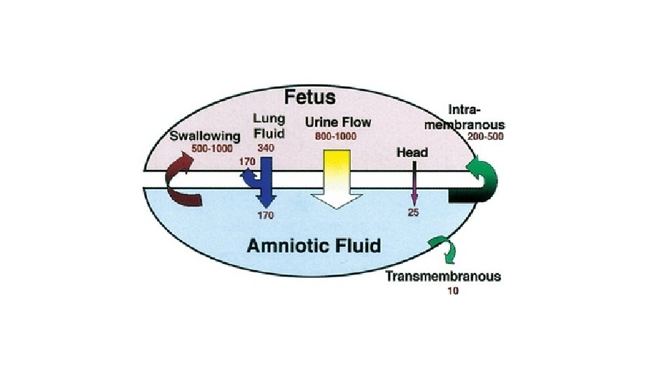
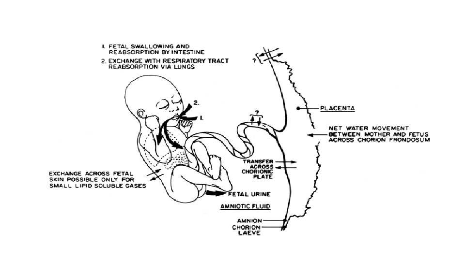
Factors determining for amniotic fluid volume • Transudation from maternal circulation through the placental surface & fetal membranes • Intramembranous flow across fetal vessels on the placental surfaces • Trans-membranous flow across amniotic membrane • Fetal urination • Fetal swallowing • Fetal lung field secretion • Transcutaneous flow-across fetal skin (prior to its keratinization)
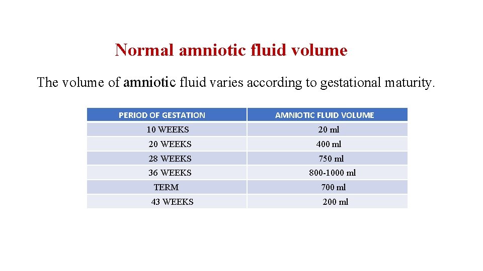
Normal amniotic fluid volume The volume of amniotic fluid varies according to gestational maturity. PERIOD OF GESTATION AMNIOTIC FLUID VOLUME 10 WEEKS 20 ml 20 WEEKS 400 ml 28 WEEKS 750 ml 36 WEEKS 800 -1000 ml TERM 700 ml 43 WEEKS 200 ml
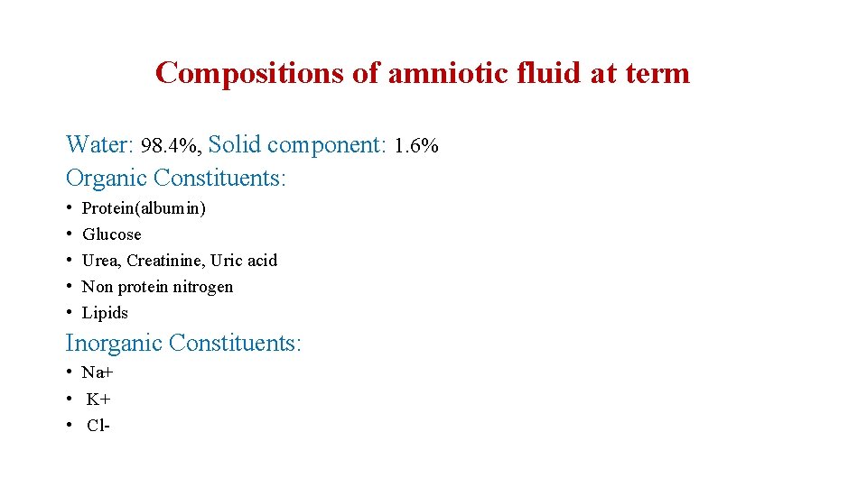
Compositions of amniotic fluid at term Water: 98. 4%, Solid component: 1. 6% Organic Constituents: • • • Protein(albumin) Glucose Urea, Creatinine, Uric acid Non protein nitrogen Lipids Inorganic Constituents: • Na+ • K+ • Cl-
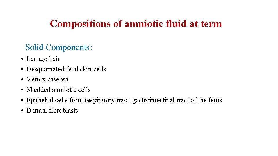
Compositions of amniotic fluid at term Solid Components: • • • Lanugo hair Desquamated fetal skin cells Vernix caseosa Shedded amniotic cells Epithelial cells from respiratory tract, gastrointestinal tract of the fetus Dermal fibroblasts
Physical features of amniotic fluid p. H: - slightly alkaline (around 7) Specific gravity: - 1. 007 -1. 010 Osmolarity: -Hypotonic. Osmolarity( 260 m. Osm/L) ** osmolarity of fetal plasma approx. 280 m. Osm/L Color: - Colorless in early pregnancy Pale straw color near term
Abnormal appearance of amniotic fluid Golden yellow: - In Rh incompatibility patients Greenish yellow: - In post-maturity patients Dark maroon: - Due to altered blood in accidental hemorrhage. Dark brown: - In presence of retained dead fetus Blood stained: - In abruption, vasa previa.
What are the functions of amniotic fluid? Functions during pregnancy: • Protects fetus from trauma • Allows space for fetal movement which is necessary for musculo-skeletal development. • Permits fetal swallowing-essential for GIT development • Inhalation into respiratory tract-helps in growth and differentiation of respiratory tract as amniotic fluid has growth factors in it. • Maintains fetal temperature • Prevents the adhesion formation between fetal parts and amniotic sac. Functions during labor: -

What are the functions of amniotic fluid? Functions during pregnancy: Functions during labor: • Amniotic fluid forms a hydrostatic wedge(bag of membranes) to help in cervical dialatation. • During uterine contractions, amniotic fluid in intact membranes prevents interference with placental circulation. • Protects fetus from ascending infections by its bacteriostatic properties.
Measurement of amniotic fluid Direct method by DYE DILUTION TECHNIQUE: Injection of a small quantity of dye (amino hippurate) into the amniotic cavity under USG guidance. Then amniotic fluid is sampled to determine the dye concentration to find out the total amniotic fluid volume. Disadvantages: Invasive procedure Risk of fetal injury Chorioamnionitis Rupture of membrane

Measurement of amniotic fluid Sonographic assessment: Single deepest pocket (SDP) • The largest pocket of liquor avoiding umbilical cord and fetal extremity is measured • Normal range in 2 cm to 8 cm.

Measurement of amniotic fluid Sonographic assessment: Amniotic fluid index • Maternal abdomen is divided in 4 quadrants by drawing two perpendicular lines through umbilicus. • Measured by sum of single deepest vertical pocket from each quadrant. • Normal range 5 cm to 24 cm • Color doppler is used to pick-up the cord free pocket.

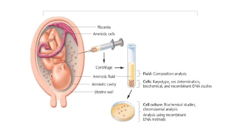
Clinical significance of amniotic fluid • Diagnosis of fetal genetic disorders: By doing cytogenic-analysis of amniocytes obtained by amniocentesis to diagnose chromosomal-abnormalities using CMA/ Karyotyping/ FISH. • Diagnosis of congenital infections: e. g. CMV infection by PCR or viral culture • Screening for congenital malformations: By doing biochemical analysis for -Alpha-feto protein estimation -Acetylcholinesterase level estimation
Clinical significance of amniotic fluid • Assessment of fetal well-being: By measuring the amniotic fluid index As it is component a Biophysical profile. • Assessment of fetal lung maturity: By measuring Lecithin-sphingomyelin ratio If it is >2. 0; it indicates fetal lung maturity If it is less, fetus is susceptible to respiratory distress syndrome. By assessing the phosphatidyl-glycerol in amniotic fluid
Clinical significance of amniotic fluid • Assessment of severity of Rh isoimmunization: Measurement of bilirubin is an indication of degree of haemolysis occurring in utero, therefore an indication of danger of anaemia in the fetus. • Amniotic fluid testing for fetal age determination: At 36 weeks, fetal kidneys excrete >2. 0 mg/dl creatinine This test now has been replaced by ultrasound measurements. ** the above mentioned two tests are not used now-a-days.
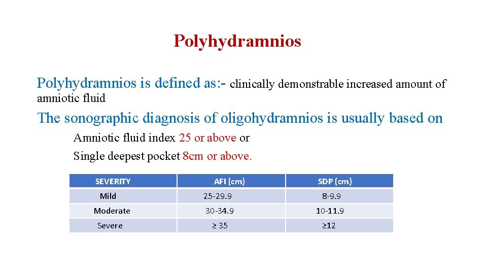
Polyhydramnios is defined as: - clinically demonstrable increased amount of amniotic fluid The sonographic diagnosis of oligohydramnios is usually based on Amniotic fluid index 25 or above or Single deepest pocket 8 cm or above. SEVERITY Mild AFI (cm) SDP (cm) 25 -29. 9 8 -9. 9 Moderate 30 -34. 9 10 -11. 9 Severe ≥ 35 ≥ 12
Etiology of polyhydramnios • • Idiopathic Maternal causes Placental causes Fetal causes
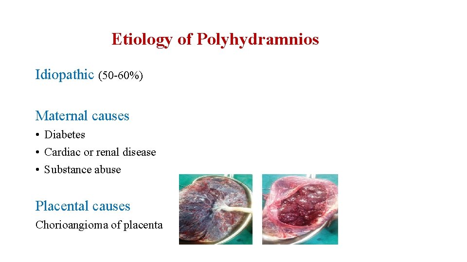
Etiology of Polyhydramnios Idiopathic (50 -60%) Maternal causes • Diabetes • Cardiac or renal disease • Substance abuse Placental causes Chorioangioma of placenta
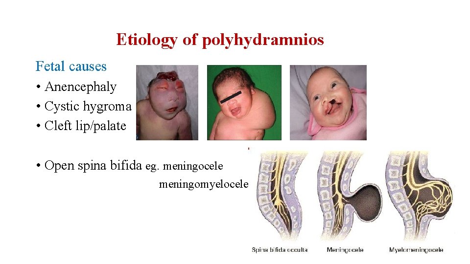
Etiology of polyhydramnios Fetal causes • Anencephaly • Cystic hygroma • Cleft lip/palate • Open spina bifida eg. meningocele meningomyelocele
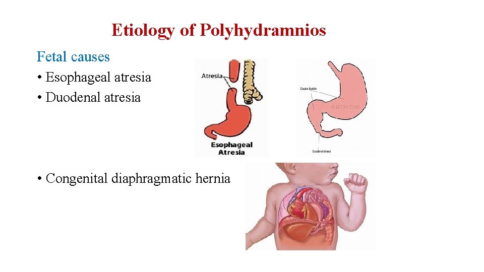
Etiology of Polyhydramnios Fetal causes • Esophageal atresia • Duodenal atresia • Congenital diaphragmatic hernia
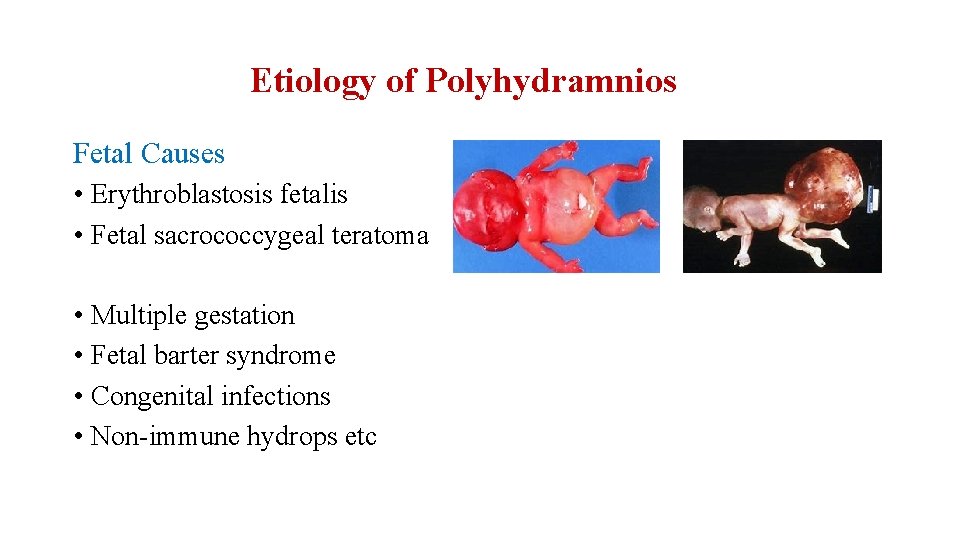
Etiology of Polyhydramnios Fetal Causes • Erythroblastosis fetalis • Fetal sacrococcygeal teratoma • Multiple gestation • Fetal barter syndrome • Congenital infections • Non-immune hydrops etc
Differential diagnosis • • Multiple pregnancy Pregnancy with large ovarian cyst Pregnancy with fibroid Maternal ascites Concealed abruption Full bladder Hydatidiform mole
Types of Polyhydramnios Acute Polyhydramnios Chronic Polyhydramnios • Is very rare • Sudden onset • Severe abdominal pain is common symptom • Most common type • Is gradual in onset

Complications of Polyhydramnios Fetal Complications Maternal Complications • • Cardiorespiratory embarrassment • Maternal mirror syndrome seen Unstable lie Malpresentation Cord prolapse PROM Placental abruption Premature labor High perinatal mortality rate especially with hydrops fetalis • • Premature labor Increased LSCS rate Retained placenta, PPH Subinvolution of uterus, Puerperal sepsis • Maternal morbidity
Diagnosis of Polyhydramnios Clinical features: • • • Breathlessness Palpitation Swelling of the lower extremities, vulva and abdominal wall Heart burn/ Indigestion Varicose vein in legs and vulva
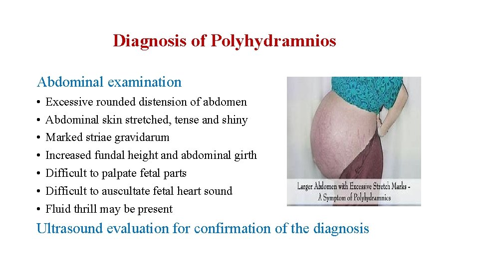
Diagnosis of Polyhydramnios Abdominal examination • • Excessive rounded distension of abdomen Abdominal skin stretched, tense and shiny Marked striae gravidarum Increased fundal height and abdominal girth Difficult to palpate fetal parts Difficult to auscultate fetal heart sound Fluid thrill may be present Ultrasound evaluation for confirmation of the diagnosis
Management of Polyhydramnios 1. Treatment according to underlying pathology 2. Treatment according to symptoms 3. Role of Indomethacin • • Impairs fetal lung fluid production Enhances absorption Increases fluid movement across fetal membranes Reduce fetal urinary production SIDE EFFECTS- premature closure of fetal ductus arteriosus -not used after 35 weeks
Management of Polyhydramnios 4. Amnioreduction -around 1000 -1500 ml fluid withdrawn -procedure is done slowly -done weekly or semiweekly SIDE EFFECTS- PROM -Preterm labor -Abruption
Oligohydramnios is defined as: - clinically demonstrable decreased amount of amniotic fluid. The sonographic diagnosis of oligohydramnios is based on Amniotic fluid index <5 cm or Single deepest pocket <2 cm
Etiology of oligohydramnios • • • Fetal causes Maternal causes Placental causes Drugs Idiopathic Iatrogenic
Etiology of Oligohydramnios Fetal Causes • • • PROM Chromosomal abnormalities -Trisomy 18, Triploidy, Turner syndrome Post term pregnancy Fetal growth restriction Intrauterine fetal death Congenital anomalies -renal agenesis -renal dysplasia -posterior urethral valve -bladder outlet obstruction -amniotic band syndrome -cardiac defects- septal defects, Fallot’s tetralogy -CNS defects- microcephaly
Etiology of oligohydramnios Maternal causes Drugs induced Uteroplacental insufficiency Hypertension Pre-eclampsia PGs synthesis inhibitors ACE inhibitors Idiopathic Placental causes Amnion nodosum Abruption Twin-twin transfusion syndrome Iatrogenic Amniocentesis, CVS
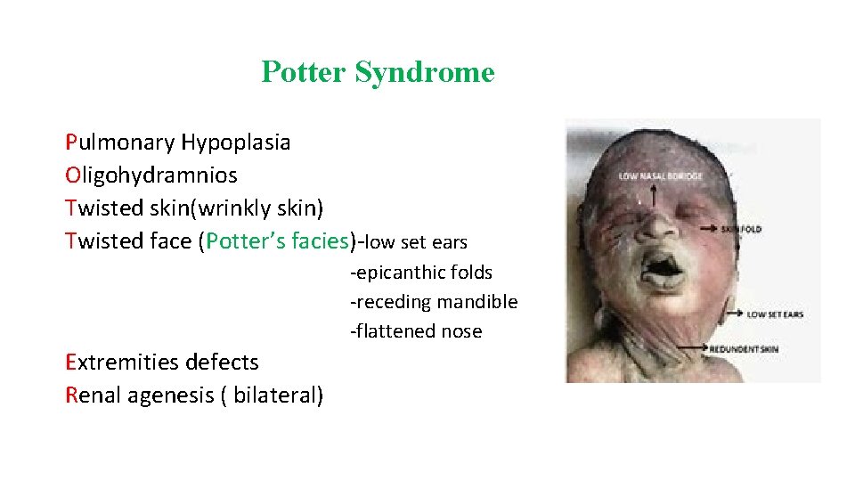
Potter Syndrome Pulmonary Hypoplasia Oligohydramnios Twisted skin(wrinkly skin) Twisted face (Potter’s facies)-low set ears -epicanthic folds -receding mandible -flattened nose Extremities defects Renal agenesis ( bilateral)
Potter sequence When the abnormalities same as potter syndrome result from another etiology (not due to renal agenesis) such as bilateral multicystic dysplastic kidney, autosomal recessive polycystic kidney disease, it is called Potter sequence.
Complications of Oligohydramnios Fetal effects Maternal effects EARLY PROBLEMS • Pulmonary hypoplasia • Limb deformities like talipes • Potter’s facies • Amniotic adhesions or bands causing • Prolonged labor(d/t uterine intertia and dysfunctional labor) • Higher chances of operative delivery d/t malpresentations • Increased maternal morbidity deformities like amputation of digits • Fetal growth restriction, IUD LATE PROBLEMS • Cord compression • Meconium aspiration syndrome
Diagnosis of Oligohydramnios History: H/O of leaking P/V Post term H/O drugs intake Examination: Uterus-small for date Malpresentation USG: Decreased AFI & SDP
Management of Oligohydramnios Directed to the causes • • Drug induced- omit drug PROM- antibiotics, steroid Post urethral valve- VESICO AMNIOTIC SHUNT Amnio infusion ** oligohydramnios associated with FGR requires antepartum fetal surveillance and doppler and proper timing of delivery
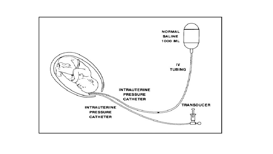
Amnioinfusion Instillation of sterile fluid into the amniotic cavity to increase liquor volume is called amnioinfusion Indications • In variable decelerations • In prophylaxis for women with oligohydramnios • To dilute / wash-out thick meconium Procedure • 500 ml bolus of NS/RL is instilled within 30 minutes f/b continuous infusion to be given at the rate of 3 ml/min. • Amnioinfusion is stopped after infusion of 1000 ml or delivery imminent or any complication arises
Amnioinfusion Complications: - Uterine overdistension Uterine hypertonicity Fetal bradycardia Umbilical cord prolapse Amniotic fluid embolism Contraindications: - Severe preeclampsia Classical cesarean section Maternal cardiac disease

POLY-OLY SYNDROME/ TTTS Seen in monochorionic twin pregnancies. Antenatal criteria for defining TTTS: • • • Same sex fetuses Monochorionicity with placental vascular anastomosis Weight difference between twins greater than 20% Hydramnios in the larger twin, oligohydramnios in the smaller twin Stuck smaller twin Hemoglobin difference >5 g/dl
POLY-OLY SYNDROME Quintero staging system Stage I- discordant amniotic fluid volume but urine is still visible sonographically within donor twin’s bladder. Stage-II- Stage I + but urine is not visible within donor’s bladder. Stage III- Stage II + abnormal doppler studies of umbilical artery, ductus venosus or umbilical vein. Stage IV- ascites or frank hydrops in either twin. Stage V- it is most severe form with demise of either twin.
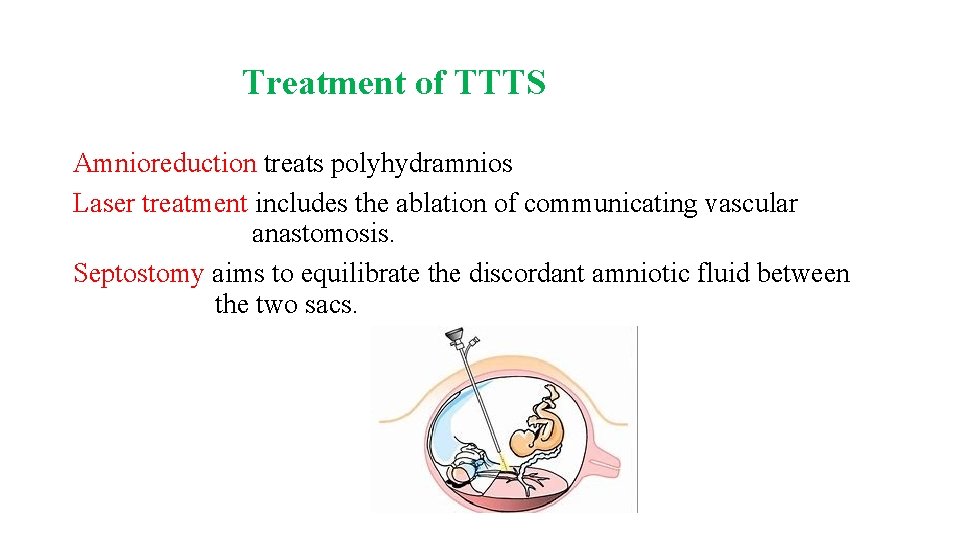
Treatment of TTTS Amnioreduction treats polyhydramnios Laser treatment includes the ablation of communicating vascular anastomosis. Septostomy aims to equilibrate the discordant amniotic fluid between the two sacs.
- Slides: 50