Hemodynamic Monitoring and Circulatory Assist Devices Chapter 66


/stroke volume index (SVI)")

 and pulmonary vascular resistance (PVR) •")


 Copyright © 2014 by Mosby, an imprint of")


 Monitoring • Calculates continuous CO and CI • Used to")
 Monitoring Copyright © 2014 by Mosby, an imprint of Elsevier")

 • Distal")
 • Inserted via")
 -Pulmonary HTN, embolism, & edema, -Valvular disease")


 • PA catheter with thermal filament")

 • Inject saline or D 5")


 • Noninvasive method of obtaining CO and")


 • Decrease cardiac work and improve organ perfusion when drug")


 • Most common CAD • Benefits • ↓ventricular workload •")
 • Consists of: • Sausage-shaped balloon • Pump that inflates")

 • Balloon inserted into femoral artery and placed in thoracic")
 Copyright © 2014 by Mosby, an imprint of Elsevier Inc.")

 • Complications of IABP therapy • Vascular injuries • Thrombus")
 • Complications of IABP • Mechanical complications • Improper timing")

 • To decrease risks of IABP therapy • Frequent assessments")
 • Short- and long-term support • Allows more mobility than")
 • VADs can • Be implanted (e. g. , peritoneum)")

 • Indications for VAD therapy • Failure to wean from")


- Slides: 83
Hemodynamic Monitoring and Circulatory Assist Devices Chapter 66 Copyright © 2014 by Mosby, an imprint of Elsevier Inc.
Hemodynamic Monitoring • Measurement of pressure, flow, and oxygenation within the cardiovascular system • Assesses heart function, fluid balance, and effects of drugs on CO Copyright © 2014 by Mosby, an imprint of Elsevier Inc.
Hemodynamic Monitoring • Invasive and noninvasive measurements • Systemic and pulmonary arterial pressures • Central venous pressure (CVP) • Pulmonary artery wedge pressure (PAWP) • Cardiac output (CO)/cardiac index (CI) Copyright © 2014 by Mosby, an imprint of Elsevier Inc.
Hemodynamic Monitoring • Invasive and noninvasive measurements • Stroke volume (SV)/stroke volume index (SVI) • Stroke volume variation (SVV) • O 2 saturation of arterial blood (Sa. O 2) • Mixed venous oxygenation saturation (Sv. O 2) Copyright © 2014 by Mosby, an imprint of Elsevier Inc.
Hemodynamic Monitoring Terminology • CO: volume of blood pumped by heart in 1 minute • CI: CO adjusted for body surface area (BSA) • SV: volume ejected with each heartbeat • SVI: SV adjusted for body size Copyright © 2014 by Mosby, an imprint of Elsevier Inc.
Hemodynamic Monitoring Terminology • Systemic vascular resistance (SVR) and pulmonary vascular resistance (PVR) • Opposition to blood flow by systemic and pulmonary vasculature • Preload, afterload, and contractility determine SV Copyright © 2014 by Mosby, an imprint of Elsevier Inc.
Hemodynamic Monitoring Terminology • Preload • Volume of blood within ventricle at end of diastole • PAWP: reflects left ventricular enddiastolic pressure • CVP: reflects right ventricular enddiastolic pressure Copyright © 2014 by Mosby, an imprint of Elsevier Inc.
Hemodynamic Monitoring Terminology • Afterload • Forces opposing ventricular ejection • SVR and arterial pressure indices of left ventricular afterload • PVR and pulmonary arterial pressure indices of right ventricular afterload Copyright © 2014 by Mosby, an imprint of Elsevier Inc.
Hemodynamic Monitoring Terminology • Vascular resistance • Systemic and pulmonary • Reflect afterload • Contractility • Strength of ventricular contraction • No direct clinical measure Copyright © 2014 by Mosby, an imprint of Elsevier Inc.
Case Study i. Stockphoto/Thinkstock • A. J. , a 78 -year-old male, is admitted to the ICU in acute decompensated heart failure. • Health care provider decides to insert an arterial line and pulmonary artery catheter to facilitate treatment decisions. Copyright © 2014 by Mosby, an imprint of Elsevier Inc.
Case Study i. Stockphoto/Thinkstock • What equipment will you collect in preparation for insertion of these lines? Copyright © 2014 by Mosby, an imprint of Elsevier Inc.
Components of Pressure Monitoring System Copyright © 2014 by Mosby, an imprint of Elsevier Inc.
Case Study i. Stockphoto/Thinkstock • Why is it important to zero reference the transducer and perform a dynamic response test during initial setup of the equipment for A. J. ? • Where is the phlebostatic axis located? Copyright © 2014 by Mosby, an imprint of Elsevier Inc.
Principles of Invasive Pressure Monitoring • Equipment must be referenced and zero balanced to environment and dynamic response characteristics optimized • Referencing: positioning transducer so zero reference point is at level of atria of heart or phlebostatic axis Copyright © 2014 by Mosby, an imprint of Elsevier Inc.
Phlebostatic axis @ intersection of 4 th intercostal space, mid AP diameter
Identification of the Phlebostatic Axis Copyright © 2014 by Mosby, an imprint of Elsevier Inc.
Principles of Invasive Pressure Monitoring • Zeroing: confirms that when pressure within system is zero, monitor reads zero • Done by opening reference stopcock to room air • With initial setup and periodically thereafter Copyright © 2014 by Mosby, an imprint of Elsevier Inc.
Dynamic Response Test (Square Wave Test) Copyright © 2014 by Mosby, an imprint of Elsevier Inc.
Types of Invasive Pressure Monitoring • Arterial pressure monitoring • Various indications when continuous BP measurements useful • 20 -gauge, 2 -inch nontapered Teflon catheter into peripheral artery • Suture in place • Immobilize insertion site Copyright © 2014 by Mosby, an imprint of Elsevier Inc.
Arterial Pressure Monitoring Copyright © 2014 by Mosby, an imprint of Elsevier Inc.
Case Study i. Stockphoto/Thinkstock • When planning care for A. J. , what nursing interventions will you perform to prevent complications related to his arterial line? Copyright © 2014 by Mosby, an imprint of Elsevier Inc.
Arterial Pressure Monitoring • High- and low-pressure alarms • Risks/complications • Hemorrhage • Infection • Thrombus formation • Neurovascular impairment • Loss of limb Copyright © 2014 by Mosby, an imprint of Elsevier Inc.
Arterial Pressure Monitoring • Continuous flush irrigation system • Delivers 3– 6 m. L of saline/hour • Maintains line patency • Limits thrombus formation • Assess neurovascular status distal to arterial insertion site hourly Copyright © 2014 by Mosby, an imprint of Elsevier Inc.
Arterial Pressure-Based CO (APCO) Monitoring • Calculates continuous CO and CI • Used to assess patient’s ability to respond to fluids • Uses arterial waveform characteristics and patient demographic data to calculate SVV, CCO/CCI, and SV/SVI every 20 seconds Copyright © 2014 by Mosby, an imprint of Elsevier Inc.
Arterial Pressure-Based CO (APCO) Monitoring Copyright © 2014 by Mosby, an imprint of Elsevier Inc.
Pulmonary Artery Pressure Monitoring • Guides management of patients with complicated cardiopulmonary problems • PA diastolic (PAD) pressure and PAWP → cardiac function and fluid volume status • Allows for precise manipulation of preload Copyright © 2014 by Mosby, an imprint of Elsevier Inc.
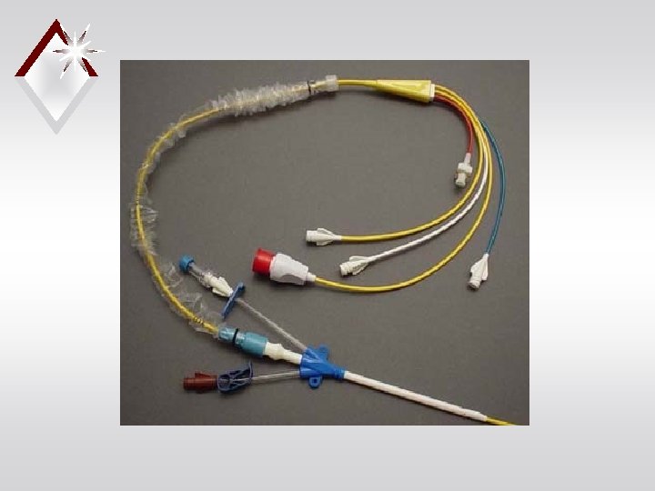
Pulmonary Artery Pressure Monitoring • PA flow-directed catheter (e. g. , Swan-Ganz) • Distal lumen port in PA • Balloon inflated to measure PAWP • Two proximal lumens to measure CVP, inject fluid for CO, draw blood, administer fluids or drugs • Thermistor port distally Copyright © 2014 by Mosby, an imprint of Elsevier Inc.
How do we measure Hemodynamics? • Pulmonary Artery Catheters (Swan Ganz) • Inserted via internal jugular vein • Line floated through right chambers of heart • End of catheter rests in proximal PA
Indications for PA Monitoring Diagnostics… -Shock (staging) -Pulmonary HTN, embolism, & edema, -Valvular disease -Cardiac Tamponade Evaluation of… -Volume status -Treatment modalities (i. e. gtt titration)
Contraindications for PA caths • Right side heart tumor or thrombus • Tricuspid or pulmonic valve prosthesis or endocarditis
PA Cath Insertion Risks • Dysrhythmias d/t RV irritation • PA rupture • Valve damage • Infection, Bleeding • Pneumo or hemothorax • Embolization • Catheter migration
PA Catheter Copyright © 2014 by Mosby, an imprint of Elsevier Inc.
Pulmonary Artery Pressure Monitoring • Specialized features • Atrial electrode • Fiberoptic sensor for mixed venous O 2 saturation • Continuous measurement of right ventricular volume and EF • Continuous CO monitoring • Additional ports for IV access Copyright © 2014 by Mosby, an imprint of Elsevier Inc.
PA Waveforms During Insertion Copyright © 2014 by Mosby, an imprint of Elsevier Inc.
Case Study i. Stockphoto/Thinkstock • After insertion of the PA line, the health care provider orders PAWP and CO measurement every 4 hours. • How will you obtain the PAWP measurement? Copyright © 2014 by Mosby, an imprint of Elsevier Inc.
Pulmonary Artery Pressure Monitoring • When measurements are obtained: • PA: at end expiration • PAWP: slowly inflate balloon with air until PA waveform changes to PAWP waveform • Do not inflate for more than four respiratory cycles or 8– 15 seconds Copyright © 2014 by Mosby, an imprint of Elsevier Inc.
Pulmonary Artery Pressure Monitoring Copyright © 2014 by Mosby, an imprint of Elsevier Inc.
Effect of Overinflated Balloon Copyright © 2014 by Mosby, an imprint of Elsevier Inc.
Copyright © 2014 by Mosby, an imprint of Elsevier Inc.
PA Catheter provides… • Continuous monitoring of… -CVP -PAP • Intermittent measure of… -PAWP
PA Waveforms Correlate with EKG Rhythm Tracing
Respiratory Considerations when Reading Waveforms
Central Venous Pressure Monitoring • Measurement of right ventricular preload → reflects fluid volume • Obtained from: • Central venous catheter • PA catheter • Similar to PAWP waveforms Copyright © 2014 by Mosby, an imprint of Elsevier Inc.
Central Venous Pressure Waveforms Copyright © 2014 by Mosby, an imprint of Elsevier Inc.
Measuring Cardiac Output • Continuous cardiac output (CCO) • PA catheter with thermal filament located in right atrium • Senses change in temperature of blood as it passes through right ventricle • Measures every 30– 60 seconds • Reflects average CO for past 3– 6 minutes Copyright © 2014 by Mosby, an imprint of Elsevier Inc.
Case Study i. Stockphoto/Thinkstock • A. J. ’s pulmonary catheter does not have the capability to obtain continuous CO measurements. • How will you perform an intermittent bolus thermodilution CO assessment for A. J. ? Copyright © 2014 by Mosby, an imprint of Elsevier Inc.
Measuring Cardiac Output • Intermittent bolus thermodilution (TDCO) • Inject saline or D 5 W into proximal lumen of PA catheter • Thermistor sensor detects differences in blood temperature and calculates CO • Uses average of three measurements Copyright © 2014 by Mosby, an imprint of Elsevier Inc.
Cardiac Output Monitor
Measuring Cardiac Output Copyright © 2014 by Mosby, an imprint of Elsevier Inc.
Measuring Cardiac Output • SVR, SVRI, SV, and SVI calculated when CO is measured • ↑ SVR • Indicates vasoconstriction • ↓ SVR • Indicates vasodilation Copyright © 2014 by Mosby, an imprint of Elsevier Inc.
Venous Oxygen Saturation • PA and CVP catheters can be used • CVP measures central venous oxygen saturation (Scv. O 2) • PA measures mixed venous oxygen saturation (Sv. O 2) • Determines adequacy of tissue oxygenation Copyright © 2014 by Mosby, an imprint of Elsevier Inc.
Venous Oxygen Saturation • Sv. O 2/Scv. O 2 reflect balance between oxygenation of arterial blood, tissue perfusion, and tissue oxygen consumption. • Assess hemodynamic status and response to treatment/activity • Normal 60%– 80% Copyright © 2014 by Mosby, an imprint of Elsevier Inc.
Venous Oxygen Saturation • ↓ In Sv. O 2/Scv. O 2 • ↓ Arterial oxygenation • Low CO • Low hemoglobin level • ↑ Oxygen consumption or extraction Copyright © 2014 by Mosby, an imprint of Elsevier Inc.
Venous Oxygen Saturation • ↑ In Sv. O 2/Scv. O 2 • May indicate clinical improvement (e. g. , improved arterial oxygen saturation) • Worsening clinical condition (e. g. , sepsis) Copyright © 2014 by Mosby, an imprint of Elsevier Inc.
Case Study i. Stockphoto/Thinkstock • What nursing interventions will you plan to prevent complications related to A. J. ’s pulmonary artery catheter? Copyright © 2014 by Mosby, an imprint of Elsevier Inc.
Complications with PA Catheters • Infection and sepsis • Asepsis for insertion and maintenance • Change flush bag, pressure tubing, transducer, and stopcock every 96 hours • Air embolus (e. g. , disconnection) • Monitor for balloon integrity • Luer-Lok connections; alarms on Copyright © 2014 by Mosby, an imprint of Elsevier Inc.
Complications with PA Catheters • Pulmonary infarction or PA rupture • Do not inflate balloon with >1. 5 ml • Monitor waveforms continuously • Maintain continuous flush system • Ventricular dysrhythmias • Monitor during insertion and removal • Also for migration of PA catheter Copyright © 2014 by Mosby, an imprint of Elsevier Inc.
Case Study i. Stockphoto/Thinkstock • A. J. ’s PA diastolic and PCWP are elevated. • What does this mean? Copyright © 2014 by Mosby, an imprint of Elsevier Inc.
Noninvasive Arterial Oxygenation Monitoring • Pulse oximetry • Continuous method of determining arterial oxygenation (Sp. O 2) • Normal 95%– 100% • Accurate measurements may be difficult—consider forehead or earlobe • Used to evaluate effectiveness of O 2 therapy Copyright © 2014 by Mosby, an imprint of Elsevier Inc.
Noninvasive Hemodynamic Monitoring • Impedance cardiography (ICG) • Noninvasive method of obtaining CO and assessing thoracic fluid status • Based on concept of impedance • Uses four sets of external electrodes • Measures change in impedance in ascending aorta and left ventricle • Calculates CO, SV, and SVR Copyright © 2014 by Mosby, an imprint of Elsevier Inc.
Nursing Management • Obtain baseline observational data • General appearance • Level of consciousness • Skin color/temperature • Vital signs • Peripheral pulses • Urine output Copyright © 2014 by Mosby, an imprint of Elsevier Inc.
Nursing Management • Correlate baseline data with data obtained from biotechnology (e. g. , ECG; arterial, CVP, PA, and PAWP pressures; Sv. O 2/Scv. O 2) • Monitor trends Copyright © 2014 by Mosby, an imprint of Elsevier Inc.
Circulatory Assist Devices (CADs) • Decrease cardiac work and improve organ perfusion when drug therapy fails • Provide interim support when: • Recovering from acute injury • Stabilizing before surgical repair • Awaiting cardiac transplant Copyright © 2014 by Mosby, an imprint of Elsevier Inc.
Case Study i. Stockphoto/Thinkstock • Despite aggressive medical care, A. J. ’s heart failure does not respond to medical treatment. Copyright © 2014 by Mosby, an imprint of Elsevier Inc.
Case Study i. Stockphoto/Thinkstock • The health care provider decides to insert an intraaortic balloon pump (IABP). • What is the purpose of the IABP? • How does it work? Copyright © 2014 by Mosby, an imprint of Elsevier Inc.
Intraaortic Balloon Pump (IABP) • Most common CAD • Benefits • ↓ventricular workload • ↑myocardial perfusion • Augment circulation • Temporary use only Copyright © 2014 by Mosby, an imprint of Elsevier Inc.
Intraaortic Balloon Pump (IABP) • Consists of: • Sausage-shaped balloon • Pump that inflates and deflates balloon • Control panel for synchronizing balloon inflation to cardiac cycle • Fail-safe features Copyright © 2014 by Mosby, an imprint of Elsevier Inc.
IABP Machine Copyright © 2014 by Mosby, an imprint of Elsevier Inc.
Intraaortic Balloon Pump (IABP) • Balloon inserted into femoral artery and placed in thoracic aorta • Confirm placement with x-ray • Inflate balloon with helium in conjunction with ECG Copyright © 2014 by Mosby, an imprint of Elsevier Inc.
Intraaortic Balloon Pump (IABP) Copyright © 2014 by Mosby, an imprint of Elsevier Inc.
IABP Timing Copyright © 2014 by Mosby, an imprint of Elsevier Inc.
Intraaortic Balloon Pump (IABP) • Complications of IABP therapy • Vascular injuries • Thrombus and embolus formation • Thrombocytopenia • Peripheral nerve damage • Ischemia to periphery, kidneys, bowel • Infection Copyright © 2014 by Mosby, an imprint of Elsevier Inc.
Intraaortic Balloon Pump (IABP) • Complications of IABP • Mechanical complications • Improper timing of balloon inflation • Balloon leak • Malfunction of balloon or console Copyright © 2014 by Mosby, an imprint of Elsevier Inc.
Case Study i. Stockphoto/Thinkstock • When updating the plan of care for A. J. , what interventions will you include related to the IABP? Copyright © 2014 by Mosby, an imprint of Elsevier Inc.
Intraaortic Balloon Pump (IABP) • To decrease risks of IABP therapy • Frequent assessments • Keep patient immobile and limited to side-lying or supine positions with HOB <45 degrees • Wean from IABP by gradually reducing assist ratio Copyright © 2014 by Mosby, an imprint of Elsevier Inc.
Ventricular Assist Devices (VADs) • Short- and long-term support • Allows more mobility than IABP • Inserted into path of flowing blood to augment or replace action of ventricle • Internal or external • Left, right, or biventricular Copyright © 2014 by Mosby, an imprint of Elsevier Inc.
Ventricular Assist Devices (VADs) • VADs can • Be implanted (e. g. , peritoneum) or positioned externally • Provide biventricular support Copyright © 2014 by Mosby, an imprint of Elsevier Inc.
Schematic Diagram of Biventricular Assist Device Copyright © 2014 by Mosby, an imprint of Elsevier Inc.
Ventricular Assist Devices (VADs) • Indications for VAD therapy • Failure to wean from bypass • Failure after MI • Bridge while awaiting transplant • Cannula sites depend on type of device used Copyright © 2014 by Mosby, an imprint of Elsevier Inc.
Nursing Management Ventricular Assist Devices • Similar to care for patient with an IABP • Frequent assessments and observe for complications • Patient may be mobile and will require an activity plan • In-depth teaching if discharged to home Copyright © 2014 by Mosby, an imprint of Elsevier Inc.
Nursing Management Circulatory Assist Devices • Goal • Recovery through ventricular improvement • Heart transplantation • Artificial heart implantation • Many patients die or choose to terminate device, causing death • Emotional support for patient and caregiver essential Copyright © 2014 by Mosby, an imprint of Elsevier Inc.
Audience Response Question The nurse is caring for a patient with a pulmonary artery catheter. If the pulmonary artery waveform is blunted, the nurse will suspect which problem? a. The catheter balloon is overinflated. b. The catheter is occluded by a thrombus. c. The catheter is wedged in a pulmonary artery. d. The catheter has migrated to the right ventricle. Copyright © 2014 by Mosby, an imprint of Elsevier Inc.