Focal Segmental Glomerulosclerosis Sandeep G Huilgol Introduction 20


")


- Slides: 33
Focal Segmental Glomerulosclerosis Sandeep G Huilgol
Introduction • 20% of cases in Children and 40% cses in Adults • Progressive glomerular scarring early in the disease course • Glomerulosclerosis is both focal, involving a minority of glomeruli, and segmental, affecting a portion of the glomerular globe.
• It is typically characterized by Proteinuria, accompanied by hypoalbuminemia, hypercholesterolemia, and peripheral edema – i. e. Nephrotic Syndrome. • Approximately 75 to 90% of children and 50 to 60% of adults with focal segmental glomerulosclerosis have the nephrotic syndrome at presentation.
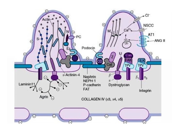
• Now considered a part of a complex of disorders – PODOCYTOPATHIES. • Podocyte injury leads to effacement of the podocyte foot processes. • Change in podocyte shape occurs due to rearrangement of the actin cytoskeleton • E. g. Minimal Change disease and FSGS.
Mechanisms of Disease Podocyte cell changes ↓ Perturbation of podocyte attachment to GBM (α 3β 1 integrin, dystroglycan, podoplanin) ↓ GBM denudation and collapse of capillary loops ↓ Deposition of hyaline material, adherence to Bowman’s capsule and formation of synechiae ↓ Sclerosis (GBM-like material)
Podocyte injury Causes of podocyte injury • Immunologic circulating factors (recurrence of FSGS after Tx) • Genetic mutations (nephrin, podocin, α-actinin-4, WT 1) • Hyperfiltration / stretch (remnant kidney, obesity, reflux) • Ischemia (ageing, hypertensive nephrosclerosis) • Viral infection (HIV, Parvovirus B 19, Simian virus 40) • Toxic agents (heroin, pamidronate)
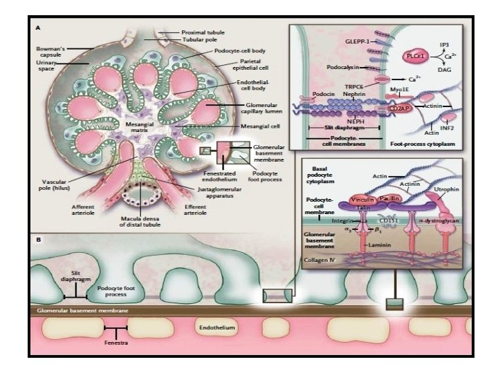
• The normal glomerular capillaries are widely patent and have intact foot processes (or pedicels) along their outer aspect. • Podocytes that are targeted by cellular stresses, such as permeability factors (external causes) or disease causing mutations (intrinsic defects), respond by the reorganization of their actin cytoskeleton, leading to foot-process effacement. • This change in cell shape forms a sheet of undifferentiated cytoplasm over the surface of the glomerular basement membrane.
• If the inciting injurious factors are long-standing or the podocyte is exposed to second hits, a critical level of cell stress is reached and the injured or dying podocyte detaches from the glomerular basement membrane. • Because podocytes are unable to repair by cell division, attrition of a finite number of podocytes leads to sclerosis of the underlying glomerular capillaries, which become obliterated by matrix. • At these sites, adhesions to Bowman’s capsule may form, and parietal cells often migrate onto the tuft, where they lay down loose matrix material. • In the early stages, the sclerotic lesions are typically segmental, involving a portion of the glomerular tuft.
Indirect evidences of immunological basis • Ability to modulate proteinuria by immunoadsorption • Potential disease recurrence minutes after renal transplantation. • Therapeutic reduction in proteinuria by plasmapheresis. • Serum samples from patients with focal segmental glomerulosclerosis cause increased permeability to albumin in isolated glomeruli and induce foot-process effacement and proteinuria when injected into rats.
Suspected plasma factors: • Cardiotrophin-like cytokine 1, a member of the interleukin-6 family, has permeability activity in a plasma fraction with a molecular weight of less than 30 k. D • Elevated serum levels of soluble urokinase receptor (>3000 pg per milliliter) have been identified in up to two thirds of patients with primary focal segmental glomerulosclerosis but not in those with minimal change disease.
• Increased serum levels of soluble urokinase receptor before renaltx- associated with an increased risk of recurrent disease in the allograft. • Circulating soluble urokinase receptor induces footprocess effacement through the activation of podocyte β 3 integrin, and its effect can be blocked in animal models by neutralizing antibodies targeting soluble urokinase receptor. • The cellular source and stimulants of soluble urokinase receptor in patients with focal segmental glomerulosclerosis are unknown.
Viral infections • Viruses can act on the podocyte either by direct infection or by the release of inflammatory cytokines that interact with podocyte receptors. • The best studied - human immunodeficiency virus type 1 (HIV-1), which directly infects podocytes and tubular epithelial cells. • Evidence supports HIV-1 entry by transfer from infected T cells to tubular epithelial cells through virologic synapses formed during cell adhesion, independent of CD 4. • HIV-1 can persist in the kidney epithelium despite antiretroviral therapy and normalization of peripheral CD 4 counts. • HIV-1 gene expression by infected renal epithelium in turn induces dysregulation of host genes.
• In vivo and in vitro models have identified viral genes nef and vpr as particularly important in HIVAN pathogenesis. • Nef, a virulence factor, contains a proline-rich motif that interacts with the SH 3 domain of the Src family kinases. • Parvovirus B 19 is another virus that can infect podocytes and tubular cells, leading to collapsing focal segmental glomerulosclerosis. • Other viruses associated with this disease, such as simian virus 40, cytomegalovirus, and Epstein–Barr virus, are less well characterized.
Adaptive FSGS • Maladaptive responses may arise through a reduction in the number of functioning nephrons or through mechanisms that place hemodynamic stress on an initially normal nephron population (e. g. , in morbid obesity, cyanotic congenital heart disease, and sickle cell anemia) • Biopsy samples obtained from such patients often show enlarged glomeruli, perihilar sclerosis, and relatively mild degrees of foot-process effacement.
• Reflex vasodilatation of both the afferent and efferent arterioles follows a marked reduction in renal mass, causing elevation in the flow rate in the glomerular capillaries. • Because the reduction in vascular resistance is greater in the afferent arteriole than in the efferent arteriole, glomerular hydrostatic pressure rises, producing glomerular hypertension. • These responses cause an elevation in the single-nephron glomerular filtration rate. • Glomerular volume and surface area increase, placing mechanical strain on podocytes that stretch to cover the expanding tuft. • Some hypertrophied podocytes detach, producing denuded patches of glomerular basement membrane. • These sites become covered by parietal cells, leading to the formation of a synechia to Bowman’s capsule and a nidus for the development of segmental sclerosis.
• Although this scenario is the initiating step in the adaptive forms of focal segmental glomerulosclerosis, it may supervene in the later stages of other forms of the disease. • The loss of a critical number of nephrons promotes the activation of (RAS), exacerbating proteinuria and setting the stage for progressive glomerulosclerosis regardless of the initial cause. • Angiotensin II also has direct proapoptotic effects on podocytes. • Excessive protein uptake by podocytes induces podocyte TGFβ, which promotes apoptosis and leads to endoplasmic reticulum stress, cytoskeletal reorganization, and dedifferentiation.
• Drugs that are aimed at the inhibition of RAS (such as angiotensin-converting– enzyme [ACE] inhibitors and angiotensinreceptor blockers) lower intraglomerular filtration pressures through the inhibition of angiotensin II–mediated vasoconstriction of the efferent arteriole. • ACE inhibition also augments bradykinin, which contributes to efferent arteriolar dilatation. • The resulting reduction in proteinuria exerts a protective effect on podocytes and tubular cells
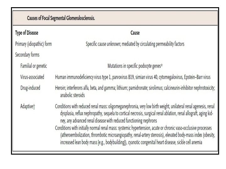
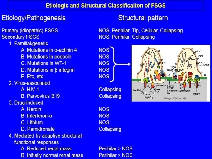
Primary : Idiopathic • Genetic disorders – Slit diaphragm proteins: NPHS 1, NPHS 2, CD 2 AP – Cell membrane–associated proteins: TRPC 6, PTPRO, – LAMB 2, ITGB 4, CD 151, ITGA 3 – Cytosolic or cytoskeletal proteins: ACTN 4, PLCE 1, – MYH 9, INF 2, MYO 1 E, ARHGAP 24 – Nuclear proteins: WT 1, SMARCAL 1 – Mitochondrial components: mt. DNA-A 3242 G, COQ 2, – COQ 6 – Lysosomal protein: SCARB 2 • Circulating pathogenic factor(s)
Secondary • Virus-associated: HIV, parvovirus B 19 • Medication-associated: interferon , , or , lithium, bisphonates, anabolic steroids • Adaptation to reduced kidney mass: oligomeganephronia, unilateral kidney agenesis, kidney dysplasia, cortical necrosis, reflux nephropathy, surgical kidney ablation, chronic allograft nephropathy, advanced chronic kidney disease with reduced functioning nephrons, sickle cell anemia • Initially normal kidney mass: diabetes, hypertension, obesity, cyanotic congenital heart disease • Nonspecific pattern of FSGS caused by kidney scarring in glomerular disease
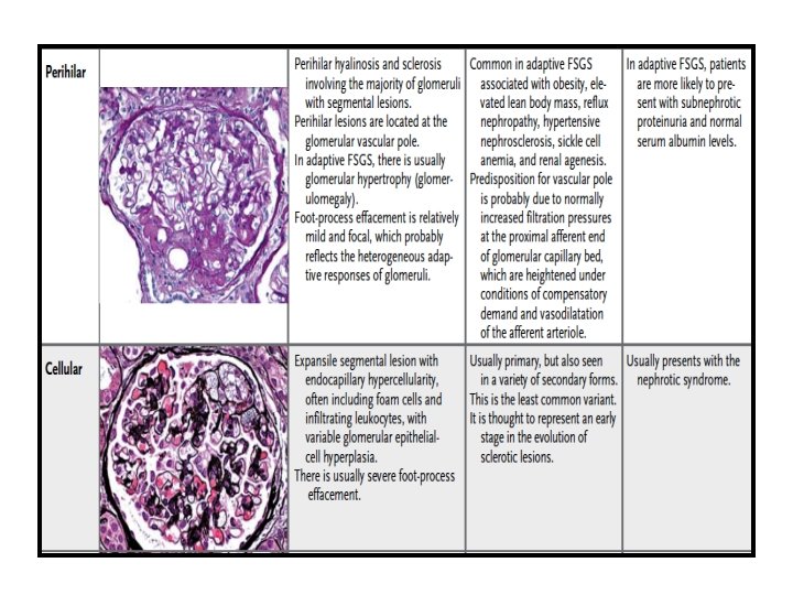
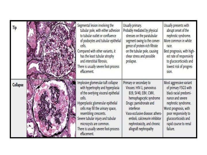
Pathological Classification- (Columbia)
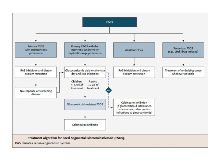
Treatment - KDIGO • Corticosteroid and immunosuppressive therapy be considered only in idiopathic FSGS associated with clinical features of the nephrotic syndrome. • Prednisone be given at a daily single dose of 1 mg/kg (maximum 80 mg) oralternate-day dose of 2 mg/kg (maximum 120 mg). • Initial high dose of corticosteroids be given for a minimum of 4 weeks; continue high-dose corticosteroids up to a maximum of 16 weeks, as tolerated, or until complete remission has been achieved, whichever is earlier.
• Corticosteroids be tapered slowly over a period of 6 months after achieving complete remission. • CNIs be considered as first-line therapy for patients with relative contraindications or intolerance to high-dose corticosteroids (e. g. , uncontrolled diabetes, psychiatric conditions, severe osteoporosis).
Therapy for SR FSGS • Cyclosporine: 3– 5 mg/kg/d: in two divided doses (initial target levels 12 5 to 175 ng/ml ; in case of a remission continue treatment for 1 year then try to slowly taper cyclosporine: reduce cyclosporine dose by 25% every 2 months. • If no remission by 6 months, discontinue cyclosporine treatment. Or • Tacrolimus: 0. 1– 0. 2 mg/kg/d in two divided doses (initial target levels 5– 10 ng/ml And • Prednisone: 0. 15 mg/kg/d for 4– 6 months, then taper off over 4– 8 weeks.
THANK YOU