
z Dr Abeera SarfrazDr Muhammed Zubair KIDNEYS z

z Dr Abeera Sarfraz/Dr Muhammed Zubair KIDNEYS
z INTRODUCTION § The two kidneys function to excrete most of the waste products of metabolism. They play a major role in controlling the water and electrolyte balance within the body and in maintaining the acid–base balance of the blood. The waste products leave the kidneys as urine, which passes down the ureters to the urinary bladder, located within the pelvis. The urine leaves the body in the urethra.
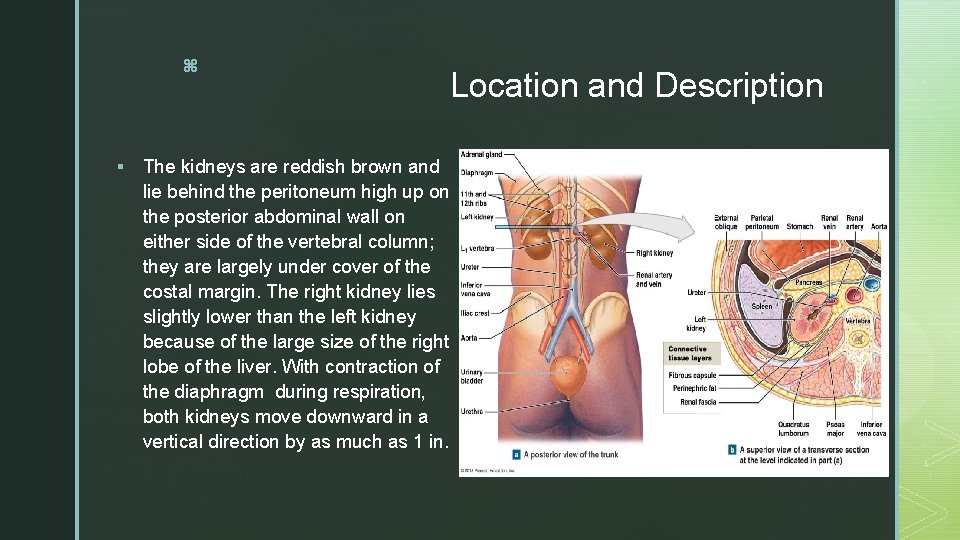
z § The kidneys are reddish brown and lie behind the peritoneum high up on the posterior abdominal wall on either side of the vertebral column; they are largely under cover of the costal margin. The right kidney lies slightly lower than the left kidney because of the large size of the right lobe of the liver. With contraction of the diaphragm during respiration, both kidneys move downward in a vertical direction by as much as 1 in. Location and Description
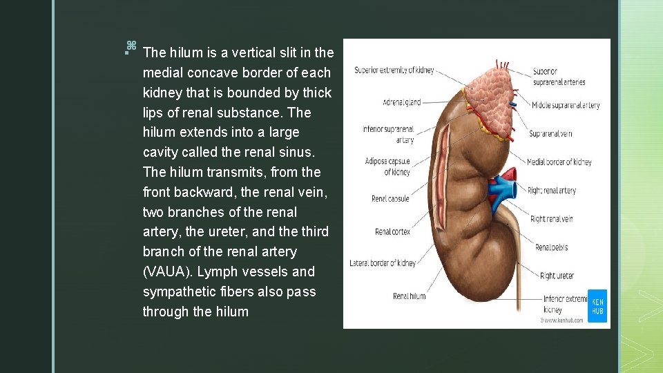
z § The hilum is a vertical slit in the medial concave border of each kidney that is bounded by thick lips of renal substance. The hilum extends into a large cavity called the renal sinus. The hilum transmits, from the front backward, the renal vein, two branches of the renal artery, the ureter, and the third branch of the renal artery (VAUA). Lymph vessels and sympathetic fibers also pass through the hilum
z Coverings § The kidneys have the following coverings: § Fibrous capsule: This surrounds the kidney and is closely applied to its outer surface. § Perirenal fat: This covers the fibrous capsule. § Renal fascia: This is a condensation of connective tissue that lies outside the perirenal fat and encloses the kidneys and suprarenal glands; it is continuous laterally with the transversalis fascia. § Pararenal fat: This lies external to the renal fascia and is often in large quantity. It forms part of the retroperitoneal fat. The perirenal fat, renal fascia, and pararenal fat support the kidneys and hold them in position on the posterior abdominal wall.
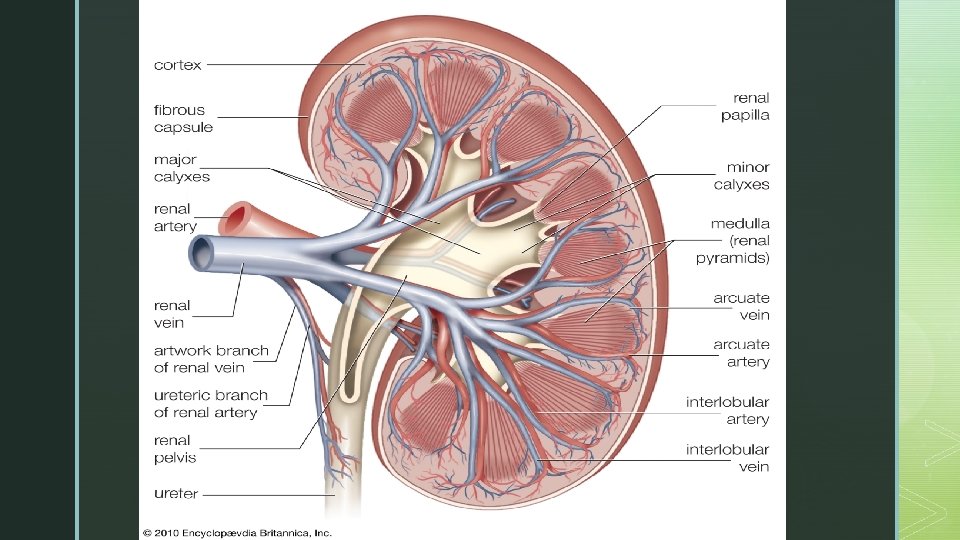
z Renal Structure § Each kidney has a dark brown outer cortex and a light brown inner medulla. The medulla is composed of about a dozen renal pyramids, each having its base oriented toward the cortex and its apex, the renal papilla, projecting medially (Fig. 7. 64 B). The cortex extends into the medulla between adjacent pyramids as the renal columns. Extending from the bases of the renal pyramids into the cortex are striations known as medullary rays. The renal sinus, which is the space within the hilum, contains the upper expanded end of the ureter, the renal pelvis. This divides into two or three major calyces, each of which divides into two or three minor calyces (see Fig. 7. 64 B). The apex of the renal pyramid, the renal papilla, indents each minor calyx.
z Important Relations(Right Kidney) § Note that many related structures are directly in contact with the kidneys, whereas visceral layers of peritoneum separate others. § Anteriorly: The suprarenal gland, the liver, the second part of the duodenum, and the right colic flexure. § Posteriorly: The diaphragm, the costodiaphragmatic recess of the pleura, the 12 th rib, and the psoas, quadratus lumborum, and transversus abdominis muscles. The subcostal (T 12), iliohypogastric, and ilioinguinal nerves (L 1) run downward and laterally
z Important Relations(Left Kidney) § Anteriorly: The suprarenal gland, the spleen, the stomach, the pancreas, the left colic flexure, and coils of the jejunum § Posteriorly: The diaphragm, the costodiaphragmatic recess of the pleura, the 11 th (the left kidney is higher) and 12 th ribs, and the psoas, quadratus lumborum, and transversus abdominis muscles. The subcostal (T 12), iliohypogastric, and ilioinguinal nerves (L 1) run downward and laterally
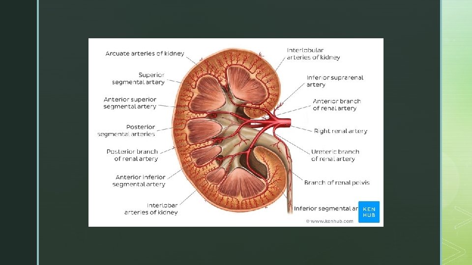
z Blood supply § The renal artery arises from the aorta at the level of the second lumbar vertebra. Each renal artery usually divides into five segmental arteries that enter the hilum of the kidney. They are distributed to different segments or areas of the kidney. Lobar arteries arise from each segmental artery, one for each renal pyramid. Before entering the renal substance, each lobar artery gives off two or three interlobar arteries. The interlobar arteries run toward the cortex on each side of the renal pyramid. At the junction of the cortex and the medulla, the interlobar arteries give off the arcuate arteries, which arch over the bases of the pyramids. The arcuate arteries give off several interlobular arteries that ascend in the cortex. The afferent glomerular arterioles arise as branches of the interlobular arteries. § The renal vein emerges from the hilum in front of the renal artery and drains into the inferior vena cava.
z Lymphatic Drainage § § § Lymph drains to the lateral aortic lymph nodes around the origin of the renal artery. Nerve Supply The nerve supply is the renal sympathetic plexus. The afferent fibers that travel through the renal plexus enter the spinal cord in the 10 th, 11 th, and 12 th thoracic nerves.
z Clinical Notes § Renal Mobility § The kidneys are maintained in their normal position by intra-abdominal pressure and by their connections with the perirenal fat and renal fascia. Each kidney moves slightly with respiration. The right kidney lies at a slightly lower level than the left kidney, and the lower pole may be palpated in the right lumbar region at the end of deep inspiration in a person with poorly developed abdominal musculature. Should the amount of perirenal fat be reduced, the mobility of the kidney may become excessive and produce symptoms of renal colic caused by kinking of the ureter. Excessive mobility of the kidney leaves the suprarenal gland undisturbed because the latter occupies a separate compartment in the renal fascia.
z Kidney Trauma § The kidneys are well protected by the lower ribs, the lumbar muscles, and the vertebral column. However, a severe blunt injury applied to the abdomen may crush the kidney against the last rib and the vertebral column. Depending on the severity of the blow, the injury varies from a mild bruising to a complete laceration of the organ. Stab or gunshot wounds usually cause penetrating injuries and often involve other viscera. Because 25% of the cardiac outflow passes through the kidneys, renal injury can result in rapid blood loss.
z Kidney Tumors § Malignant tumors of the kidney have a strong tendency to spread along the renal vein. The left renal vein receives the left testicular vein in the male, and this may rarely become blocked, producing left-sided varicocele. § Renal Pain § Renal pain varies from a dull ache to a severe pain in the flank that may radiate downward into the lower abdomen. Renal pain can result from stretching of the kidney capsule or spasm of the smooth muscle in the renal pelvis. The afferent nerve fibers pass through the renal plexus around the renal artery and ascend to the spinal cord through the lowest splanchnic nerve in the thorax and the sympathetic trunk. They enter the spinal cord atthe level of T 12. Pain is commonly referred along the distribution of the subcostal nerve (T 12) to the flank and the anterior abdominal wall.
z Transplanted Kidneys § The iliac fossa on the posterior abdominal wall is the usual site chosen for transplantation of the kidney. The fossa is exposed through an incision in the anterior abdominal wall just above the inguinal ligament. The iliac fossa in front of the iliacus muscle is approached retroperitoneally. The kidney is positioned and the vascular anastomosis constructed. The renal artery is anastomosed end to the internal iliac artery, and the renal vein is anastomosed end to side to the external iliac vein
z § Polycystic Kidney A hereditary disease, polycystic kidneys can be transmitted by either parent. It may be associated with congenital cysts of the liver, pancreas, and lung. Both kidneys are enormously enlarged and riddled with cysts. Failure of union between the developing convoluted tubules and collecting tubules is the most likely cause. The accumulation of urine in the proximal tubules results in the formation of retention cysts. § Pelvic Kidney § In pelvic kidney, the kidney is arrested in some part of its normal ascent; it usually is found at the brim of the pelvis (Fig. 7. 70). Such a kidney may present with no signs or symptoms and may function normally. However, should an ectopic kidney become inflamed, it may— because of its unusual position—give rise to a mistaken diagnosis.
z Horseshoe Kidney § When the caudal ends of both kidneys fuse as they develop, the result is horseshoe kidney. Both kidneys commence to ascend from the pelvis, but the interconnecting bridge becomes trapped behind the inferior mesenteric artery so that the kidneys come to rest in the low lumbar region. Both ureters are kinked as they pass inferiorly over the bridge of renal tissue, producing urinary stasis, which may result in infection and stone formation. Surgical division of the bridge corrects the condition. § Unilateral Double Kidney § The kidney on one side may be double, with separate ureters and blood vessels. In unilateral double kidney, the ureteric bud on one side crosses the midline as it ascends, and its upper pole fuses with the lower pole of the normally placed kidney. Here again, angulation of the ureter may result in stasis of the urine and may require surgical treatment.
z Rosette Kidney § Both kidneys may fuse together at their hila, and they usually remain in the pelvis. The two kidneys together form a rosette. This is the result of the early fusion of the two ureteric buds in the pelvis. § Supernumerary Renal Arteries § Supernumerary renal arteries are relatively common. They represent persistent fetal renal arteries, which grow in sequence from the aorta to supply the kidney as it ascends from the pelvis. Their occurrence is clinically important because a supernumerary artery may cross the pelviureteral junction and obstruct the outflow of urine, producing dilatation of the calyces and pelvis, a condition known as hydronephrosis. § § Double Pelvis Double pelvis of the ureter is usually unilateral The upper pelvis is small and drains the upper group of calyces; the larger lower pelvis drains the middle and lower groups of calyces. The cause is a premature division of the ureteric bud near its termination.
- Slides: 19