DIAGNOSIS AND MANAGEMENT OF ACUTE MYELOID LEUKEMIA IN


 and AML (FAB M 0) have")


(p 13; q 13) [")


(q 22; q")
 � t(10; 11) �")


 Response to")


")


 in first induction did")
 study in adults showed that 4")
,")


,")


- Slides: 51
DIAGNOSIS AND MANAGEMENT OF ACUTE MYELOID LEUKEMIA IN CHILDREN AND
Absence of published recommendations specific for pediatric AML motivated an international group of pediatric hematologists and oncologists to develop : Evidence- based and Expert opinion based consensus recommendations Recommendations for specific subgroups are also included
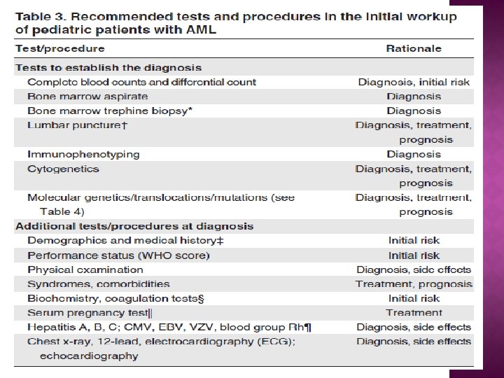
WHO 2008 CLASSIFICATION AND PEDIATRIC AML � The minimal diagnostic requirements in childhood AML are morphology with cytochemistry, immunophenotyping, karyotyping, FISH, and specific molecular genetics in the bone marrow, or peripheral blood � investigation of CNS involvement at diagnosis is not practiced routinely in adults but is considered necessary in children because specific treatment is required in case of CNS involvement � In the event of a dry tap or of a suspected underlying myelodysplastic syndrome (MDS), a bone marrow trephine biopsy has to be performed as well
MORPHOLOGY � The morphologic classification of AML is based on the lineage associated phenotype (undifferentiated, myeloid, monoblastic, erythroblastic, or megakaryoblastic) and defined according to the FAB classification � Morphologic studies reveal the percentages of undifferentiated, granulated or atypical blasts, intracellular structures, such as Auer rods, and presence of myelodysplasia. � Cytochemistry v v confirms lineage affiliation and classifies: myeloid (MPO - positive) and monoblastic differentiation (NSE positive )
DIFFERENTIATION BETWEEN AML AND MDS � Differentiating between AML and advanced MDS may be difficult in children with a low percentage of blasts. � In adults, a blast threshold of 20% is used to differentiate between these diseases, but in children blast percentages between 20% and 30% may be seen in MDS (refractory anemia with excess of blasts in transformation). � AML-specific genetics, hyperleukocytosis, extramedullary disease, and progression within a short time frame (2 -4 weeks) are supportive of AML rather than MDS
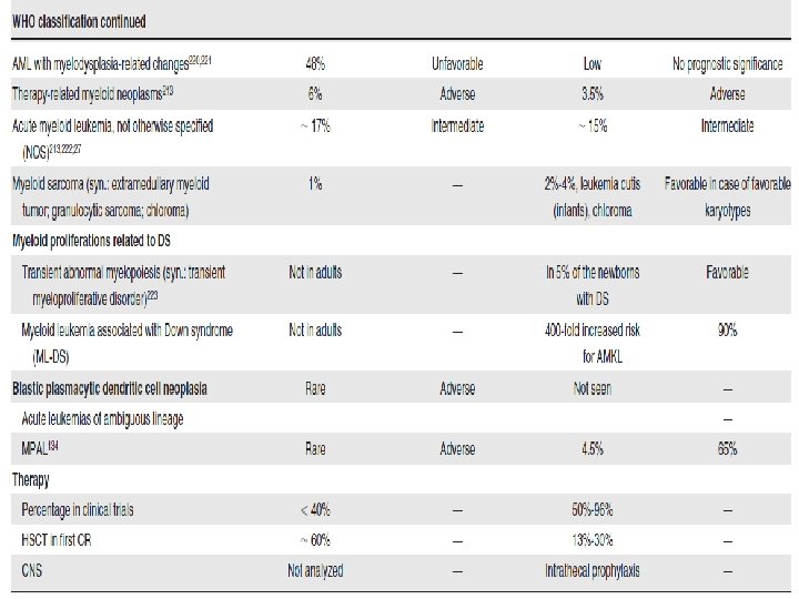
AML should be diagnosed even if the blast threshold of 20% is not reached. Children with Down syndrome t(15; 17), t(8; 21), inv(16), t(16; 16)
� Acute megakaryoblastic leukemia (AMKL, FAB M 7) and AML (FAB M 0) have to be confirmed by immunophenotyping, � The presence of myelofibrosis frequently associated with AML-M 7, may lead to an underestimation of blasts by both morphology and immunophenotyping.
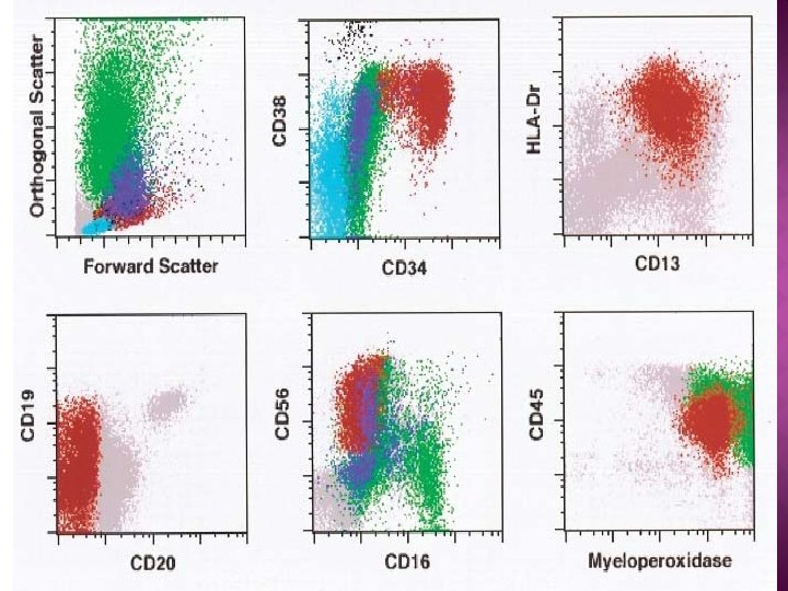
IMMUNOPHENOTYPING � AML - M 0 (negative MPO activity by cytochemistry, but positive by for myeloid markers, such as MPO [proenzyme] and/or CD 13, CD 33, CD 117) � FAB M 7 (positive for Platelet markers, such as CD 41 and/or CD 61)
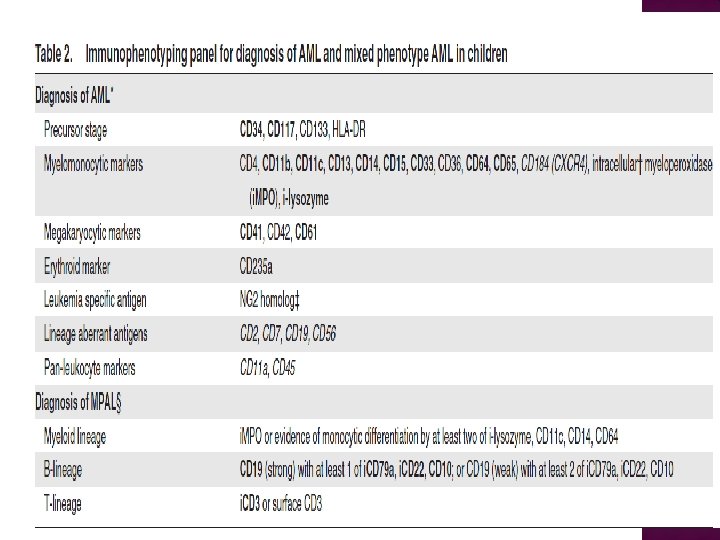
• Immunophenotyping does not usually substitute for morphologic classification of FAB criteria • According to the currently used WHO 2008 classification, markers essential to assign lineage affiliations include: • MPO • Lysozyme • CD 11 c • CD 14 • CD 64 • i(intracellular)CD 3 • CD 19 • i. CD 22 • i. CD 79 a • CD 10
MIXED PHENOTYPE ACUTE LEUKEMIA Includes : � biphenotypic leukemia, � bilineage leukemia with distinctly differentiated blast populations, � undifferentiated leukemia without any lineage commitment
� At present, there is no standardization of antibody panels used for immunophenotyping among the large trial groups � upcoming standards suggest the use of multicolor Monoclonal antibody combinations that include CD 45 to enable optimal gating and analysis of the blast population within the complex context of residual hematopoiesis
RECOMMENDED PANEL FOR AML The mandatory minimal panel required to fulfill WHO and EGIL criteria for AML includes : � CD 34, CD 117, CD 11 b, CD 11 c, CD 13, CD 14, CD 15, CD 33, CD 64, CD 65, i. MPO, i-lysozyme, CD 41, and CD 61; � MPAL: CD 19, i. CD 79 a, i. CD 22, CD 10, and i. CD 3.
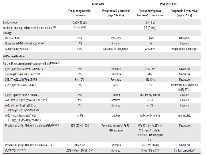
cytogenetics can detect structural and numerical cytogenetic abnormalities in 70%-80% of children with AML. � Conventional � Certain fusion genes, products from translocations, or loss of chromosome material can only be reliably detected using FISH. � The most frequent chromosomal abnormalities in children with AML include : q t(8; 21)(q 22; q 22), q inv(16)(p 13, q 22) (referred as core binding factor [CBF]AML),
ARE MORE PREDOMINANT IN PEDIATRIC AML ARE � t(1; 22)(p 13; q 13) [ RBM 15(OTT)-MKL 1] � the cryptic abnormalities t(7; 12)(q 36; p 13)[ETV 6(TEL)HLXB 9(MNX 1], which are strongly associated with a +19 � t(5; 11)(q 35; p 15. 5)/NUP 98 -NDS 1, predominantly found in cytogenetically normal AML (CN-AML) � t(9; 11)(p 22; q 23)[MLL-MLLT 3], � Monosomy t(10; 11), t(6; 11) 7, monosomy 5/5 q deletions, aberrations of 12 p are rare events (seen in 3%-5% of patients) that occur in
Monosomal karyotypes, which are associated with poor prognosis in adults, are extremely rare in children Trisomies 8 und 21 are often associated with additional aberrations. Cytogenetic abnormalities correlate strongly with age: 50% of infants have MLL-rearranged AML, whereas CBFAML occur typically in older children.
� Routine evaluation should include the evaluation of prognostically relevant genetic aberrations by cytogenetics/FISH, including at least the following fusion genes at diagnosis: � RUNX 1 -RUNX 1 T 1 [t(8, 21)] � CBFB-MYH 11[inv(16)] � PML-RARA � MLL rearrangements. � Other rare fusion genes mentioned in Table 4 should be traced to determine adverse risk patients.
MOLECULAR GENETICS AML is thought to result from at least 2 classes of mutations: � type I mutations inducing proliferation, such as abnormalities in tyrosine kinases, � type II mutations, inducing maturation arrest, comprising most of the translocations. � In CN-AML, several mutations, such as NPM 1, FLT 3, WT 1, and biallelic CEPBA mutations, are clinically relevant and should be included in standard diagnostics
� Mutations in the WT 1 gene are found mainly in CN-AML and are often associated with FLT 3 -ITD mutations � The frequency of activating mutations of tyrosine kinase receptor genes, such as FLT 3 increases with age. � FLT 3 mutations predominantly occur in CN-AML, t(15; 17) and t(5; 11) � Point mutations in the activating loop domain of the FLT 3 receptor are mutually exclusive of FLT 3 -ITD mutations (frequency 2%-8% in children)
� C-KIT mutations occur in 25% of children with CBFAML, but in only 5%-8% of those with other leukemia types � MLL-PTDs � Mutations are rare in childhood AML in genes involved in the RAS-RAF-ERK signal transduction pathway occur in 5%-21% of children with AML, more frequently in those with CBF-AML, and in young children with MLL- rearranged. AML
ﺳﻄﻮﺡ ﺍکﺴپﺮﺷﻦ ﺍﺑﻨﺮﻣﺎﻝ ﺩﺭ ژﻦ ﻫﺎ � In addition to mutations aberrant expression levels of genes have recently been reported in both adults and children; however, the biologic and clinical relevance might differ � BAALC and ERG overexpression is associated with CN- AML, � EVI 1 expression, and inv(3), rarely occurs in children but is mainly found in association with t(6; 11) and M 6/7
RECOMMENDATION Routine evaluation should include the evaluation of a prognostically relevant and potentially targetably selected set of molecular genetic markers FLT 3 -ITD, WT 1, C-KIT, CEBPA (double mutation), NPM 1, and further specific MLL-abnormalities with Favorable or very poor prognosis (eg, MLLAF 1 Q, AF 6, AF 10)
PROGNOSTIC SIGNIFICANCE � The most relevant factors are genetic abnormalities and treatment response, with differences between adult and childhood AML. � In the AML- (BFM) , age could not be used as an independent prognostic factor in infants and adolescents � Very high blast counts at diagnosis are associated with an increased risk of early death and nonresponse, but
PROGNOSIS ACCORDING CYTOGENETIC � As in adult AML, CBF-AML and t(15; 17)(q 22; q 21) in children are highly predictive of a favorable outcome. � Translocation t(1; 11)[MLL-MLLT 11]is a newly described translocation associated with favorable outcome in childhood AML � Although prognosis of different MLL fusions is heterogeneous
CYTOGENETICS INDICATING AN ADVERSE OUTCOME � -7 � t(6; 11) � t(10; 11) � t(7; 12) � t(6; 9) � t(5; 11) and � other rare abnormalities, such as 12 p � Adverse cytogenetics described in adult. AML, such as 5 q- , inv(3)(q 21 q 26. 2) or t(3; 3), are very rare in children.
� Intermediate risk factors include normal and other karyotypes. � However, CN-AML has been shown to be a heterogeneous disease and the clinical outcome highly dependent on the presence of additional molecular aberrations
MOLECULAR GENETICS � In CN-AML, single-gene mutations are of specific interest, especially the NPM 1 and biallelic CEPBA mutations, as they are associated with favorable outcome. � In contrast, a FLT 3 -ITD mutant /wild-type ratio of > 0. 4 has been associated with adverse outcome � Coincidentally occuring translocations such as t(5; 11) or mutations such as WT 1 or NPM 1, can modify the prognostic relevance of the FLT 3 -ITD
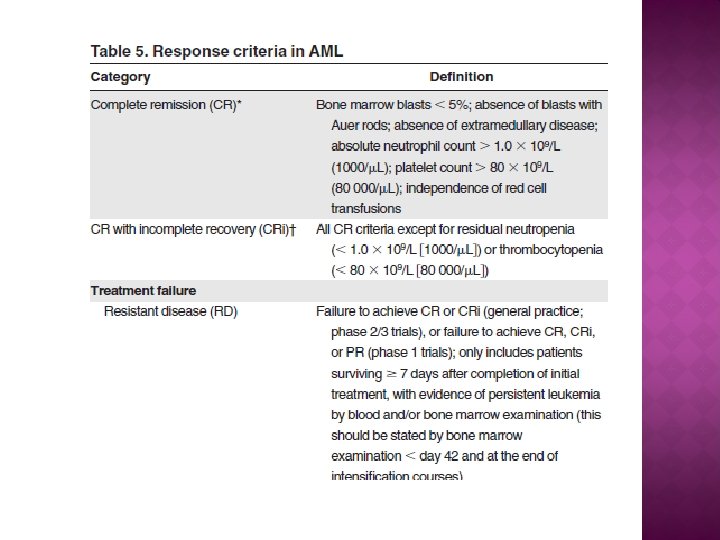
RESPONSE AND PROGNOSIS THE 2 MOST IMPORTANT INDICATORS OF OUTCOME � 1) Response to the first course of treatment and � 2) cytogenetics and molecular genetics. Both are independent prognostic factors and are usually essential elements of the risk group classification � Most study groups evaluate treatment response morphologically in the bone marrow after the first (eg, on day 15 or day 28) and second induction courses. � This may be challenging in hypoplastic bone marrows. � Blast after cell reduction until day 15 and treatment response
MONITORING OF RESIDUAL DISEASE � Residual disease can be monitored by morphology, immunophenotyping, and quantification of molecular aberrations and gene expression levels � Depending on the method and the informative marker used, a single approach may not meet features of all patients.
MRD ASSESSMENT BY IMMUNOPHENOTYPING � can be done in up to 96% of children with AML � heterogeneity of leukemia-associated immunophenotypes and frequent antigen shifts over time limits the sensitivity and specificity of immunophenotypic detection of MRD � Current technologic advances, such as 6 -color flow cytometry, may overcome any limitations.
MRD assessment by fusion genes o. The high specificity and sensitivity (up to 105) of real-time quantitative PCR of AML fusion genes of RUNX 1(AML 1)-RUNX 1 T 1(ETO), CBFB-MYH 11, PML-RARA, and MLLT 3(AF 9)-MLL lend themselves to MRD monitoring but are applicable in only 35% of pediatric patients. o. Importantly, the kinetics of relapse differs between genetic subtypes with a median time from molecular to clinical relapse between 2 and 8 months. ospecific mutations, such as NPM 1, FLT 3 -ITD, or GATA 1 s, have been established in childhood AML,
Management o. Children with AML should be treated within controlled clinical trials. Treatment of childhood AML requires an intensive anthracycline- and cytarabine-based therapy using at least 4 or 5 courses.
Induction 2 courses of induction therapy o. Standard induction therapy comprises 3 days of an anthracycline(eg, daunorubicin at least 60 mg/m 2, idarubicin 10 -12 mg/m 2, or the anthracenedione mitoxantrone 10 -12 mg/m 2) and 7 -10 days of cytarabine (100 -200 mg/m 2 continuously or twice daily intravenously; o. Although a third drug, such as etoposide or 6 thioguanine, is commonly included in induction, their benefit has not been proven.
Anthracyclines o higher doses of anthracyclines improve outcome in children and adults. However, toxicity, especially acute and late cardiotoxicity, o. Cumulative dosages > 300 mg/m 2 have been associated with significant later cardiac toxicity. o. Anthracyclines with a low cardiac exposure, such as liposomal anthracyclines, o. Cardioprotection with dexrazozane was another option to reduce cardiotoxicity during anthracycline exposure.
Dosage of cytarabine. oinduction ØThe use of high-dose cytarabine (Hi-DAC) in first induction did not improve the CR rate or survival in adults or children. ØOne or 2 courses of induction therapy comprising 3 days of an anthracycline and 7 -10 days of cytarabine should be applied.
Consolidation/intensification. ØThe Cancer and Leukemia Group B (CALGB) study in adults showed that 4 courses of Hi. DAC (3 g/m 2 per every 12 hours on days 1, 3, and 5) were superior to 4 courses of lower-dose (100 mg/m 2 continuous intravenously on days 1 -5) cytarabine. Øshow that relapse rates can be reduced by introducing intensive chemotherapy courses that include Hi. DAC.
Additional agents. ØOther drugs that have been used during induction include aclarubicin, amsacrine (adults), mitoxantrone(children and adults), and 2 chlorodeoxyadenosine (children). ØIt is not clear whether these agents improve early treatment response, event-free survival, or overall survival compared with daunorubicin plus cytarabine at equivalent doses.
Postremission strategies Consolidation/intensification ØIn most pediatric studies, 2 to 5 courses of chemotherapy with non–cross-resistant drug combinations ØHigh-dose cytarabine.
HSCT. Autologous HSCT. v there is a role for auto-HSCT in relapsed APL without detectable MRD. Allogeneic HSCT. v. Auto-HSCT is not recommended for children with AML in first CR. Allo-HSCT in first CR is not beneficial in childhood. AMLwith favorable risk factors. In other risk groups, the benefit of allo. HSCT must be balanced against toxicity. Allo-HSCT in second CR is generally considered.
CNS-directed therapy o. CNS involvement at diagnosis and at relapse is seen in 5%-10% of pediatric patients with AML. o. Factors associated with CNS leukemia include hyperleukocytosis, monocytic leukemia [FAB M 4 or M 5, including M 4 eo with inv(16)], MLL gene rearrangement, and younger age.
o. CNS treatment has varied from intrathecal chemotherapy (single-agent cytarabine or methotrexate, or triple cytarabine, methotrexate, and hydrocortisone) alone or given in combination with cranial radiotherapy ohe optimal number of intrathecal treatments (range 4 -12) remains unknown.
o. In contrast to ALL, CNS positivity is not a crucial factor within the AML risk group stratification because it does not affect overall survival. o. However, those with CNS involvement (as defined in “Diagnostic procedures and initial workup”) relapse more frequently in the CNS.
Hematopoietic growth factors as priming agents Sensitization of leukemic cells with hematopoietic growth factors(priming), such as G-CSF and GM-CSF, has been studied predominantly in adults with the aim of increasing cytotoxicity of chemotherapy.
Relapsed and primary refractory. AML o. Approximately 5% of children with AML have refractory disease and 30% experience relapse. o. Bone marrow is the most common site of relapse, o. Comparing fludarabine/cytarabine/G-CSF with the addition of liposomal daunorubicin showed a second CR rate of 59% and 69%, respectively,
New therapy approaches ØNew compounds, such as epigenetically active agents, tyrosine kinase inhibitors, and antibodymediated treatment, Antibody-targeted drugs: üGemtuzumab ozogamicin (GO), a calicheamicinconjugated CD 33 antibody, has shown promising results in children with üpart of patients in the intermediate-risk group; however, there was no benefit to those in the adverse-risk group.
Tyrosine kinase inhibitors AML patients with activating FLT 3 or KIT mutations are candidates for targeted therapy. combining sorafenib and conventional chemotherapy in childhood AML, with some evidence of efficacy limited to patients with FLT 3 ITD.