Chapter 2 Epithelial Tissue Guanhua Huo Department of

 means \"on\"")
, Øonly a small amount of intercellular")
: lines both the outside (skin) and")


![C. Simple columnar epithelium Top view: polygonal [pə'lɪɡənl] ; side: column-like, prismatic form Special](https://slidetodoc.com/presentation_image_h2/1ea28d958e5f96d5d295319ab1b35f8d/image-17.jpg "C. Simple columnar epithelium Top view: polygonal [pə'lɪɡənl] ; side: column-like, prismatic form Special")
 are viewed")


")


 enter bloodstream directly. To")

")

; EM: 9+2 microtubules connected to the")


 Location: around the apex of cells Structure: belt-shaped")


- Slides: 67
Chapter 2 Epithelial Tissue Guanhua Huo Department of Histology & Embryology BMU
I. OUTLINE v Epithelial Tissue is commonly named Epithelium: In Greek ("epi") means "on" or "upon", and ("thēlē") means "nipple“ v Epithelium: is made up of cells closely packed v and ranged in one or more layers; forms the covering or lining of all internal and external body surfaces, as well as cavitys and lumen of organs.
1. Construction features Ø have more cells(arranged tightly), Øonly a small amount of intercellular substance, Øranged in one or more layers; Free (apical) surface –apical pole Ø the cell have polarity Basal surface -basal pole basement membrane: provides structural support for the epithelium and also binds it to neighbouring structures. Øno blood vessels-avascular: nutrients and precursors of products of the epithelial cells diffuse across BM and are taken up through the baso-lateral surface of the epithelial cells through the basement membrane Ørich in nerve endings: conea, epidermis 2. Distribution features line cavities and surfaces of structures throughout body surface-covering ep. inner surface of various kinds of tubes, cavities and sacs
3. Classification A. lining epithelium (covering epi. ): lines both the outside (skin) and the inside cavities and lumen of bodies. B. glandular epithelium myoepithelium C. special epithelium 4. Function germinal epi. sensory epi. protection: physical / chemical / microorganism /desiccation secretion: mainly for glandular epi. absorption: small intestine / renal tubule excretion: sweat gland / renal tubule others: Sensation/ produce germinal cells, transcellular transport
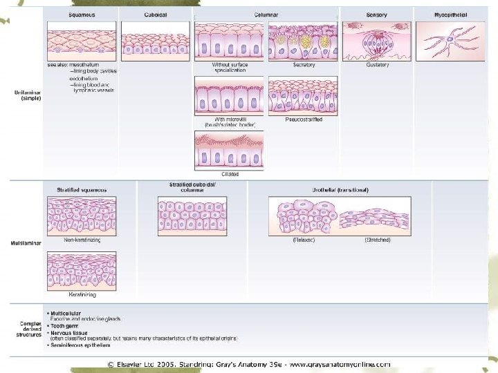
II. Covering epithelium 1. Principles of classification: Tissues are generally classified by the morphology of their cells, and the number of layers they are composed of.
Simple epi. Stratified epi.
2. Classification & structure Simple epi. s. squamous epi. s. cuboidal epi. s. columnar epi. pseudostratified ciliated columnar epi. st. squamous epi. Stratified epi. st. columnar epi. transitional epi.
A. simple squamous epithelium v v Top view: scales-like; have an irregular shape with a serrated border side view: flat
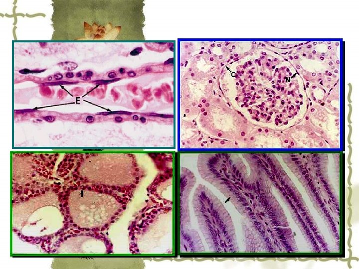
Location: Endothelium: inner lining of blood vessels, heart, and lymphatic vessels Mesothelium: walls of pericardia, pleura, and peritoneal cavities, secrete a lubricating fluid, called serous['sɪrəs] fluid Others: walls of alveoli of the lungs , parietal [pə'raɪətəl] layer of Bowman's capsule some renal tubule
alveoli of the lungs
parietal layer visceral layer Renal Corpuscle: Note the flattened cells of the parietal layer of Bowman's capsule.
B. Simple cuboidal epithelium v v Top view: hexagonal; side view: square Location: thyroid follicles; renal tubules
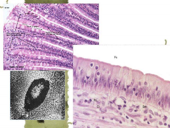
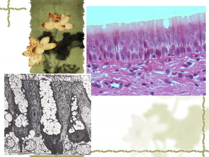
C. Simple columnar epithelium Top view: polygonal [pə'lɪɡənl] ; side: column-like, prismatic form Special structure: striated ['straɪeɪtɪd] border, brush-like border Cytoplasmic projections Location: gastrointestinal tract; gall bladder; uterus
D. Pseudostratified ciliated columnar epithelium Taller simple epithelial cells (see columnar, below) are viewed in cross section with several nuclei appearing at different heights, they can be confused with stratified epithelia. This kind of epithelium is therefore described as "pseudostratified" epithelium. fusiform cell pyramidal cell Ø 4 types of ell, Nu. at different levels
b c c f
Location: respiratory passages & epididymis.
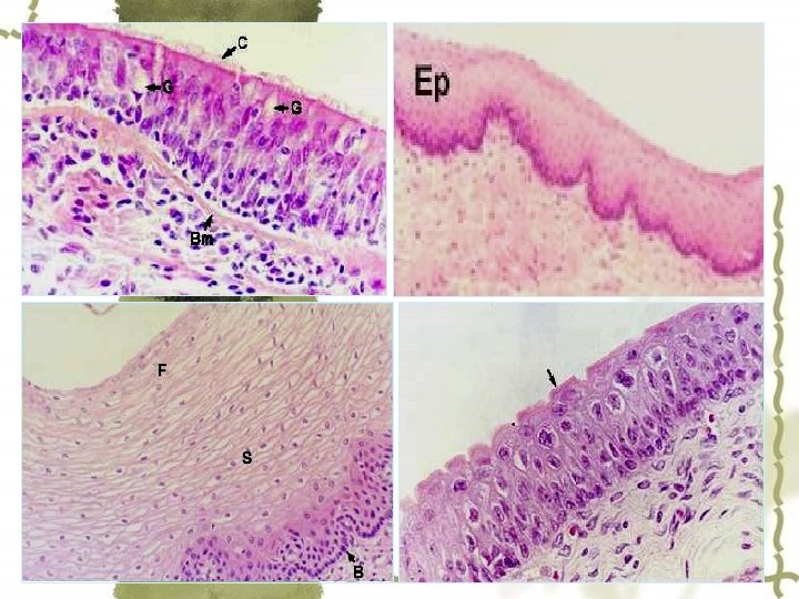
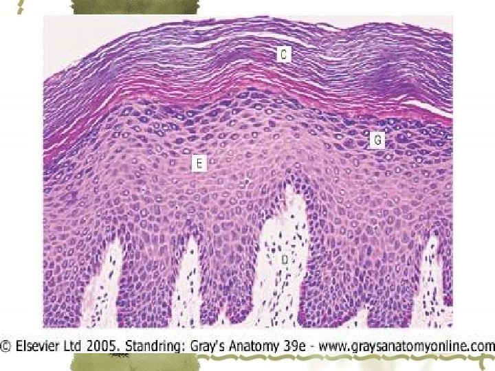
E. Stratified squamous epithelium Basal layer: low columnar on B. M. ; Middle layers: polygonal and larger; Top layers: flat / largest and scale-like platycyte polygonal cell basal layer cell B. M.
Location: Nonkeratinized: mouth / pharynx/ esophagus/ vagina; Keratinized: epidermis
stratified columnar epithelium Columnar cells + Layers of polygonal cells Location: ocular conjunctiva, large ducts of salivary glands
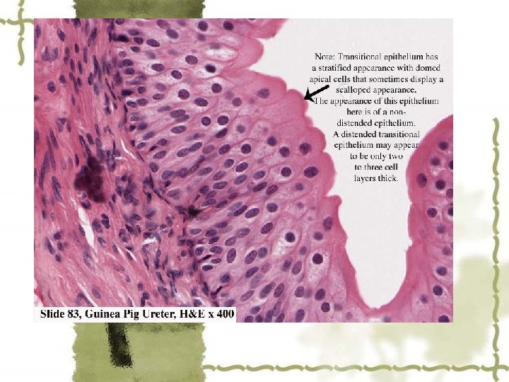
F. Transitional epithelium Different functional state has different cell layer & cell-shape. surface layer: cap cell Contracted Distended
Location: urinary bladder Full state: 2 -3 layers empty state: 5 -6 layers
III. Glandular epithelium & gland 1. Definition Ø Glandular epithelia. : The epithelia specialized for secretion Ø Glands: The organs composed mainly of glandular epi. 2. Classification Ø Exocrine gland Ø Endocrine gland
3. Development Derived from the localized covering epithelium retain obliterate The agglomerated cells form anastomosing cords interspersed between dilated blood capillaries. The others, cells line a vesicle or follicle filled with noncellular material.
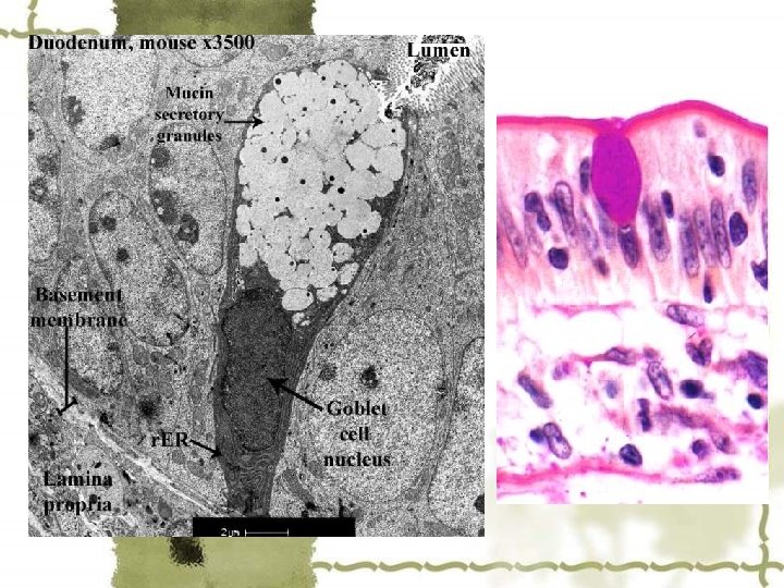
4. Exocrine gland v Classification:Depend on number of cells Unicellular glands: goblet cell Multicellular glands secretory unit duct system
v Section of large intestine showing goblet cells secreting mucus to the extracellular space. The mucus precursor stored in the cytoplasm of the goblet cells is also stained in a dark color. PAS-PT stain. Medium magnification.
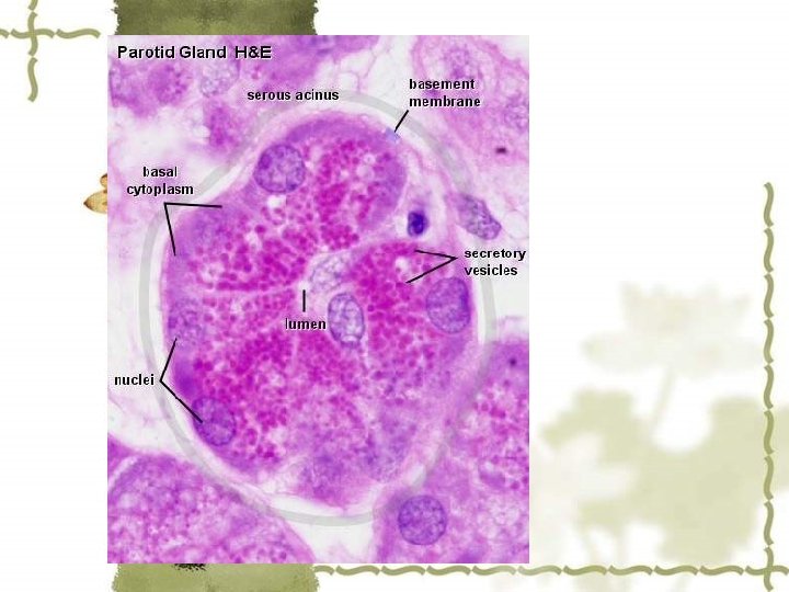
Apical portion: Large, lightly staining granules containing strongly hydrophilic glycoproteins-mucin. mucous acini
Parotid Gland, Human and Sublingual Gland, Human - H&E serous acini
Electron micrograph of a pancreatic cell. Note the nucleus, mitochondria, Golgi complex, secretory (zymogen) granules in various stages of condensation, and rough endoplasmic reticulum. x 13, 000.
Ø Depend on shape: Tubular glands Alveolar glands Tubulo-alveolar glands
Ø Depend on function: Serous glands, mucous glands, mixed glands
5. Endocrine gland Consist of endocrine cells; the secretions (hormone) enter bloodstream directly. To be discussed more detail late.
IV. Special structure of epithelium 1. Cellular free surface cell coat microvillus cilium A. Cell coat: extracellular glycoprotein layer
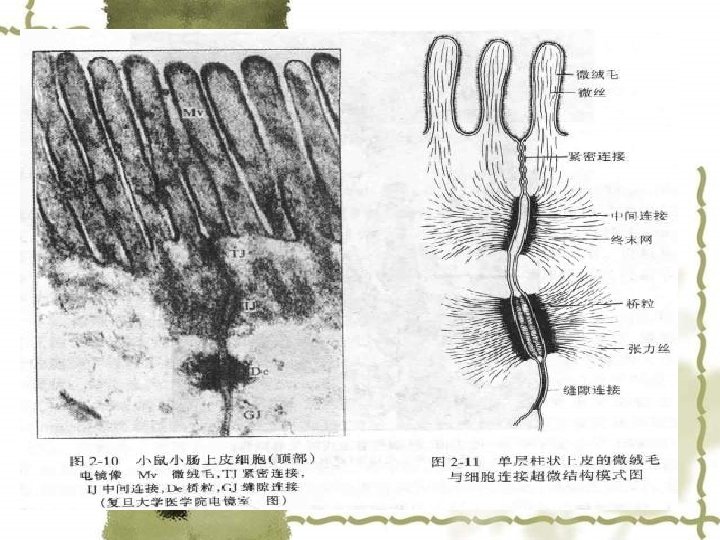
B. Microvilli LM: striated borders brush-like borders EM: finger-like projections (cell -membrane & cytoplasm) (0. 1/1. 4 μm) microfilaments (actin filaments): interconnect terminal web Function: increase the surface area of absorption
Cell coat Electron micrograph of the apical region of an intestinal epithelial cell. Note the terminal web composed of a horizontal network that contains mainly actin microfilaments. The vertical microfilaments that constitute the core of the microvilli are clearly seen. An extracellular cell coat (glycocalyx) ['glaɪkoʊ'kælɪks] is bound to the plasmalemma of the microvilli. x 45, 000.
C. Cilium LM: elongated & mobile projections(0. 2/510μm); EM: 9+2 microtubules connected to the basal body (the centrioles)
inner dynein arm microtubule radial spoke central microtubule Cilia have a rapid back-and-forth movement- outer dynein arm permit a current of fluid or particular matter to be propelled in one direction over the ciliated epithelium--ATP
Ciliary ultrastructure, Left, Normal cilium from a healthy individual in which both inner and outer dynein arms can clearly identified. Right, the absence of outer and inner dynein arms in a patient with primary ciliary dyskinesia [dɪskɪ'ni: ʒə].
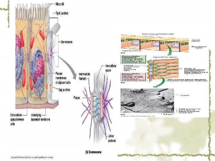
2. Lateral surfaces A. B. C. D. Tight junction Intermediate junction Desmosome Gap junction
A. Tight junction (zonula occludens ) Location: around the apex of cells Structure: belt-shaped anastomosing network of ridges formed by fused protein in adjacent cellular membrane Function: form a seal & a barrier to prevention. Zonula: form a band completely encircling occludens: membrane fusion that close off the intercellular space
B. Intermediate junction, zonula adherens Location: around adjacent cells, just below T. J. Structure: a gap: 15 -20 nm, amorphous material (glycoprotein) electron-dense material: on the cytoplasmic faces filaments: numerous, being continuous terminal web Function: hold adjacent cells Firmly, maintain the cell shape, and pass cellular contract forces
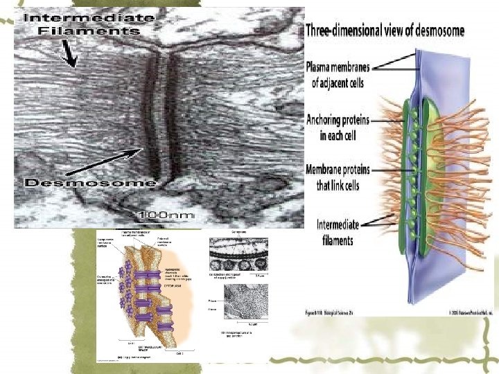
C. Desmosome, macula adherens Location: along the intercellular faces, widely distributed, plate-shaped Structure: a gap: 20 -30 nm, filled with extracellular material & an intermediate line attechment plaque: electron dense, on inner surface of cell-membrane tonofillaments: a great number, inserted into the plaque, hairpin loop-like Function: “spot weld [weld] ” type attachment, provide a firm adhesion
D. gap junction Location: deep part Structure: a narrow gap: 2 -3 nm, spanned by connexons in opposing cell membranes connexons(D=7 -8 nm): 6 subunits of protein , a central channel (2 nm); Function: communication (transfer the impulses; passion & molecules) ; rich in cardiac muscles and neurons.
E. Junction complex 2 or more types of junction present collectively in same area
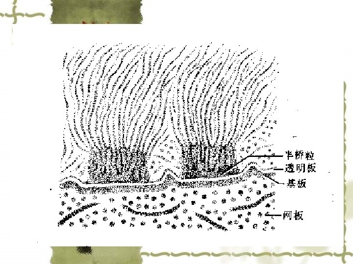
3. Basal face A. Basement membrane B. Plasma membrane infolding C. Hemi-desmosome
A. Basement membrane Location: between epi. & underlying C. T. 50 -300 nm thick, PAS +, argyrophilia
Structure: Basal lamina: glycoprotein; produced by epi. cells. Reticular lamina: reticular fibers + matrix, formed by fibroblasts in C. T. Main chemical components: polysaccharide & glycoprotein (Laminin, type Ⅵ collagen etc. ) Basal Lamina lucida lamina Lamina densa reticular lamina
lamina lucida lamina densa basal lamina reticular lamina Function: support, connection, serve as a semi-permeable membrane, facilitate the proliferation and differentiation of cells
B. Hemidesmosome Function: enhance the cell adhesion to the B. M. hemidesmosome basal lamina reticular lamina
Hemidesmosome & Desmosome
C. Plasma membrane infolding Structure: infolding membrane mitochondria, Location: renal tubules Function: increase the basal surface area & enhance the reabsorption of water & ions
Proximal tubule kidney. Basal membrane is highly infolded. Note fenestrated capillary at right.