
Epithelial and membrane transport G Ogweno Types of
Epithelial and membrane transport G Ogweno
Types of epithelial tissues • Surface linning • Glandular • Cell shape: squamaous, cuboidal, columnar • Layers: simple, stratified, pseudostratified • Examples: skin, intestine, respiratory, uroepithelium

Epithelial polarity • Apical surface: microvilli absorptive, cilia motile • Lateral junctional complexes • Basal: basal lamina, basement membrane, basal infoldings for ion transport, hemidesmosomes for fixation • Distribution of enzymes, transporters, Antigen detection, signal transduction
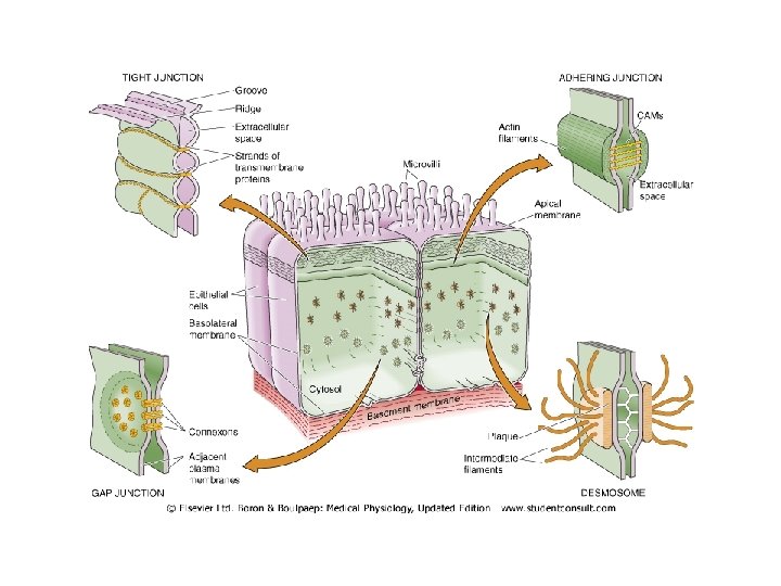
Epithelial junctional complexes. Tight • Also called zonula occludens • Integral membrane proteins- occludins, claudins • Determines permeability characteristics& cell migration • On apical side-continuous belt, divides into apical and basal domains (fencing) • Prevents Para cellular transport and binds cells together; barrier; gating • Common intestinal and skin
Epithelial junctions: adhering • • Also zonula adherens Belt just below tight junctions Cell-cell contact by CAMs Cytoskeleton-actin
Epithelial junctions: Gap • • 2 nm gap between adjacent cells Hollow cylindrical structures-connexons Form channels-Interecellular Point of electrical coupling : low -resistance communication, • may be gated-responsive to physiological stimuli • Nexus in smooth muscles; intercalated disc in cardiac muscles
Epithelial junctions: desmosomes • Also called spot, macula adherens, button like between adjacent cells • Intermediate filaments-tonofilaments cytoskeleton of adjacent cells • Gap between of 25 -30 nm • Provide structural support to physical stress • Prevent shearing forces e. g epidermis
Functions of epithelia • • Body barrier e. g microorganisms Prevents water loss/dessication Maintenance of internal environment Regulated vectorial transport
Types of capillaries • Type 1: continuous-10 -15 nm clefts, much tighter in BBB. found pulmonary, brain, testis • Type 2: fenestrated-thin fenestrated. small intestine, exocrine glands, muscles • Type 3: discontinuous-large gaps, can pass cells, sinusoids e. g liver, bone marrow
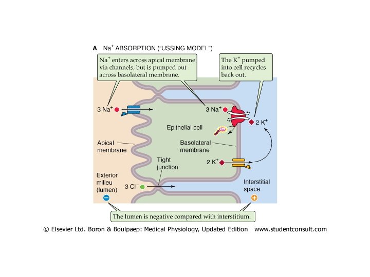
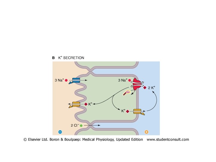
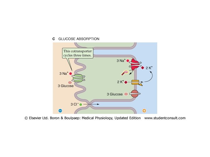
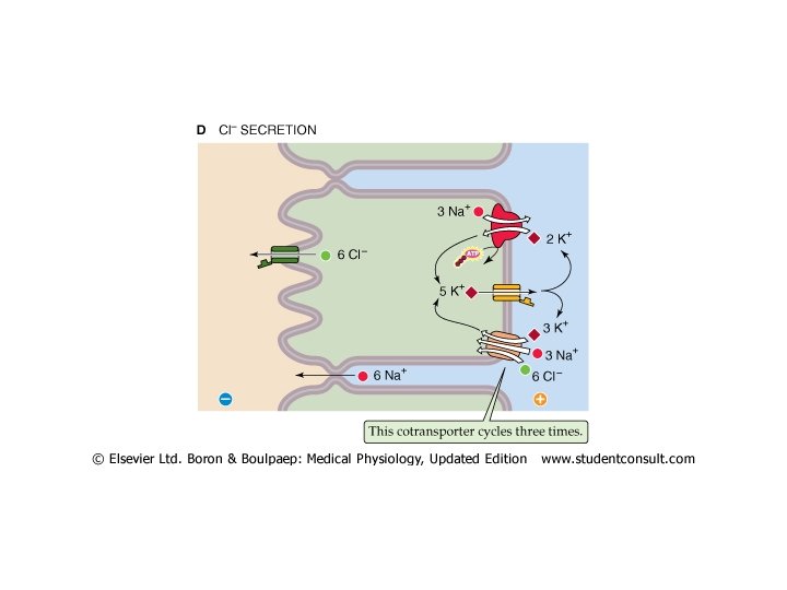
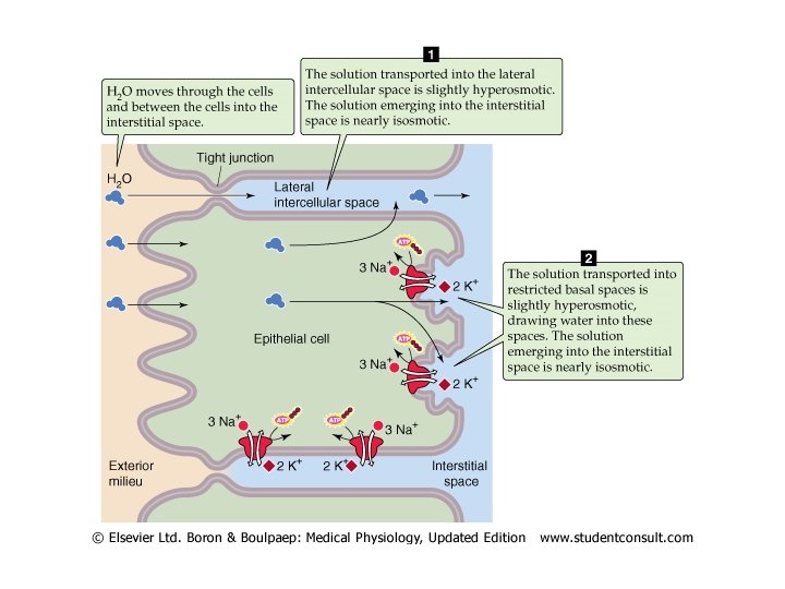
Epithelial vectorial transport • • • Function of membrane transport protein polarity Apical- absorption or reabsorption; basolateral-secretion Create ionic gradients Both Transcellular(tight) and paracellular (leaky) transport depending on epithelium Are specific for ions-cations vs anions Transport proteins(channels, carriers&pumps) and kekyness determine direction and type of solutes transport Active transport is transcellular while paracelluler is passive Na+/K+ ATPase exclusively basolateral except in choroid plexus apical K+ through channels recycled Inwardly directed Na+ gradient driving force for apical Na+ channels, secondary active transport for other solutes e. g Na+glucose. Na-H+ exchanger
Epithelial membrane solute transporters • Placed either on apical or basolateral membranes control absorption or secretion • Na+: basolateral Na+/K+ ATPase pumps out, creates inward gradient, Cl- paracellular • K+: apical channel eflux; basal Na+/K+ • Glucose: apical secondary active transport(SGLT), basal carrier exit(Glut) • Cl-: secondary active uptake basal Na+/K+/Clcontransporter, passive cl-apical exit through channels e. g CFTR; Na+ paracellularly into lumen
Transepithelial water movement • Passive, driven by osmotic pressure gradient • Transepithelial(transcelluler): aquaporins • Paracelluler(junctional): follows Na. Cl gradient(secretion/reabsorption)& solvent drag • Some eipthelia have high solute-transport and low water permeability • Hydraulic conductivity varies • Isotonic movement occurs due to constitutive expression of aquaporins and localized interepithelial osmotic gradient
Regulation of epithelial transport • Controlled by Neural/hormonal signals • Neural: enteric&sympathetic • Hormonal: Aldosterone • Paracrine: gastric histamine for HCl Mechanisms: 1. Gene expression: Synthesis/degradation of specific transporters 2. Change in activity of membrane transporters/post-translational modification of pre-existing transport proteins(channel gating) 3. Recruitment/Retrieval of transporters from membrane by endocytosis or insertion into membrane from intracelluler vesicular pool 4. Change in paracellular(leak) pathway 5. Luminal supply of transported species
- Slides: 19