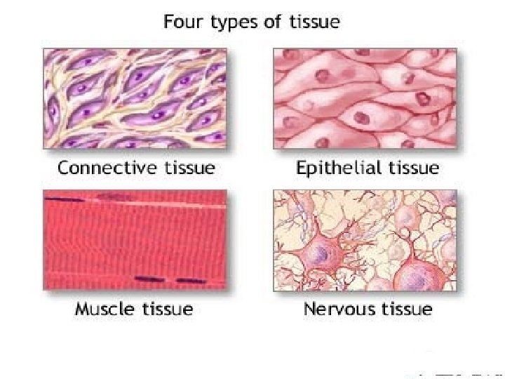
ANIMAL TISSUE TYPES OF ANIMAL TISSUES EPITHELIAL TISSUE


 EXOCRINE GLANDS: Products")


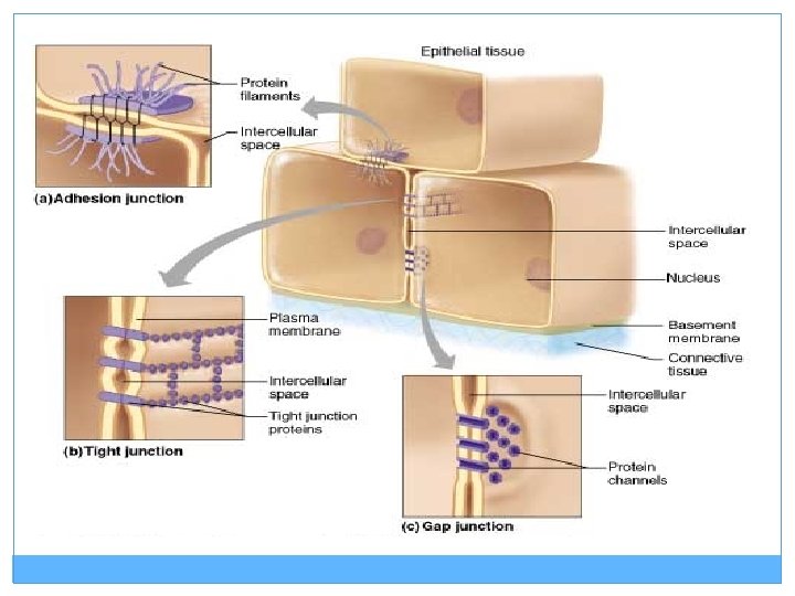
 TIGHT JUNCTION: Plasma")


")


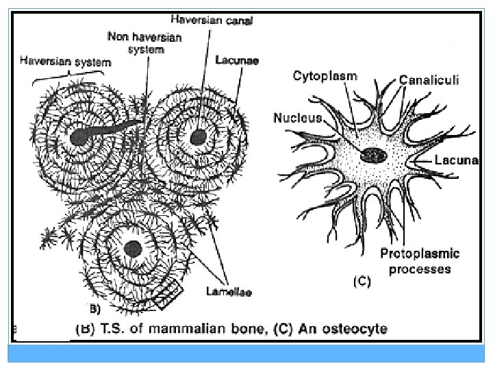
 are found in Lacunae within the matrix. Intercellular")
. Bone cells (osteocytes) present in spaces")


 SKELETAL MUSCLE (STRIATED,")
 Cells are spindle shaped or fusiform. Prominent nucleus present in")


- Slides: 51
ANIMAL TISSUE
TYPES OF ANIMAL TISSUES
EPITHELIAL TISSUE General Characteristics: q. Found throughout the body, covers all body surfaces both inside and out q Cells arranged compactly with very little extracellular material. q Attached to underlying connective tissue by non-cellular nonliving basement membrane. q Usually has no vascular tissue - blood supply.
EPITHELIAL TISSUE
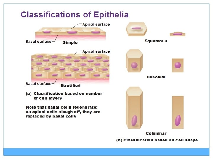
DIFFERENCES BETWEEN SIMPLE AND COMPOUND EPITHELIUM SIMPLE EPITHELIUM COMPOUND EPITHELIUM 1. There is a single layer of cells. 1. There are several layer of cells. 2. All the cells directly rest on 2. Only the basal layer of cells the basement membrane rest on the basement membrane. 3. Efficient in absorption and 3. This has very little role in secretion and absorption. 4. Not efficient in protecting the underlying tissues 4. Provides protection against mechanical , chemical , thermal and osmotic stresses.
SIMPLE SQUAMOUS Thin , flat and have irregular boundaries, that fit closely into those of the neighbouring cells. Forms the inner lining of alveoli, blood vessels, and peritoneum of body cavity.
SIMPLE SQUAMOUS EPITHELIUM
SIMPLE CUBOIDAL Cells are polygonal and appear cubical in vertical section. Found in the smaller ducts of salivary and pancrease glands and in the thyroid vessicles. May bear microvilli on its free surface to increase the area of absorption ; such epithelium is called BRUSH BORDERED CUBOIDAL EPITHELIUM.
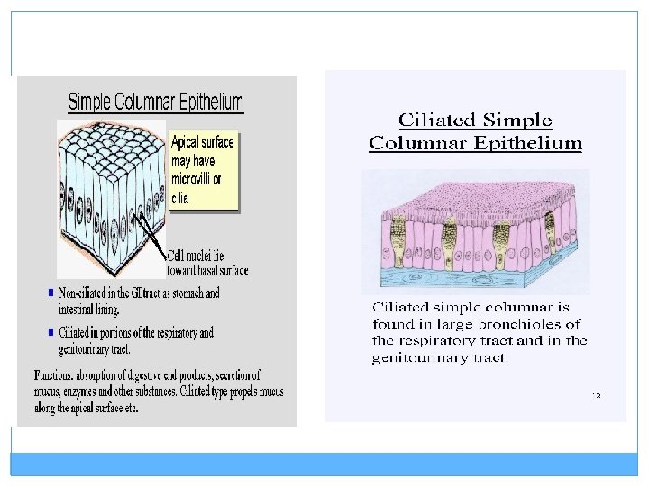
SIMPLE COLUMNAR EPITHELUM Cells are tall and pillar – like. Oval nucleus is present at the base of the cell. Can have microvilli Function: Secretion and Absorption. Found in Digestive tract and uterus. Contains goblet cells to secrete mucus.
COLUMNAR CELLS AND CILIATED COLUMNAR CELLS WITH CILIA
CILIATED EPITHELIUM Consist of columnar or cuboidal epithelial cells with cilia on their free surface. Cilia helps in the movement of substances / particles or mucus in a particular direction. Found in the fallopian tubes, bronchioles and nasal passage.
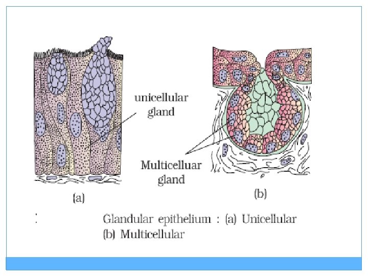
GLANDULAR EPITHELIUM v. Cells are specialized to produce and secrete substances. v. They form GLANDS. v. Normally cuboidal or columnar.
TYPES OF GLANDULAR EPITHELIUM UNICELLULAR: v Consists of isolated glandular cells v Eg: Goblet cells of alimentary canal. MULTICELLULAR: v Consists of cluster of cells. v Eg: Salivary glands
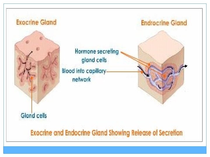
TYPES OF GLANDS (BASED ON THE MODE OF POURING THEIR SECRETIONS) EXOCRINE GLANDS: Products are released through ducts or tubes. v Secretes mucus, saliva, earwax, digestive enzymes. v ENDOCRINE GLANDS: Products are released directly into the fluid bathing the gland. v Hormones v
SALIVARY GLANDS
COMPOUND EPITHELIUM Consist of more than one layer of cells. Cover dry/outer surface of the skin, moist surface of buccal cavity, pharynx etc. Main function is to provide protection against chemical and mechanical stresses.
TYPES OF CELL JUNCTIONS (STRUCTURAL AND FUNCTIONAL LINKS BETWEEN EPITHELIAL CELLS) TIGHT JUNCTION: Plasma membranes of adjacent cells are fused at intervals. Help to stop substances from leaking across a tissue. ADHERING JUNCTION: Perform cementing function to keep neighbouring cells together. GAP JUNCTION: Facilitates the cells to communicate with each other by connecting the cytoplasm of adjacent cells for rapid transfer of ions and molecules.
CONNECTIVE TISSUE General Characteristics: ó Most abundant tissue in your body, found throughout. ó Binds structures together. ó Provides support, protection, framework, fills space, stores fat, produces blood cells, fights infection, and helps repair tissue. ó Composed of scattered cells and fibers (collagen and elastin). ó Matrix -made up of a ground substance (fluid, semisolid).
CONNECTIVE TISSUE
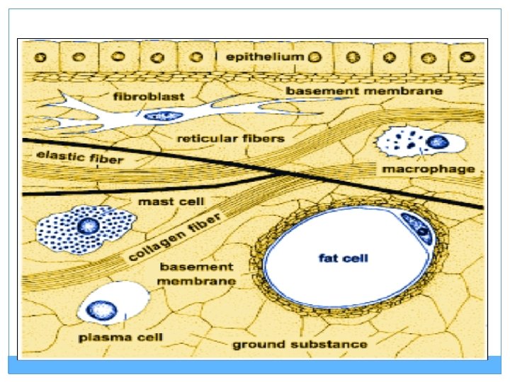
LOOSE CONNECTIVE TISSUE (CELLS AND FIBERS LOOSELY ARRANGED IN A SEMI –FLUID GROUND SUBSTANCE) AREOLAR TISSUES: Present beneath the skin. Contains fibroblasts, macrophage and mast cells. Serves as a support frame work for epithelium
AREOLAR TISSUES
LOOSE CONNECTIVE TISSUE ADIPOSE TISSUES: • Located beneath skin. Function as storage cells for lipids. Adipose cells contain a large vacuole which in the live cell contains lipids. Cell nucleus and cytoplasm are pushed out to edge of cell membrane.
DENSE CONNECTIVE TISSUE Dense Connective Tissue: Fibers and fibroblast are compactly packed. a. Dense regular connective tissue -Tendons and ligaments b. Dense irregular connective tissue Dermis of skin, submucosa of digestive tract
1. DENSE REGULAR CONNECTIVE TISSUE Consists of bundles of collagen fibers and fibroblasts forms tendons, ligaments. Function = provide strong attachment between various structures
Tendons and ligaments
DENSE IRREGULAR CONNECTIVE TISSUE consists of randomly-arranged collagen fibers and a few fibroblasts Found in fasciae, dermis of skin, joint capsules, and heart valves Function = provide strength
SPECIALISED CONNECTIVE TISSUE CARTILAGE: Cells (chondrocyte) are found in Lacunae within the matrix. Intercellular material of cartilage is solid and resists compression.
BONE Ground of matrix is Solid (Calcium carbonate). Bone cells (osteocytes) present in spaces called lacunae. Has blood supply and nerves running through the Haversian canal systems. T. S OF BONE
BLOOD Fluid connective tissue. Consist of plasma and blood cells. Blood cells: Erythrocytes , Leukocytes, Platelets
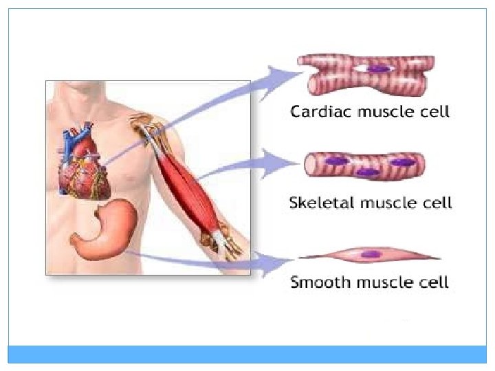
MUSCLE TISSUE
SKELETAL MUSCLE SMOOTH MUSCLE CARDIAC MUSCLE
MUSCLE TISSUE ( Tissue with cells having fibers specialized for contraction) SKELETAL MUSCLE (STRIATED, VOLUNTARY) Parallel elongated cells (fibers) multinucleated and each cell is the length of the muscle.
SMOOTH MUSCLE (VISCERAL, INVOLUNTARY) Cells are spindle shaped or fusiform. Prominent nucleus present in the centre. Striations absent. Present in wall of blood vessels , stomach, intestine.
CARDIAC MUSCLE Branched and cylindrical. Communication junctions called intercalated discs are present at the junction between branches of two different cardiac muscle fiber.
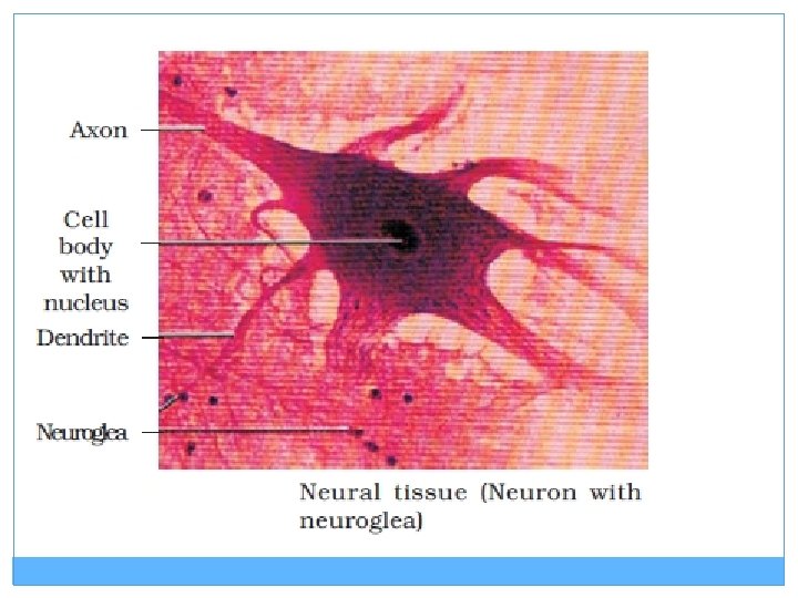
NERVOUS TISSUE Neuron- Structural and functional units of nervous system. Cells specialized to polarize and depolarize. Consists of: Cell body, axon, dendrites and nucleus. Neuroglia cells – gives protection and support. Make up for more than one half the volume of neural tissue in our body.
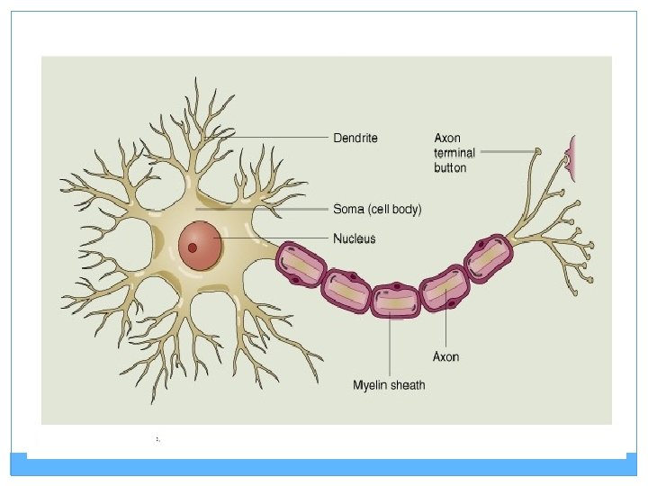
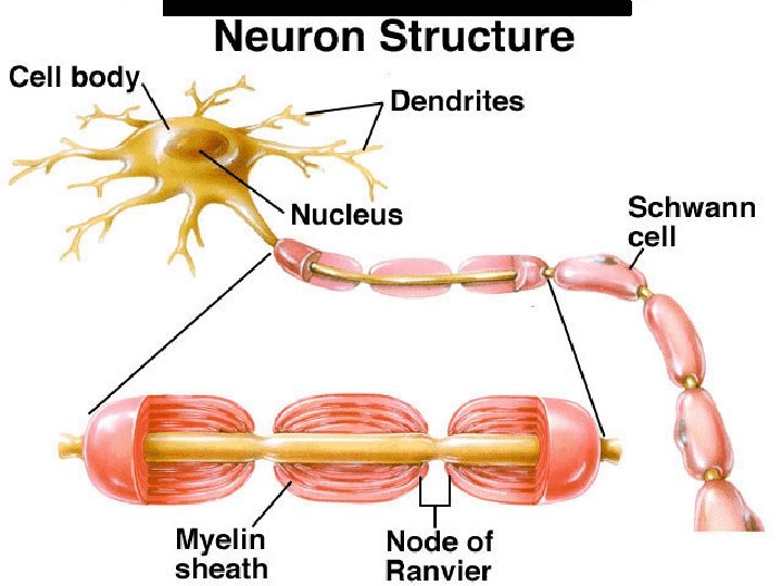
STRUCTURE OF NEURON: Dendrite : Function: receive incoming stimuli. Cell Body or Soma : Function: Directs impulses from the dendrites to the axon. Nucleus : Control center of the Soma. Axon: Pathway for the nerve impulse (electrical message) from the soma to the opposite end of the neuron. Myelin Sheath : An insulating layer around an axon. Made up of Schwann cells. Nodes of Ranvier : Gaps between schwann cells.
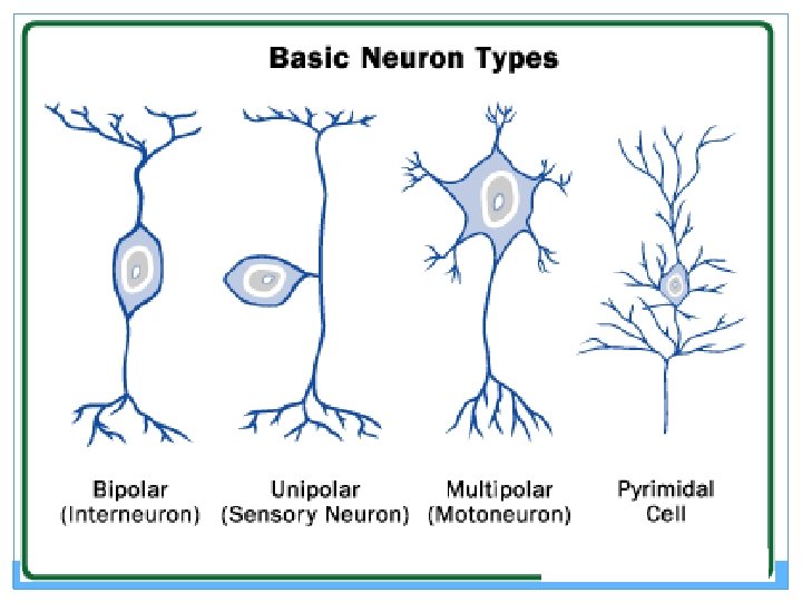
Types of Neurons