OVARY AND MAMMARY GLAND Dr Iram Tassaduq OVARIES


 cells 3 -")


- Slides: 46
OVARY AND MAMMARY GLAND Dr Iram Tassaduq
OVARIES • Ovarian ligament • Suspensory ligament • Mesovarium
FUNCTIONS OF OVARIES 1. Ova production 2. Hormone production
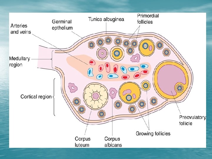
STRUCTURE OF OVARY • Covered with germinal epithelium • Tunica Albuginea • Cortex (outer layer) • Medulla (inner core)
GERMINAL EPITHELIUM • Surface epithelium having a single layer of columnar or cuboidal cells
GERMINAL EPITHELIUM
TUNICA ALBUGENIA • Dense connective tissue present between epithelium and cortex
CORTEX
CORTEX • Outer zone • Contains follicles and smooth muscle fibres • Only primordial and primary follicles before puberty
MEDULLA • Inner zone • Contains loose , connective tissue, blood vessels , lymphatics and nerves
MEDULLA OF OVARY
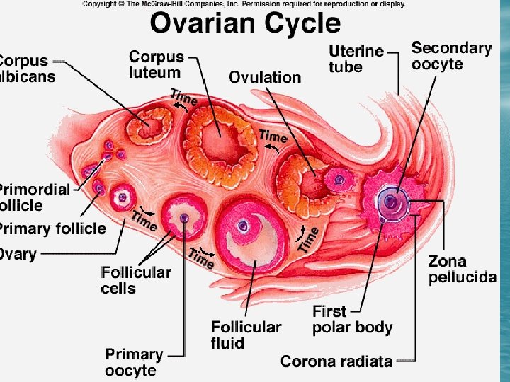
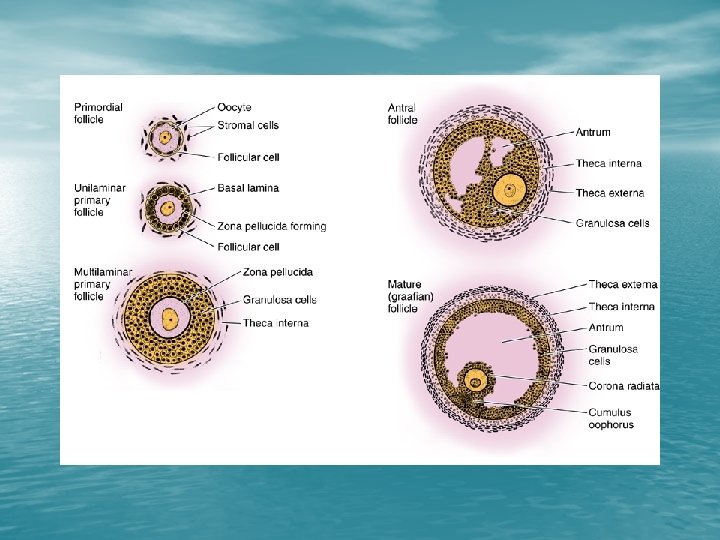
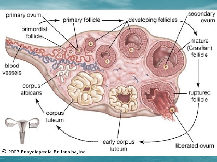
TYPES OF OVARIAN FOLLICLES • Primordial Follicle • Growing Follicle/Antral Follicles 1. Primary Follicle 2. Secondary Follicle • Mature /Graffian Follicle
PRIMORDIAL FOLLICLE • Earliest stage of development • Independent of gonadotrophin stimulation • Oocyte is 30 micrometer
PRIMORDIAL FOLLICLE
PRIMORDIAL FOLLICLE
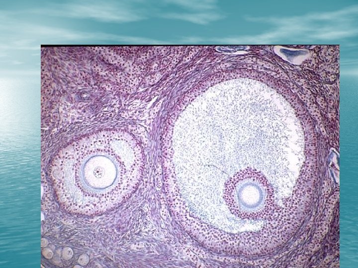
PRIMARY FOLLICLE • Changes in oocyte , follicular cells and stroma • Follicular cells become cuboidal • Zona pellucida appears between oocyte and follicles • Oocyte size 50 to 80 micrometer • Basal lamina separates the follicle from stroma
PRIMARY FOLLICLE
PRIMARY FOLLICLE • 1 - primary follicle 2 - follicular (granulosa) cells 3 - oocyte 4 - zona pellucida 5 - primordial follicle 8 - interstitial connective tissue 9 - theca
LATE PRIMARY FOLLICLE • Stratum • • • Granulosum Theca Folliculi Cortical granules develop Microvilli from oocyte develop
Stratum Granulosum • Stratified • • epithelium appear due to proliferation of follicle cells Outermost layer is columnar Gap junctions between granulosa cells
Theca Folliculi • Stromal cells forming sheath of connective tissue. It is subdivided into two layers 1. Theca interna 2. Theca externa
COMPOSITION OF THECA FOLLICULI • Theca interna the inner vascular layer is formed of cuboidal secretory cells, fibroblasts, collagen bundles and LH receptors • Theca externa consists of smooth muscle cells and collagen fibers
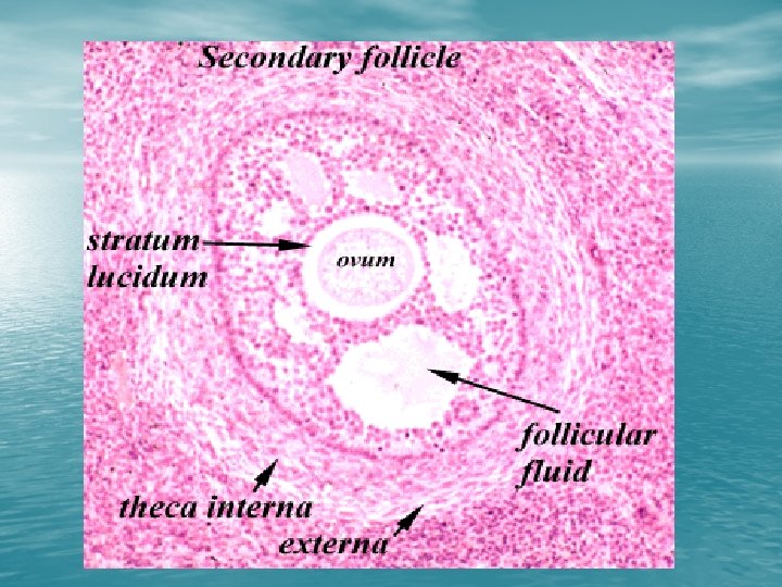
SECONDARY FOLLICLE • Few relative to number of primary follicles • Produce follicular fluid • Rapid enlargement • Cumulus Oophorus
Secondary Follicle
Tertiary or Graffian Follicle convert Androgens to Estrogens cause Follicular growth and sensitization of Gonadotropes A surge of FSH and LH Granulosa cells
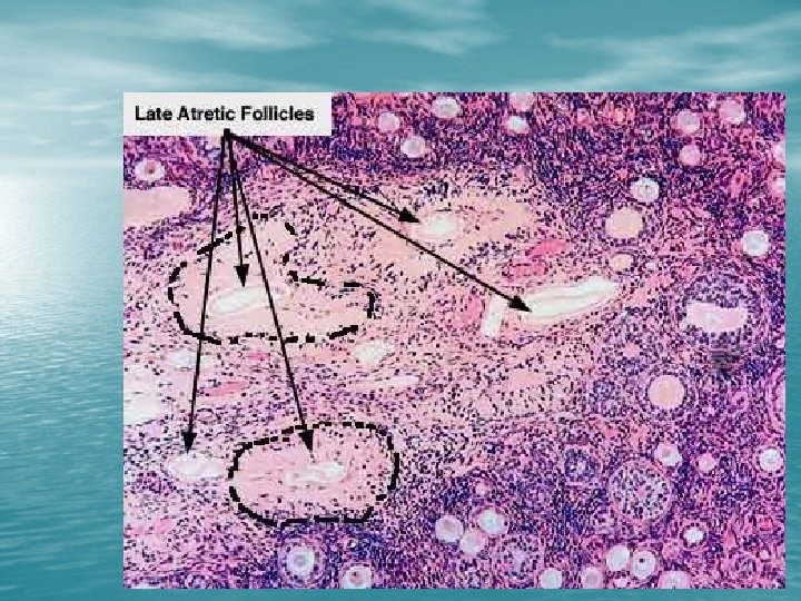
ATRETIC FOLLICLE • Degeneration of • • oocyte Break down of zona pellucida Separation and increased thickness of basement membrane
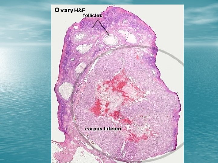
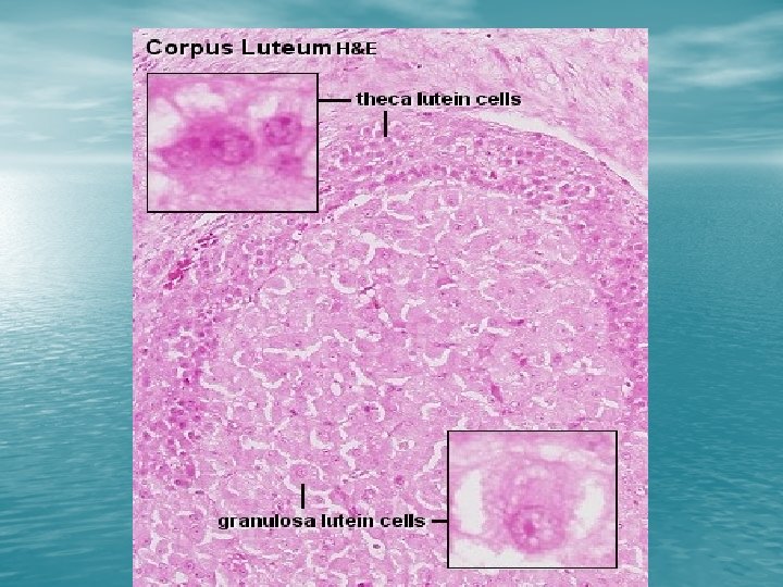
CORPUS LUTEUM • After ovulation, a temporary endocrine gland is • • constructed from what remains of the follicle Granulosa and theca interna cells hypertrophy after ovulation Blood vessels of theca interna grow into the interior of corpus luteum and provide rich vascular network Develop characteristics of steroid secreting cells The corpus luteum produces estrogen and progesterone
CORPUS LUTEUM v Theca lutein cells • Smaller in size , contain dark staining nuclei v Granulosa lutein cells • Large pale staining cells with large vesicular nuclei • Form a thick folded layer
CORPUS ALBICANS • Degenerated Corpus Luteum • large masses of amorphous eosinophilic staining material • Low cellularity. • Collagenous scar tissue that has filled the space occupied by the corpus luteum.
CORPUS ALBICANS
INACTIVE MAMMARY GLAND • Sparse glandular • tissue Consist of tubules having the appearance of ducts
MAMMARY GLAND DURING PREGNANCY