CHRONOLOGY AND MORPHOLOGY OF PRIMARY AND PERMANENT TEETH


. • Dental")
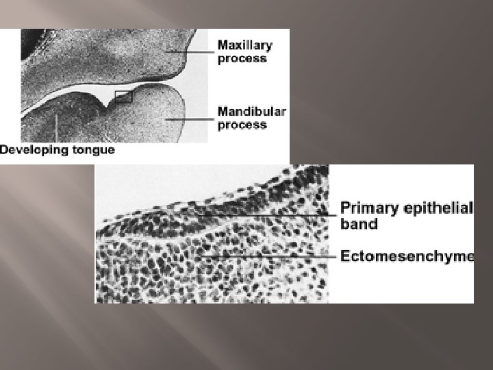
 • The enamel organ appears as a")
 • Invagination of the deeper surface of")


: period of gingival emergence until contact is achieved with the")


 Preterm and low birth weight infants")

 is typically wider than the")


- Slides: 65
CHRONOLOGY AND MORPHOLOGY OF PRIMARY AND PERMANENT TEETH
Outline � � Tooth development phases Pre-eruptive period Period of eruption of the primary dentition Static period of the primary dentition
Tooth Development Phases 1 - Initiation � The process of tooth development starts as early as 7 weeks in utero � In this phase, the locations of teeth are established with the appearance of tooth germs.
Tooth Development Phases 2 - Morphogenesis � The shape of the teeth is determined in this phase.
Tooth Development Phases 3 - Histogenesis � Differentiation of cells takes place to produce the fully formed dental tissues.
• The primary epithelial bands divides into: • Vestibular lamina (buccally). • Dental lamina (lingually). Underlying Mesenchyme The dental lamina contributes to the formation of the teeth.
Tooth germ formation � Bud stage: (initiation) • The enamel organ appears as a simple ovoid epithelial mass • Surrounded by mesenchyme. • Mesenchyme separated from the epithelium by a basement membrane.
Tooth germ formation � Cap stage (morphogenesis) • Invagination of the deeper surface of the enamel organ. • Peripheral cells start to be arranged as external and internal enamel epithelium.
Cap stage
Tooth germ formation � Bell stage � The shape of the internal enamel epithelium decides the shape of the crown.
Bell stage
Bell stage � � Cells of the inner enamel epithelium differentiate into Ameloblasts Adjacent cells from the dental papilla will differentiate into Odontoblasts • Odontoblasts produce pre-dentine and dentine. • The presence of dentine induces Ameloblasts to form enamel • Apposition of enamel and dentin will be followed by calcification
Calcification � Starts between 14 and 16 weeks of intrauterine life for primary teeth � Begins in cusp tips and incisal edges of teeth and continues cervically � Very sensitive process that takes place over a long period of time.
Calcification � Any severe systemic event during the development of teeth will result in some dental abnormality � Chronological enamel defects.
Calcification � Different teeth will show defects at different levels of the crown depending on the stage of crown formation.
Eruption � Eruption is the movement of teeth within and through the bone of the jaws and the overlying mucosa to appear in the oral cavity and contact the opposing teeth. � Emergence of the tooth is the first sign of appearance in the oral cavity.
� Pre-eruptive phase: period during which the tooth root begins its formation and begins to move towards the surface of the oral cavity from its bony vault.
� Eruptive phase (prefunctional): period of gingival emergence until contact is achieved with the opposing tooth. � Functional eruptive phase: after the tooth meets its antagonist. A dynamic unit throughout life.
Factors influencing tooth formation and eruption Why is it difficult to study and understand the process of eruption? � Tooth structure and eruption vary from one species to another � Histological studies in humans are rarely possible because of the inaccessibility of tissue for sampling and ethical considerations
Theories of eruption 1 - Root formation 2 - Hydrostatic pressure 3 - Bone remodeling 4 - Periodontal ligament
Root formation theory � The space for the growing root is accommodated by occlusal movement of the tooth crown However; � Some teeth with extensive root development fail to erupt � A study using dogs, showed that the tooth itself played no part in the eruptive process
Hydrostatic pressure theory � Studies using dogs demonstrated that the tissue pressure apical to the erupting tooth was greater than occlusally, theoretically generating an eruptive force However; � The study only compared pressure differentials but whether this pressure difference actually caused eruption is not proven
Bone remodeling theory � Bone remodeling around the tooth causes eruption However; � Animal studies showed that bony remodeling occurs around the dental follicle regardless of the presence of a tooth
Periodontal ligament theory � Strong evidence exists to show that the periodontal ligament, which is derived from the dental follicle, provides the force required for eruption mainly by fibroblast contraction However; in vitro tissue studies have limitations
In conclusion There is no evidence that one hypothesis fully explains tooth eruption, and that eruption is likely to be a multifactorial process
Basic biology of tooth eruption � 1 - Bone resorption Resorption appears to be genetically controlled and not mechanically by the eruption of the tooth � 2 - Role of the dental follicle Removing the follicle means the tooth will not erupt while leaving it and replacing the tooth with an artificial replica means the tooth will erupt 3 - Cellular events and molecules Certain molecules will recruit mononuclear cells into the � dental follicle
Control of eruption 1 - Hormonal control 2 - Systemic conditions 3 - Physical control mechanism
Hormonal control � Most eruption occurred in the late evening indicating that eruption was probably under hormonal control � Mainly due to the effects of the late evening secretion of growth hormone and thyroid hormone. � Children with growth hormone deficiency had delayed tooth eruption
Systemic conditions � � � Nutritional deficiency (extremes) Preterm and low birth weight infants Cerebral palsy Anemia Renal failure Genetic disorders Apert syndrome Cleidocranial dysostosis Down syndrome Ectodermal dysplasia Gardner syndrome Osteopetrosis
Physical control mechanism � According to the equilibrium theory teeth remained in a position within the jaws where forces acting in equal and opposite directions cancelled each other � Oral musculature, soft tissue pressures, masticatory forces, and eruptive force.
Pre-eruptive period � Upper anterior gum pad (intercanine width) is typically wider than the lower anterior gum pad � Upper anterior gum pad protrudes (Overjet) about 5 mm � Overbite about 0. 5 mm.
Pre-eruptive period � Marked palatal width increase and a decrease in the overjet over the first 6 months of postnatal life.
Pre-eruptive period � Labial frenum is usually hypertrophic but it does not hinder suckling � Retro incisal papilla is hypertrophic.
Pre-eruptive period � Palate is straight at birth. Becomes concave under the effect of growth of the alveolar bone � Tongue is relatively large.
Oral mucosa � � � Epstein pearls. Bohn nodules Dental lamina cyst.
Epstein pearls � � � Small white or greyish white lesions Present in about 80% of neonates. Formed along the midpalatine raphe.
Epstein pearls � � Considered remnants of epithelial tissue trapped along the raphe as the fetus grows. Disappear within a few weeks of life.
Bohn nodules � Formed along the buccal and lingual aspects of the dental ridge and on the palate away from the raphe.
Bohn nodules � � Considered remnants of mucous gland tissue and are histologically different from Epstein pearls. Disappear spontaneously in the early months of life.
Dental lamina cysts � � � Found on the crest of the maxillary and mandibular dental ridges. Are remnants of the dental lamina. Slough within the first few months of life.
Dental lamina cysts � � Differential diagnosis: natal teeth. No treatment necessary.
Period of eruption of the primary dentition � Commences at 6 months and is well established by 30 -36 months � Maximum growth of the jaws occurs during this period.
Deciduous Teeth � � 20 in number, 10 in each jaw. There are no premolars in the deciduous dentition. The primary molars are replaced by the permanent premolars. The permanent molars erupt distal to the primary second molars.
Nomenclature � • • • Beginning with the midline, the teeth are named as follows: Central incisor. Lateral incisor. Canine. First molar. Second molar.
Tooth numbering � • Palmer Notation Method. Children’s 20 primary teeth are lettered “A” through "E" in each quadrant. � Universal Numbering System. � FDI Two-Digit Notation. The currently accepted convention to view the FDI notation chart is from the perspective of the patient's right. •
FDI Two-Digit Notation � 1 s are central incisors, 2 s are laterals, 3 s are canines, 4 s are 1 st premolars etc. , up through 8 s which are 3 rd molars � The permanent teeth quadrants are designated 1 to 4 such that 1 is upper right, 2 is upper left, 3 is lower left and 4 is lower right � In the deciduous dentition the numbering is correspondingly similar except that the quadrants are designated 5, 6, 7 and 8.
Chronology of eruption of primary teeth � 1 st tooth at approximately 6 months � Usually the lower central incisor
Chronology of eruption of primary teeth � All eruption schedules are estimates � No two individuals are alike. � Multiple parameters: race, gender, ethnicity, familial environment, heredity.
Chronology of eruption of primary teeth Maxillary Mandibular Central incisors 6 -10 months 5 -8 months Lateral incisors 8 -12 months 7 -10 months Canines 16 -20 months First molars 11 -18 months Second molars 20 -30 months
Sequence of eruption � A then B then D then C then E � Mandibular precede maxillary most of the time.
Rhythm of eruption of primary teeth � Symmetrical groups of 4 teeth every 6 months � Teeth erupt symmetrically in both jaws, simultaneously and in pairs.
The ‘six/four’ rule for primary tooth emergence � 1. 2. 3. 4. 5. Four teeth emerge for each 6 months of age. 6 months: 4 teeth (lower & upper As) 12 months: 8 teeth (1+upper & lower Bs) 18 months: 12 teeth (2+ upper & lower Ds) 24 months: 16 teeth (3+ upper & lower Cs) 30 months: 20 teeth (4+ upper & lower Es)
Static period of the primary dentition � Period of stability of the primary teeth � 3 -6 years of age � Child has 20 primary teeth in their final and functional position � Occlusion is well established
Static period of the primary dentition � Occlusal features � Occlusion of the primary second molar � Inter-arch relationship of primary teeth
Occlusal features in the established primary dentition � Incisor teeth tend to be spaced � Primate spaces exist between upper B & C and between lower C and D
Occlusal features in the established primary dentition � Upper incisors are upright � Incisor relationship is more towards edge to edge.
Occlusal features in the established primary dentition � Long axis of primary teeth is parallel � Absence of the curve of Spee � In general, teeth in the primary dentition tend to be well aligned.
Classification of occlusion of the primary second molar � Look at the distal aspect of the primary second molar � Flush terminal plane � Mesial step � Distal step
Flush terminal plane
Mesial step
Distal step
Inter-arch relationship of primary teeth � � � Each tooth occludes with two opposing teeth except for the lower central incisors and the upper second molars. Canine is a key to occlusion in the primary dentition. Look at the long axis of the canine
Canine relationship � Long axis of the canine should be placed in the midline between the lower D and C for a class I relationship.
Inter-arch relationship � Natural wearing away of the canines is an important physiologic process that facilitates movement of the mandible � In children raised on soft food the natural wearing process may be slowed down � May have to carry out selective grinding on primary canines, especially in the presence of a unilateral crossbite.