The Nervous Tissue Categories of Tissue 1 Epithelial


 Two of the known roles of cytoskeletal components")


")


 degeneration q complete degeneration")


- Slides: 37
The Nervous Tissue
Categories of Tissue 1. Epithelial Tissue 2. Connective Tissue 3. Muscle Tissue 4. Nervous Tissue Epithelial tissue Nervous tissue Connective tissue Muscular tissue
The Nervous Tissue Contains two different varieties of cells 1. Neurons 2. Neuroglia = supporting cells
NEURON Also called nerve cells Structural unit of nervous system Character: § Excitability § conductivity Conduct messages in the form of nerve impulses from one part of the body to another = synapse
Structure of neuron 1 - Cell body 2 -Cell processes = Soma around nucleus perikaryon axon dendrites
. Perikaryon = nerve cell body contains q Nucleus : large, central, active & prominent nucleolus q Rough endoplasmic reticulum (r. ER) Nissl bodies are condensations of r. ER and free ribosomes. for synthesis of structural and transport proteins (neurotransmitters) q Golgi apparatus : near to the nucleus. Expected, since intense synthetic activity of neurotransmitters that must be packaged in vesicles. q Mitochondria : abundant for high energy requirements
q. Cytoskeleton (neurofilaments , microtubules ) Two of the known roles of cytoskeletal components include maintaining the link structure or the physical form of neurons and transporting cell components from the soma to the synapses. q. Inclusions : Ø pigment Ø residual bodies increase with age.
AXON The nerve cell processes: . Dendrite Origin From axon hillock From any part of cell Number Always single Usually multiple length long Short Thickness Thin uniform Thick near cell then tapers away Branching Does not branch except at its termination Many branches. Organelles Few No Nissl bodies No golgi Contains most of organelles Contain golgi + Nissl Surrounding structures Surrounded with sheaths Not surrounded with sheaths Direction of Away Towards Axoplasm Axolemma
Classification of neurons q Functionally q Morphologically Ø Sensory v. Size Ø Motor 1. Golgi I (long axon ) Ø Interneurons 2. Golgi II (short axon) Ø Neurosecretory v. No of Processes 1. one Process 2. two Processes 3. more Processes
According to the number of processes
Multipolar Neurons According to the shape of the cell body Stellate Pyriform Pyramidal Granule
Neuroglia Definition : Supporting cells of the nervous system Stain : ØGold chloride ØSilver stain ØImmuno-histochemical Shape : Branching cells …. Origin : All ectodermal in origin except microglia derived from blood monocytes = mesodermal in origin
Site : q. Inside CNS macroglia =astrocytes, microglia, ependymal cell , oligodendroglia q. Outside CNS= peripheral NS Schwann cell, satellite cells in ganglia, pituicytes in posterior lobe of pituitary gland.
Types of neuroglia Name of the cell Location Function v Astrocytes protoplasmic & Fibrous CNS Grey & white matter Regulate microenvironment B. B. B v Oligodendrocytes CNS White & grey matter Myelination of axon in CNS v Microglia CNS Grey & white matter Mesodermal in origin Phagocytic v Ependymal cell CNS Ventricle & central canal of spinal cord------with a ciliated simple columnar shape Assist in producing & controlling composition of CSF v Schwann cell PNS Myelination of axon Structural support v Satellite cell PNS Regulate microenvironment
Protoplasmic Fibrous Site Gray matter White matter Processes No, length, thickness, course Numerous, short thick & wavy Fewer, longer, thinner & straight GFAP Few bundles Rich in it
Neuron Large 2. Transmit nerve impulse 3. Not able to divide 1. 4. Form synapse Neuroglia 1. Small 2. Not transmit nerve impulse 3. Able to divide 4. Not form synapse Function of neuroglia 1. Supportive 2. Nutritive 3. Electrical insulation= formation of myelin sheath 4. Repair 5. Formation of blood brain barrier 6. Formation of CSF 7. Phagocytosis
Myelin Sheath Segmented protein-lipoid sheath around most long or large-diameter axons Functions : 1. Protect and electrically insulate the axon 2. Increase speed of nerve impulse transmission
Myelin Sheaths in the PNS Schwann cells wraps many times around the axon q. Myelin sheath —concentric layers of Schwann cell membrane Nodes of Ranvier ---? ? Myelin sheath gaps between adjacent Schwann cells Myelinated nerve are separated by nodes of Ranvier, at these points , the axons are bare. Impulses jump from one node to the next q. Unmyelinated Axons. Thin nerve fibers are unmyelinated. One Schwann cell may incompletely enclose 15 or more unmyelinated axons. Conduction in unmyelinated nerve is slower
v Schwann cell plasma membrane v Schwann cell Cytoplasm A Schwann cell envelopes an axon. Schwann cell nucleus v Axon Neurilemma The Schwann cell then rotates around the axon, wrapping its plasma membrane loosely around it in successive layers. Myelin sheath Myelination of a nerve fiber (axon)------? ? Unmyelinated nerve fiber (axon)
Myelin Sheaths in the CNS Formed by processes of oligodendrocytes, not the whole cells Nodes of Ranvier are present No neurilemma Thinnest fibers are unmyelinated
The Axon+ sheath = Nerve fiber • Cytoplasm is called axoplasm • Plasma membrane is called axolemma • One axon per cell arising from the axon hillock • Long , thin • No Nissl bodies • Knoblike axon terminals (synaptic knobs) • Numerous terminal branches • Occasional branches (axon collaterals) • Mostly myelinated some axons not myelinated • Generates and transmits nerve impulses (action potentials) away from the cell body
Peripheral nerves are made up of: Group of NF + Connective tissue sheath Ø Epineurium Ø Perineurium Ø Endoneurium
Peripheral nerve
Peripheral nerve (osmic acid)
SYNAPSE Def : Synapse is a specialized area of functional contact . Through synapses nerve impulses are transmitted from a presynaptic neuron to a postsynaptic cell (another neuron, muscle or gland).
Classification of synapse: According to the effect on the target neuron, synapse could be: q. Excitatory q. Inhibitory 1 -
2 - According to the components: q Axodendritic: between axon and dendrite q Axosomatic: between axon and cell body. q Axo-axonic: between two axons. q Dendrodendritic: between two dendrites. Ø Axodendritic and axosomatic synapses are common Ø axoaxonic and dendrodendritic synapses are
3 - According to mode of transmission of nerve impulse: 1 - Chemical synapse: q. Number: The most common mode of communication between neurons. q. Structure : With EM, chemical synapse is formed of:
q. Terminal bouton: It is a bulbous expansion "synaptic knob" found at the terminations of the axon. It contains numerous mitochondria and many synaptic vesicles assembled around the presynaptic membrane. These membrane - bound synaptic vesicles are filled with the neurotransmitter e. g. acetylcholine and biogenic amines. q. Presynaptic membrane: it is the electron dense membrane of the axon terminal end at the site of synapse. q. Synaptic cleft: it is a gap of 20 nm between the pre and postsynaptic membranes. Into this gap the neurotransmitter is released by exocytosis. q. Postsynaptic membrane: It contains the specific receptors for the neurotransmitter.
2 - Electrical synapse: • Number: Few in the nervous system. • Structure: It is formed of gap junctions. • Function: It allows movement of ions between neurons "electrical coupling" thus permits the direct spread of electrical current from one cell to another. • Transmission of impulse: Direct, bidirectional and do not require release of neurotransmitters.
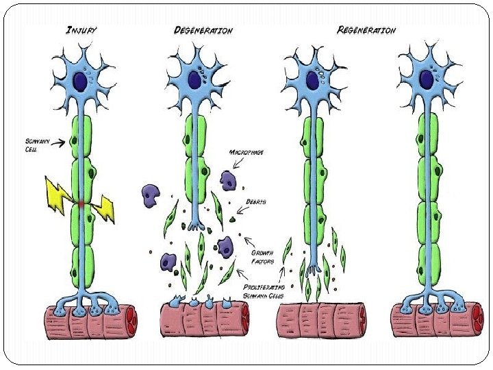
Response of neuron to injury A. Damage to the cell body - occurs as a result of injury or disease - degeneration and permanent loss of neuron leading to atrophy of the innervated muscle -The perikaryon enlarges, eccentric nucleus and chromotolysis (loss of Nissl substance) B. Damage to the axon q proximal portion q. Retrograde degeneration q incomplete degeneration -Proximal degeneration perikaryon becomes active and Nissle body reappear
Damage to the axon q distal portion q Anterograde (Wallerian) degeneration q complete degeneration of the axon and myelin sheath, fragments phagocytoced by Schwann in PNS, microglia in CNS, and blood monocytes -Schwann cells start to divide and bridge the injured site forming large number of new nerve processes sprouts, neurites, from proximal portion - neurites are guided and mylenated by Schwann cells across the injured site - if the gap is too wide or the sprouts do not reestablish contact, the sprouts grow in disorganized manner forming neuroma causing atrophy to end-organ
Nerves can be injured by: v ischaemia v. Compression v traction vlaceration vburning. Ø Damage varies in severity from: transient and quickly recoverable loss of function Ø to complete interruption and degeneration. Ø There may be a mixture of types of damage in the various fascicles of a single nerve trunk.
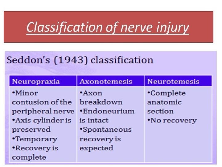
CLASSIFICATION of nerve injuries q First degree injury : transient ischaemia and neurapraxia, reversible. q Second degree injury: axonotmesis. Axonal degeneration takes place but, because the endoneurium is preserved, regeneration can, lead to complete, or near complete, recovery without the need for intervention. q Third degree injury worse than axonotmesis. The endoneurium is disrupted but the perineurial sheaths are intact and internal damage is limited. The chances of the axons reaching their targets are good, but fibrosis will limit recovery.
Thank You