Life at the Molecular Cellular and Tissue Level


- Slides: 29
Life at the Molecular Cellular and Tissue Level TOPIC: ANIMAL TISSUES - EPITHELIAL, MUSCLE & NERVE TISSUES PRESENTER: MS Z. SANDA LIFE SCIENCES PLANNER (EC)
CAPS DOCUMENT: PG. 28 WILL NOT BE COVERED IN 2020 DUE TO COVID-19
What is a Tissue? • A tissue is as a group of cells that have the same structure that work together to carry out the same function • Two or more tissues organized to carry out a particular function form organs and groups of organs with related functions make up the different organ systems.
Levels of Organization in the Human Body
Types of Animal Tissues Most animal bodies are made up of four different types of tissue: 1. Epithelial tissue 2. Connective tissue 3. Muscle tissue 4. Nervous tissue
FOUR TYPES OF ANIMAL TISSUES
Epithelial Tissue • Forms the outer layer of the body and also lines many of the body cavities where it has a protective function. • The tissue has tightly-joined, closelypacked cells. • Epithelial cells protect against mechanical injury, invasive microorganisms, and fluid loss and also provides surface for absorption, excretion and transport of molecules
Classification of Epithelial Tissues • Epithelial tissues can be classified based on the number of cell layers and the shape of cells • On the basis of the number of cell layers we have: ØSimple epithelium – made up of a single layer of cells ØCompound epithelium - has many layers of cells
Simple Epithelial Tissues • There are 5 types of simple epithelial tissues but we will look at 4 of these: • Squamous • Cuboidal • Columnar • Ciliated • (Pseudostratified)
Squamous Epithelium LOCATION: Lines the mouth , alveoli in the lungs, blood vessels STRUCTURE: Thin, large flat cells which are closely packed cells and have elliptical nuclei located mostly at the centre of the cell FUNCTION: Lines surfaces, protection and allows certain substances to pass through
Cuboidal Epithelium • LOCATION: Kidney tubules or glands (regions of the body responsible for excretion). • STRUCTURE: Cells are cuboidal in shape with rounded nuclei lying in the centre of the cell • FUNCTION: Secretion and transportation in glands, reabsorption and excretion.
Columnar Epithelium • LOCATION: Digestive tract e. g. stomach, intestines and gall bladder, reproductive organs • STRUCTURE: Cylindrical cells lying side by side with slightly elongated nuclei located at the base of the cells. Sometimes glands e. g. goblet cells are found in columnar epithelium forming glandular epithelium • FUNCTION: Provides support to other cell types. Absorbs food, water and minerals. Contains goblet cells to secrete mucous
Ciliated Epithelium • LOCATION: Trachea and bronchi of the respiratory system and in the fallopian tubes of the female reproductive tract • STRUCTURE: Ciliated columnar epithelium contain little finger-like projections called cilia. • FUNCTION: Mucous from goblet cells traps dust. Beating cilia drives out the mucous with the dust. Sense organs – helps detect stimuli
COMPOUND EPITHELIUM • Compound epithelium is made up of many layers of cells • An example is Stratified Epithelium which is made up several layers of epithelial cells of the same shape or different shape • It is found lining the skin. cc
Muscle Tissue • Enables various forms of movement, both voluntary and involuntary. • Voluntary movement means it is consciously controlled by one’s will whereas involuntary means it occurs automatically without control of one’s will Walking: a voluntary movement Peristalsis: Food movement through the alimentary can- an involuntary movement
Types of Muscle Tissue There are 3 types of Muscle Tissue: 1. Skeletal Muscle 2. Smooth Muscle 3. Cardiac Muscle
Skeletal muscle is a voluntary muscle. It is striated in appearance. has regularly arranged bundles. is anchored by tendons and is used to effect skeletal muscle movement such as locomotion and maintain posture. • The muscles have a reflex action but can also respond to conscious control. • • The voluntary movements are done with the help of skeletal muscles
Skeletal Muscle
Smooth Muscle • is an involuntary, non-striated muscle with tapered ends. It is found within the walls of blood vessels such as arteries and veins. • Smooth muscle is also found in the digestive system, urinary tract and in the trachea. • It is responsible for involuntary rhythmic contractions of peristalsis required for moving food down the alimentary canal, and for the dilation and constriction of blood vessels to control blood pressure
Smooth Muscle
Cardiac Muscle • is the major tissue making up the heart. • It is an involuntary muscle that is striated in appearance. • However, unlike skeletal muscle, cardiac muscle connects at branching, irregular angles. • The connected branches help with coordinated contractions of the heart Involuntary movement of the heart is brought about by the cardiac muscle
Cardiac Muscle
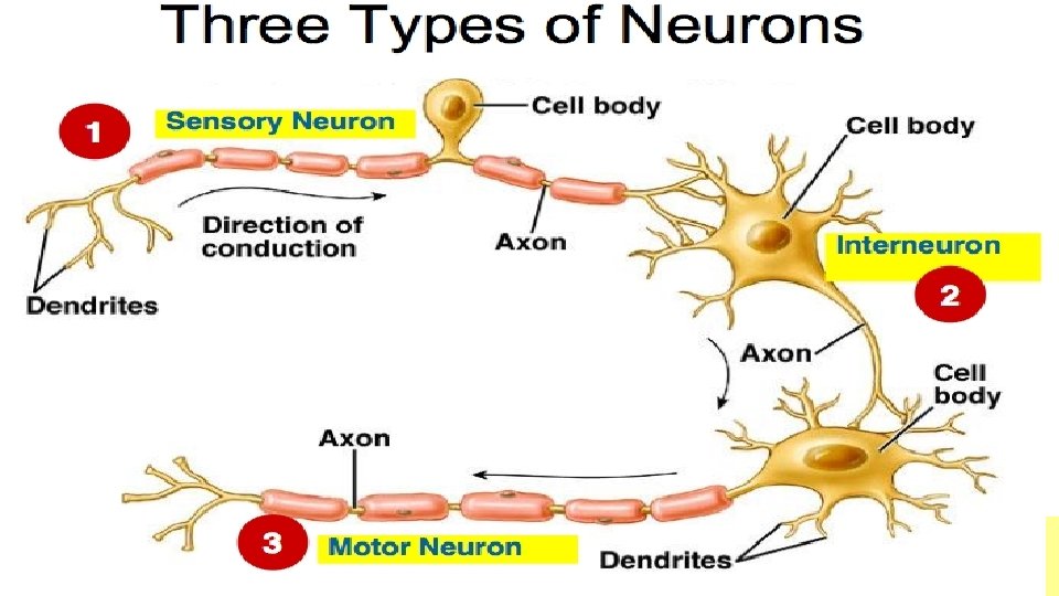
Nervous Tissue • Is responsible for the carrying of electrical and chemical signals and impulses from the brain and central nervous system to the periphery, and vice versa. • Nerves are made up of cells called neurons. • Neurons can be classified by their function, structure or their special use.
Types of Neurons • Sensory neurons carry impulses from the receptors to the central nervous system (CNS) • Motor neurons carry impulses from the CNS to the muscle cells and glands, which are effectors. • Connector Neurons: carry impulses to the motor neurons and connect neurons together.
ACTIVITY Atom • Organ Molecule Macromolecule Organelle Cell Organ System Tissue Organism
Name the four major types of tissues in the human body labelled A-D in the diagram Muscle Epithelium D Connective Nervous
Name and describe the general function of each of the following tissues protect against mechanical injury, invasive microorganisms, and fluid loss and also provides surface for absorption, excretion and transport of molecules Is responsible for the carrying of electrical and chemical signals and impulses from the brain and central nervous system to the periphery, and vice versa. enables various forms of movement, both voluntary and involuntary
Identify tissues labelled A-D in the diagrams below Squamous epithelium Cardiac Muscle Cuboidal epithelium Skeletal Muscle