EPITHELIAL TISSUE TISSUE A collection of cells together


")
 Single layered/ unilaminar/simple epithelium b) Multi layered /multilaminar/ stratified")


, Efferent ductules of")

 – location – epidermis function")


")


- Slides: 41
EPITHELIAL TISSUE
TISSUE A collection of cells together with intercellular substances that are similar in structure and function. 4 types of basic tissue : epithelial tissue connective tissue muscle tissue nerve tissue
EPITHELIAL TISSUE It is an avascular tissue composed of closely aggregated cells with little extracellular substance that are richly innervated, covers the external body surface , internal closed cavities & body tubes, is attached to an underlying basement membrane. Epithelium also forms secretory portion (parenchyma) of glands & their ducts.
Specialized epithelial cells functions as receptors of special senses (smell, taste, hearing , vision).
CHARACTERISTICS FEATURES Predominantly cellular, little extra cellular substances Rest on basement membrane A vascular , get nutrition by diffusion from adjacent tissue Very sensitive (rich innervations) Has no lymphatic drainage Regeneration takes place Develops from all 3 germ layers
Epithelial cells are adhere to one another by specialized cell junctions. Each cell has free / apical , lateral & basal surface or domain.
FUNCTIONS Protection Secretion Absorption Lubrication Transport Sensation Contractility (e. g; myoepithelial cells)
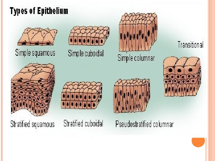
CLASSIFICATION: I. Covering epithelium: a) Single layered/ unilaminar/simple epithelium b) Multi layered /multilaminar/ stratified epithelium II. Glandular epithelium a) Exocrine glands b) Endocrine glands III. Myoepithelium IV. Neuroepithelium
COVERING EPITHELIUM 1. Unilayer Ø simple – squamous cuboidal columnar Pseudostratified– non ciliated Ø
2. Multilayer– Ø stratified squamous – keratinized non keratinized Ø Ø Ø stratified cuboidal stratified columnar transitional
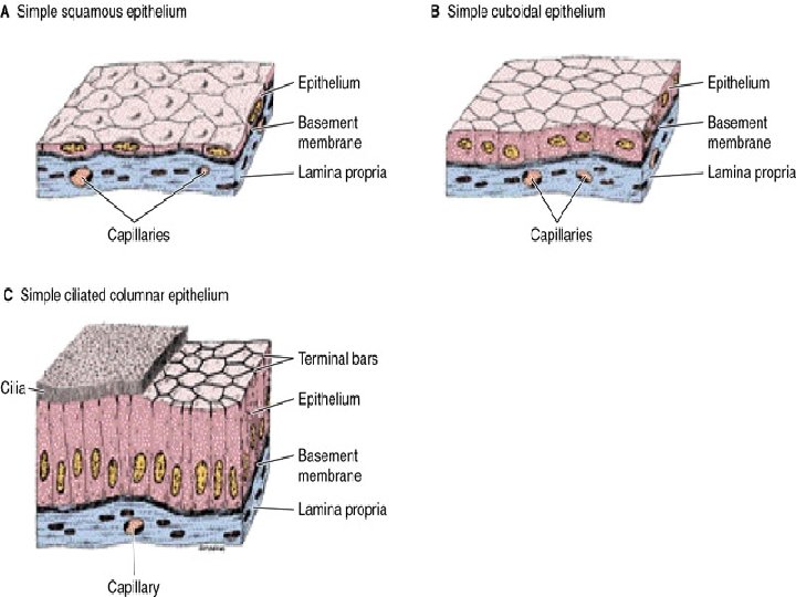
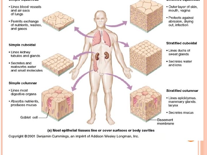
SIMPLE EPITHELIA Contains one layer of cells Types: (based on the shape of the cells) 1. Simple squamous: - width of the cell is greater than its height - nucleus is flat and centrally placed and bulges towards the surface - Locations: endothelium, mesothelium, Bowman’s capsule (kidney), Lung alveoli - Functions: exchange, barrier, lubrication
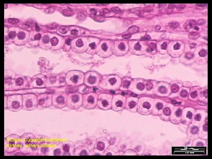
2. Simple cuboidal: - Width, depth and height of the cells are approximately same - Nucleus is round and centrally placed - Locations: Kidney tubules, germinal epithelium of ovary, small ducts of exocrine glands, thyroid follicles - Functions: absorption, secretion, barrier, conduit
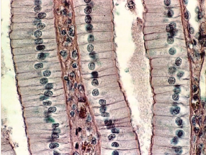
3. Simple columner: - height of the cell is more than the width - nuclei are oval in shape and either placed centrally or towards the base of the cell - Locations: Gastrointestinal tract, gall bladder, uterine tube, uterus - Functions: absorption and secretion
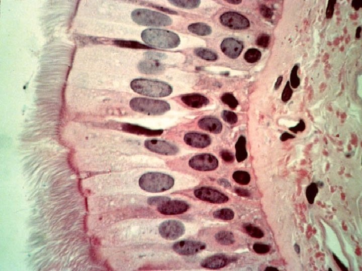
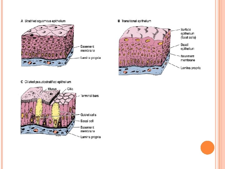
4. Pseudostratified epithelium: Characteristics: - single layered - all cells rest on the basement membrane - but the some of the cells do not reach the free or luminal surface - so the nuclei are visible at different levels thus giving the appearance of being multilayered
-Locations: trachea and bronchial tree, nasal cavity (pseudostratified ciliated columner epithelium), Efferent ductules of epididymis, Ductus deferens, part of male urethra (non ciliated) - Functions: secretion, absorption, conduit
STRATIFIED EPITHELIUM Multi layered Classified according to the cell shape of superficial layer
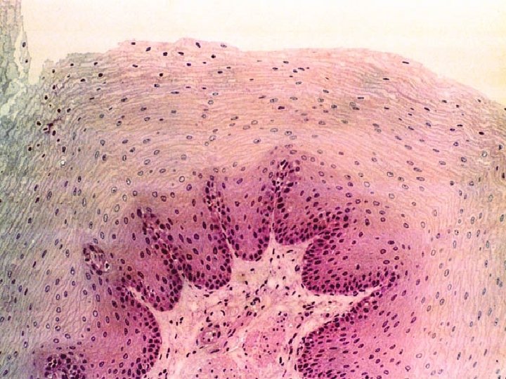
1. Stratified squamous : keratinized (rich in keratin filaments) – location – epidermis function – protection , prevents water loss
Non keratinized location – mouth, esophagus, larynx , vagina , anal canal. function – protection , secretion
- Functions: barrier conduit
3. Stratified columner: - cells of the surface layers are columner in shape - Locations: Conjunctiva, main excretory ducts of the exocrine glands, anorectal junction
Functions: barrier conduit
4. Transitional epithelium/urothelium Characteristics: - layers of cells vary from 4 -6 - the cells of surface layer are dome shaped or umbrella shaped - cells of other layers are polygonal - cells of the deepest layer are the smallest - it has the property of contractility and distensibility
- prevents reabsorption of urine due to multiple tight junctions and glycoprotein plate present at the surface cells
Locations: - Lower urinary tract (from minor calyx to upper part of urethra)
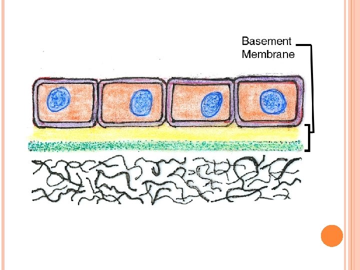
BASEMENT MEMBRANE It is a connective tissue layer Formation : formed by combination of 1. Basal lamina : a. lamina lucida b. lamina densa 2. Reticular lamina
Basal lamina: - it is the structural attachment site for overlying epithelial cells and underlying connective tissue. - seen only under electron microscope. Composition : lamina lucida & lamina densa
1. lamina lucida : Ø it lies beneath the cell membrane Ø network of fine fibrils Ø transparent Ø contain laminin , integrin , proteoglycan & glycoprotein.
2. Lamina densa : Ø it lies below the lamina lucida. Ø it is dense Ø it contain proteoglycan , type iv & type vii collagen fiber.
Reticular lamina : Ø basal lamina is attached with reticular lamina by type iii collagen fiber Ø it is attached with underlying connective tissue by type vii collagen fiber.
1. 2. 3. 4. Functions of basement membrane : Give structural support to epithelium Structural attachment site of the cell to underlying connective tissue Regulate exchange of macro molecule between cell & connective tissue Regulate cell proliferation & differentiation
Clinical importance : disrupted in malignancy.