Case Conference Hieu Vo Fellow UTHSC Outline 2



- Slides: 81
Case Conference Hieu Vo Fellow UTHSC
Outline • • • 2 Cases Pathogenesis Presentation Diagnosis Post-transplant Treatment
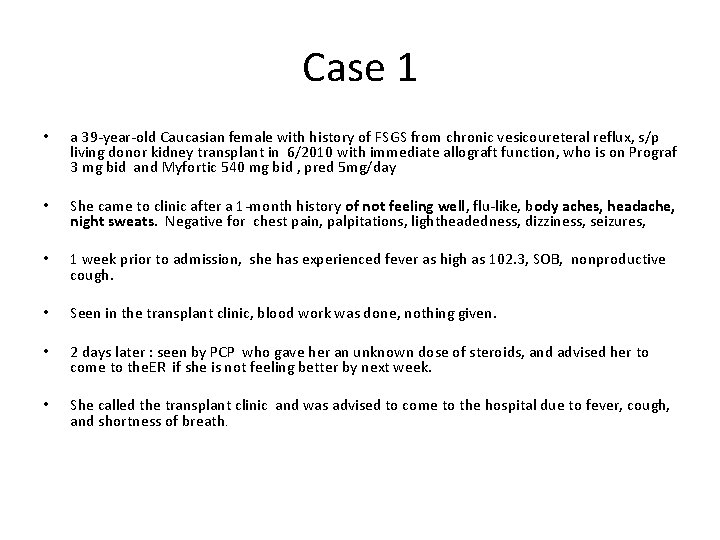
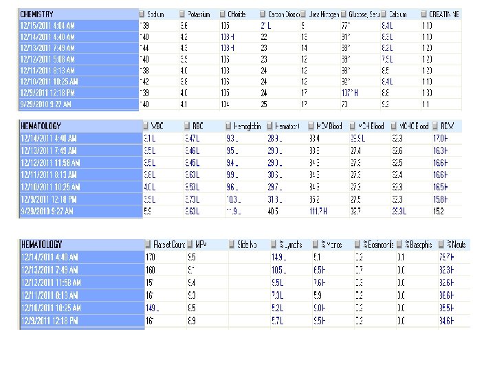
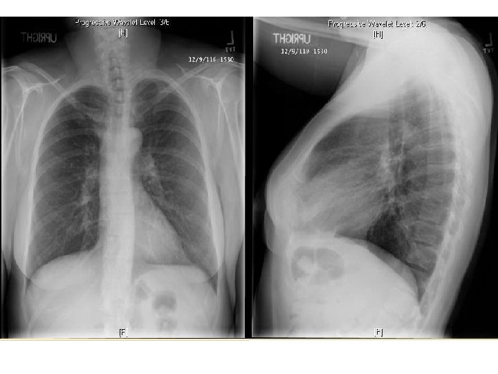
Case 1 • a 39 -year-old Caucasian female with history of FSGS from chronic vesicoureteral reflux, s/p living donor kidney transplant in 6/2010 with immediate allograft function, who is on Prograf 3 mg bid and Myfortic 540 mg bid , pred 5 mg/day • She came to clinic after a 1 -month history of not feeling well, flu-like, body aches, headache, night sweats. Negative for chest pain, palpitations, lightheadedness, dizziness, seizures, • 1 week prior to admission, she has experienced fever as high as 102. 3, SOB, nonproductive cough. • Seen in the transplant clinic, blood work was done, nothing given. • 2 days later : seen by PCP who gave her an unknown dose of steroids, and advised her to come to the. ER if she is not feeling better by next week. • She called the transplant clinic and was advised to come to the hospital due to fever, cough, and shortness of breath.
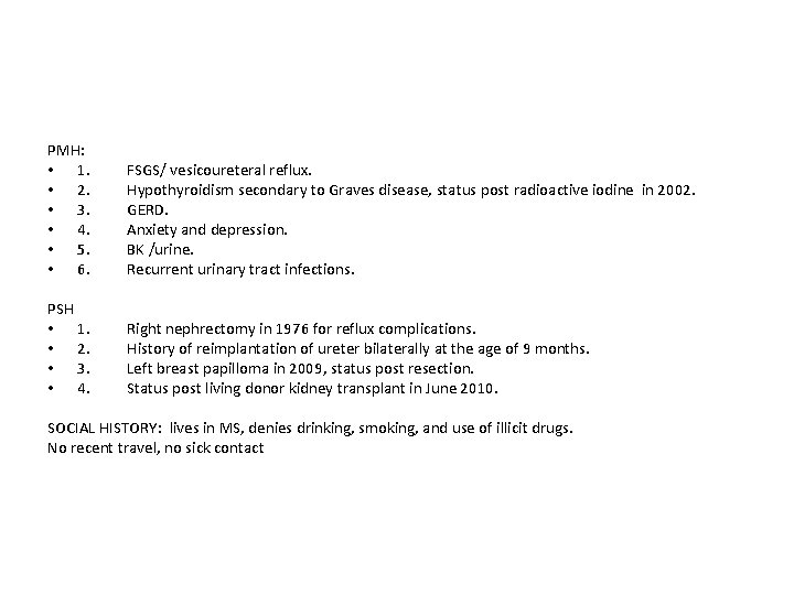
PMH: • 1. • 2. • 3. • 4. • 5. • 6. FSGS/ vesicoureteral reflux. Hypothyroidism secondary to Graves disease, status post radioactive iodine in 2002. GERD. Anxiety and depression. BK /urine. Recurrent urinary tract infections. PSH • 1. • 2. • 3. • 4. Right nephrectomy in 1976 for reflux complications. History of reimplantation of ureter bilaterally at the age of 9 months. Left breast papilloma in 2009, status post resection. Status post living donor kidney transplant in June 2010. SOCIAL HISTORY: lives in MS, denies drinking, smoking, and use of illicit drugs. No recent travel, no sick contact
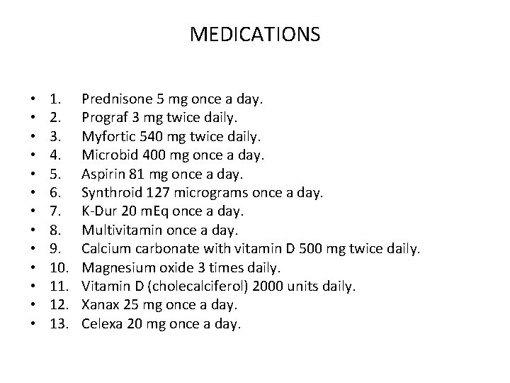
MEDICATIONS • • • • 1. 2. 3. 4. 5. 6. 7. 8. 9. 10. 11. 12. 13. Prednisone 5 mg once a day. Prograf 3 mg twice daily. Myfortic 540 mg twice daily. Microbid 400 mg once a day. Aspirin 81 mg once a day. Synthroid 127 micrograms once a day. K-Dur 20 m. Eq once a day. Multivitamin once a day. Calcium carbonate with vitamin D 500 mg twice daily. Magnesium oxide 3 times daily. Vitamin D (cholecalciferol) 2000 units daily. Xanax 25 mg once a day. Celexa 20 mg once a day.
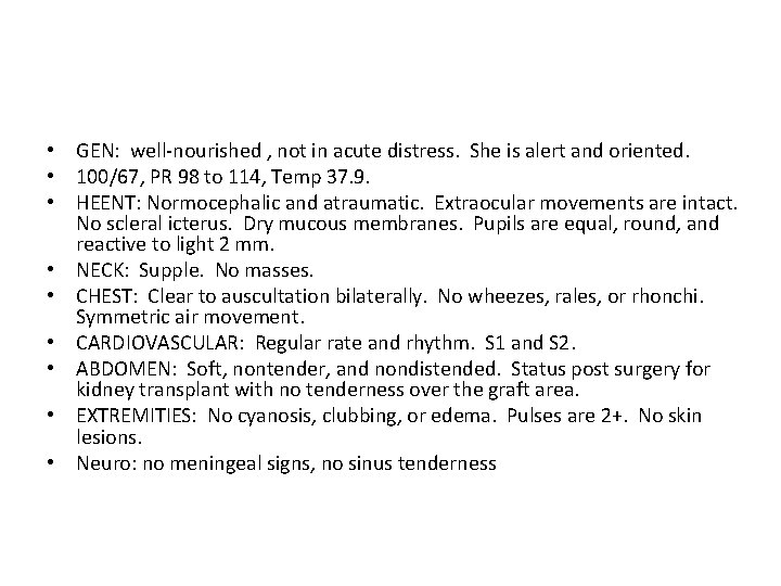
• GEN: well-nourished , not in acute distress. She is alert and oriented. • 100/67, PR 98 to 114, Temp 37. 9. • HEENT: Normocephalic and atraumatic. Extraocular movements are intact. No scleral icterus. Dry mucous membranes. Pupils are equal, round, and reactive to light 2 mm. • NECK: Supple. No masses. • CHEST: Clear to auscultation bilaterally. No wheezes, rales, or rhonchi. Symmetric air movement. • CARDIOVASCULAR: Regular rate and rhythm. S 1 and S 2. • ABDOMEN: Soft, nontender, and nondistended. Status post surgery for kidney transplant with no tenderness over the graft area. • EXTREMITIES: No cyanosis, clubbing, or edema. Pulses are 2+. No skin lesions. • Neuro: no meningeal signs, no sinus tenderness
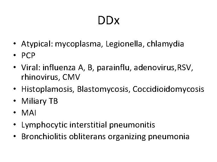
DDx • Atypical: mycoplasma, Legionella, chlamydia • PCP • Viral: influenza A, B, parainflu, adenovirus, RSV, rhinovirus, CMV • Histoplamosis, Blastomycosis, Coccidioidomycosis • Miliary TB • MAI • Lymphocytic interstitial pneumonitis • Bronchiolitis obliterans organizing pneumonia
• antibiotics : CAP vs HCAP? • ID : ok with avelox
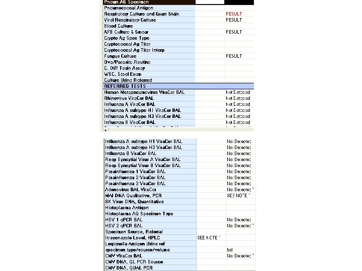
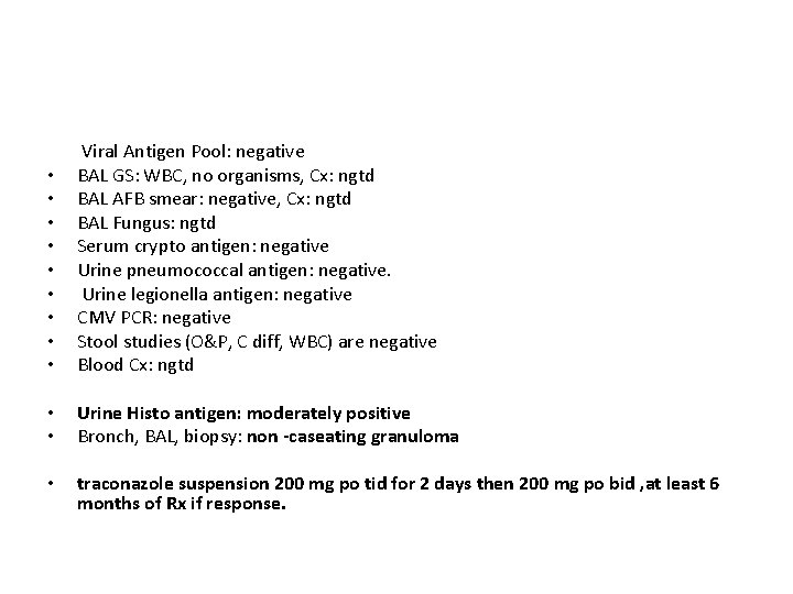
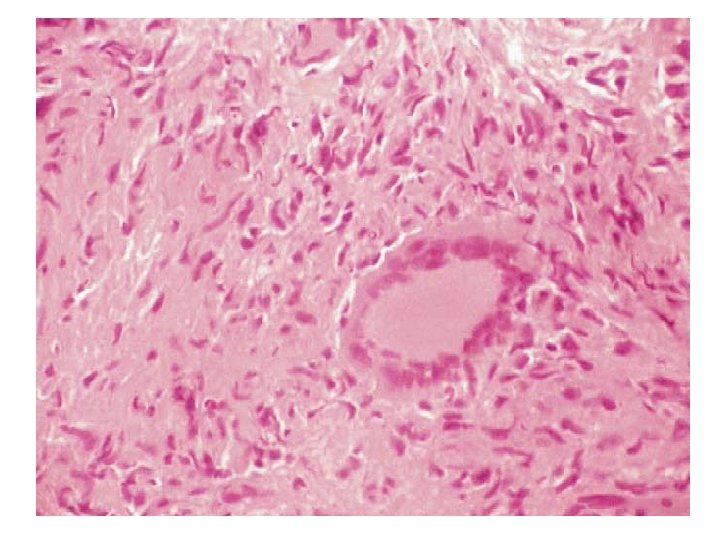
• • • Viral Antigen Pool: negative BAL GS: WBC, no organisms, Cx: ngtd BAL AFB smear: negative, Cx: ngtd BAL Fungus: ngtd Serum crypto antigen: negative Urine pneumococcal antigen: negative. Urine legionella antigen: negative CMV PCR: negative Stool studies (O&P, C diff, WBC) are negative Blood Cx: ngtd • • Urine Histo antigen: moderately positive Bronch, BAL, biopsy: non -caseating granuloma • traconazole suspension 200 mg po tid for 2 days then 200 mg po bid , at least 6 months of Rx if response.
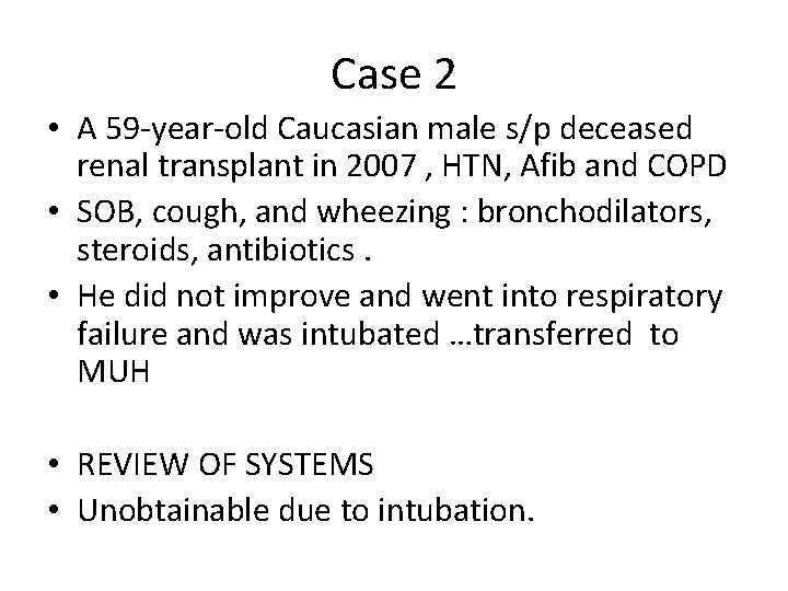
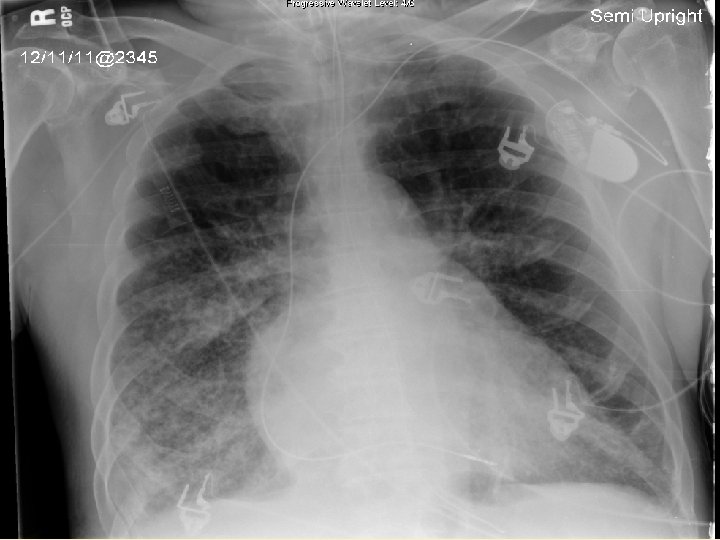
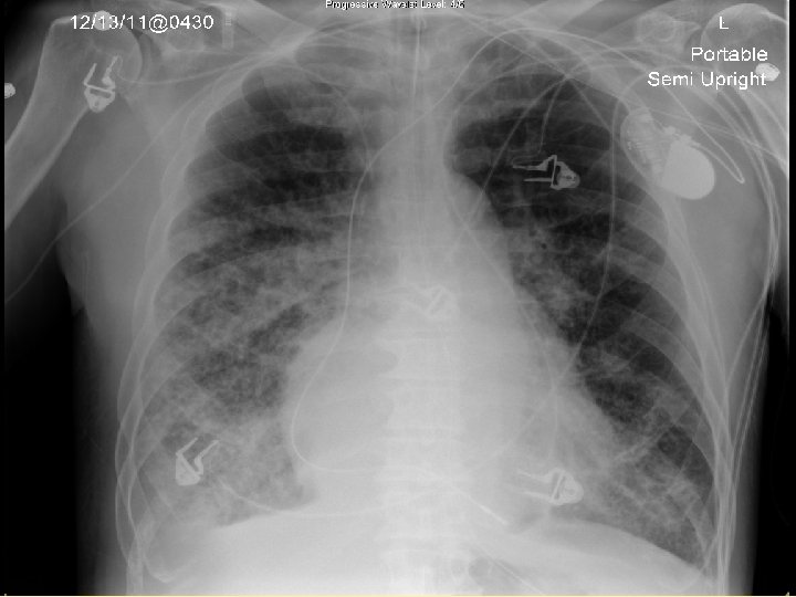
Case 2 • A 59 -year-old Caucasian male s/p deceased renal transplant in 2007 , HTN, Afib and COPD • SOB, cough, and wheezing : bronchodilators, steroids, antibiotics. • He did not improve and went into respiratory failure and was intubated …transferred to MUH • REVIEW OF SYSTEMS • Unobtainable due to intubation.
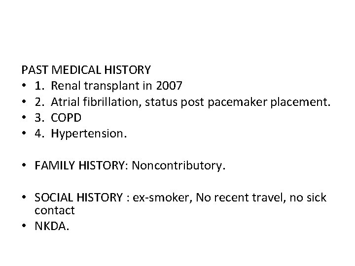
PAST MEDICAL HISTORY • 1. Renal transplant in 2007 • 2. Atrial fibrillation, status post pacemaker placement. • 3. COPD • 4. Hypertension. • FAMILY HISTORY: Noncontributory. • SOCIAL HISTORY : ex-smoker, No recent travel, no sick contact • NKDA.
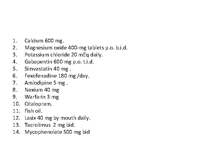
1. 2. 3. 4. 5. 6. 7. 8. 9. 10. 11. 12. 13. 14. Calcium 600 mg. Magnesium oxide 400 -mg tablets p. o. b. i. d. Potassium chloride 20 m. Eq daily. Gabapentin 600 mg p. o. t. i. d. Simvastatin 40 mg. Fexofenadine 180 mg /day. Amlodipine 5 mg. Nexium 40 mg Warfarin 3 mg Citalopram. Fish oil. Lasix 40 mg by mouth daily. Tacrolimus 2 mg bid. Mycophenolate 500 mg bid
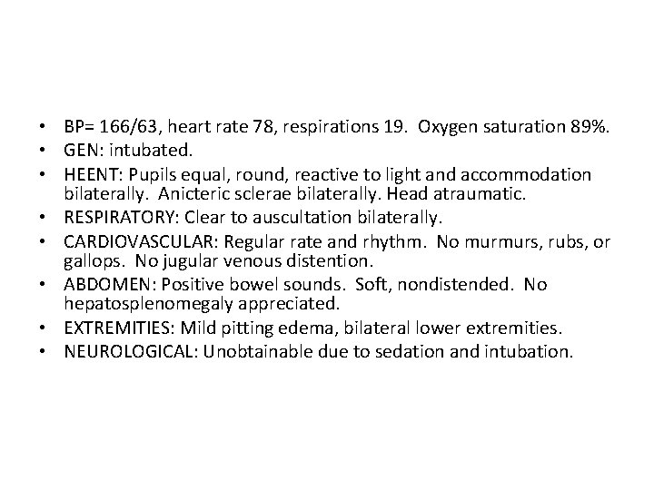
• BP= 166/63, heart rate 78, respirations 19. Oxygen saturation 89%. • GEN: intubated. • HEENT: Pupils equal, round, reactive to light and accommodation bilaterally. Anicteric sclerae bilaterally. Head atraumatic. • RESPIRATORY: Clear to auscultation bilaterally. • CARDIOVASCULAR: Regular rate and rhythm. No murmurs, rubs, or gallops. No jugular venous distention. • ABDOMEN: Positive bowel sounds. Soft, nondistended. No hepatosplenomegaly appreciated. • EXTREMITIES: Mild pitting edema, bilateral lower extremities. • NEUROLOGICAL: Unobtainable due to sedation and intubation.
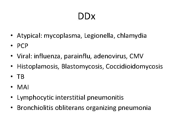
DDx • • Atypical: mycoplasma, Legionella, chlamydia PCP Viral: influenza, parainflu, adenovirus, CMV Histoplamosis, Blastomycosis, Coccidioidomycosis TB MAI Lymphocytic interstitial pneumonitis Bronchiolitis obliterans organizing pneumonia
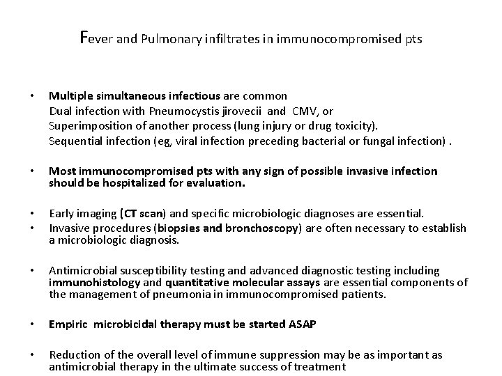
Fever and Pulmonary infiltrates in immunocompromised pts • Multiple simultaneous infectious are common Dual infection with Pneumocystis jirovecii and CMV, or Superimposition of another process (lung injury or drug toxicity). Sequential infection (eg, viral infection preceding bacterial or fungal infection). • Most immunocompromised pts with any sign of possible invasive infection should be hospitalized for evaluation. • • Early imaging (CT scan) and specific microbiologic diagnoses are essential. Invasive procedures (biopsies and bronchoscopy) are often necessary to establish a microbiologic diagnosis. • Antimicrobial susceptibility testing and advanced diagnostic testing including immunohistology and quantitative molecular assays are essential components of the management of pneumonia in immunocompromised patients. • Empiric microbicidal therapy must be started ASAP • Reduction of the overall level of immune suppression may be as important as antimicrobial therapy in the ultimate success of treatment
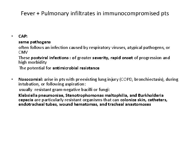
Fever + Pulmonary infiltrates in immunocompromised pts CAP: same pathogens often follows an infection caused by respiratory viruses, atypical pathogens, or CMV These postviral infections : of greater severity, rapid onset of progression and high morbidity The potential for antimicrobial resistance • Nosocomial: arise in pts with preexisting lung injury (COPD, bronchiectasis), during intubation, or following aspiration: usually resistant gram-negative bacilli or fungi: Klebsiella pneumoniae, Stenotrophomonas maltophilia, and Burkholderia cepacia are particularly resistant organisms that can colonize skin, catheters, endotracheal tubes, wound hematomas, and tracheal anastomoses •
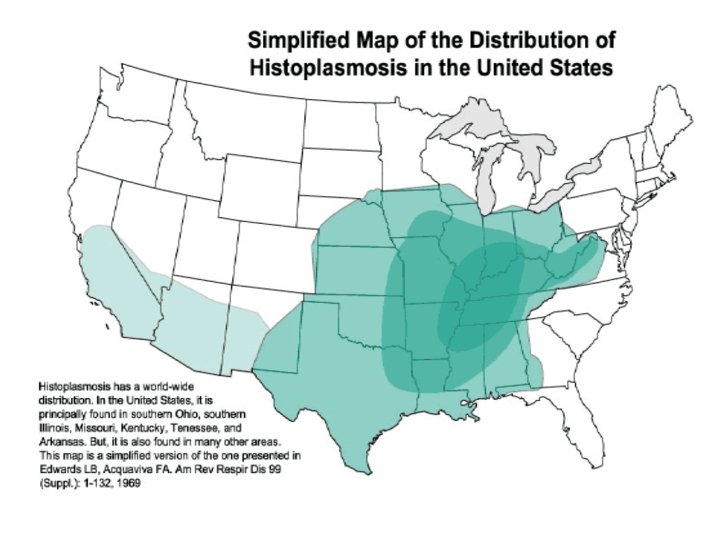
Fever and Pulmonary infiltrates in immunocompromised pts • Reactivation: CMV, strongyloidiasis, cryptococcosis, toxoplasmosis, or mycobacterial, histoplamosisis Serologic testing for Strongyloides or Toxoplasma will generally demonstrate Ig. G antibodies consistent with prior exposure: Ig. G antibodies remain detectable for life. Tuberculin skin testing : a 5 mm response • Environmental exposure: contaminated water or air supply: Legionella pneumophila and Pseudomonas aeruginosa, nontuberculous mycobacteria Soil : has been linked to infections with Aspergillus and Nocardia spp, or to Penicillium marneffei and Burkholderia pseudomallei in Southeast Asia. Travel history is important in considering the risk of certain endemic fungi (Histoplasma, Coccidioides, Blastomyces)
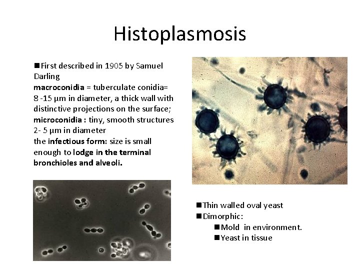
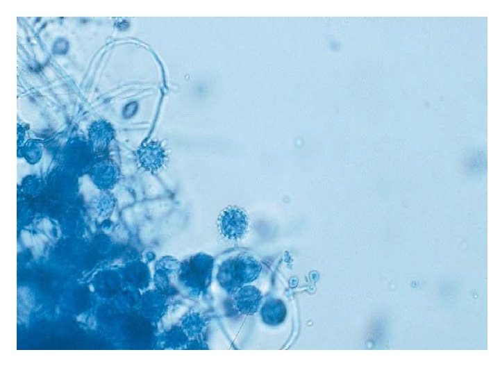
Histoplasmosis n. First described in 1905 by Samuel Darling macroconidia = tuberculate conidia= 8 -15 μm in diameter, a thick wall with distinctive projections on the surface; microconidia : tiny, smooth structures 2 - 5 μm in diameter the infectious form: size is small enough to lodge in the terminal bronchioles and alveoli. n. Thin walled oval yeast n. Dimorphic: n. Mold in environment. n. Yeast in tissue
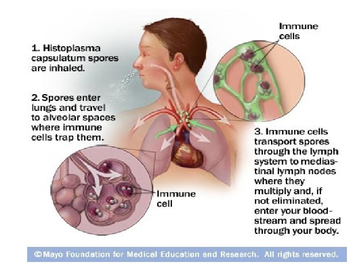
Pathogenesis • After microconidia settle into the alveoli, they bind to the CD 11 -CD 18 family of integrins and are engulfed by neutrophils and macrophages. • On exposure to 37° C, microconidia undergoes genetic, biochemical, and physical alterations that result in the production of yeast cells that are uninucleate. The duration of the phase transition ranges from hours to days. • Yeasts migrate intracellularly to local draining lymph nodes and subsequently to distant organs rich in mononuclear phagocytes (e. g. , liver and spleen). The yeasts grow readily within resting macrophages. • This transition from the mycelial to the yeast phase is the most critical determinant in the establishment of infection. This contention is supported by several findings. it is rare to find mycelial particles in tissues of humans or mammals with established infection. Rather, yeast cells are commonly detected. Exposure of H. capsulatum mycelia to p-chloromercuriphenylsulfonic acid (PCMS), a sulfhydryl inhibitor, irreversibly blocks the conversion to yeasts but does not alter growth of yeasts or mycelia. PCMS-treated mycelia fail to infect animals. • • • Activation of cellular immunity is necessary for restricting growth, and in primary infection this arm of immunity matures by 2 weeks.
Clinical Syndromes of Histoplasmosis n Acute Primary Histoplasmosis n Chronic Pulmonary Histoplasmosis n Mediastinal Granulomatosis n Fibrosing Mediastinitis n Disseminated Histoplasmosis
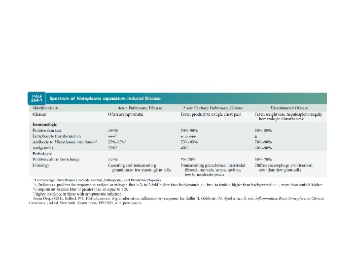
Pulmonary Histoplasmosis • ACUTE INFECTIONS : primary infection or a reinfection. Acute Primary Infection • > 90% asymptomatic or mild influenza-like • a small proportion of patients who become overtly ill. . • the typical incubation time is 7 - 21 days, most individuals manifest symptoms by day 14 • Fever , headache, nonproductive cough, chills, and chest pain • Most symptoms resolve within 10 days, but can persist for several weeks if there is an exposure to a heavy inoculum • Arthralgias, erythema nodosum, and erythema multiforme: 6%, women.
• • Contributing factors include age(>65 y, < 2), inoculum size immuno-compromised • Normal WBC • 30% of pts will have leukocytosis or leukopenia during the course of this infection. • transient increase in the serum ALP.
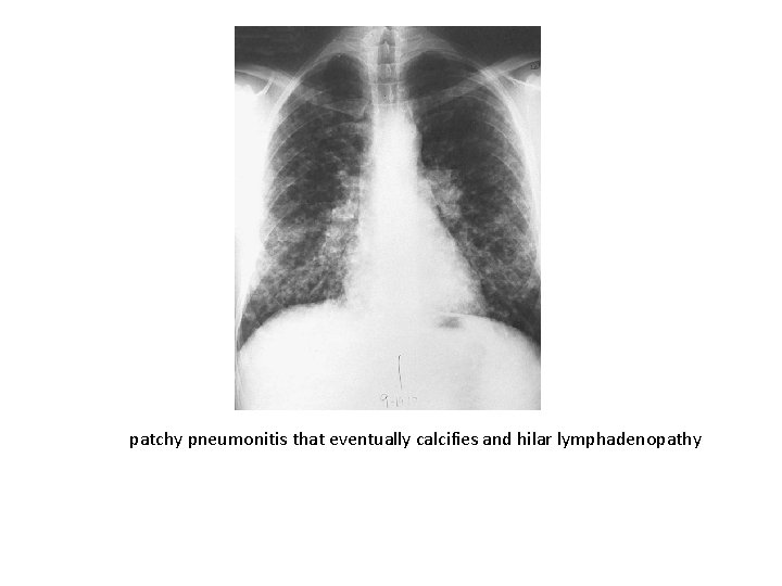
patchy pneumonitis that eventually calcifies and hilar lymphadenopathy
Bob Dylan hospitalized with chest infection May 28, 1997 Web posted at: 10: 07 p. m. EDT NEW YORK (CNN) -- Singer Bob Dylan has been hospitalized with a "potentially fatal" chest infection, according to a statement from his record label. The rock/folk legend, who turned 56 on Saturday, was admitted over the weekend with severe chest pains, the Columbia Records statement said Wednesday. He was diagnosed with histoplasmosis, which causes swelling of the sac that surrounds the heart, the statement said. Columbia did not say where he contracted it or whether the disease was associated with an underlying disorder such as HIV positivity.
Acute Reinfection • It is not uncommon for those who reside in endemic areas to be exposed more than once to H. capsulatum. Those who are reexposed to a large inoculum in heavily endemic areas present with a milder influenza-like illness. • The onset can begin within 3 days( shorter than in primary infection. ) • The characteristic CXR is one of numerous small nodules that are diffusely scattered throughout both lung fields=miliary granulomatosis. • Hilar or mediastinal lymphadenopathy is absent. • The duration of illness often is briefer than in primary infection
Acute Progressive Disseminated Histoplasmosis (acute PDH) • • • develop by reexposure to a large inoculum of the fungus or by reactivation of dormant endogenous foci. abrupt onset , extending over just a few days: Fever, malaise , weight loss, cough, diarrhea. Hepatosplenomegaly, lymphadenopathy, esp the cervical chain, in about 30%, Crackles. Jaundice is observed in a minority, oropharyngeal ulcers develop in less than 20%. DH Septicemia : – Hypotension, ARDS, hepatic, renal failure, DIC. – Usually later in the course of DH-occur in pts who did not seek attention early: 13% of AIDS pt with histo*(1990 study). Skin: papules to ulcers to E. multiforme GI: 10% of cases, diarrhea, vague, abdominal pain. May have GI bleeding, polypoid, ulcerative lesions in colon.
Acute Progressive Disseminated Histoplasmosis • • • Neuro: AMS with focal neurologic findings(10 -30%), Encephalopathy, lymphocytic meningitis Focal parenchymal lesions in brain/SC. Historically, worse prognosis.
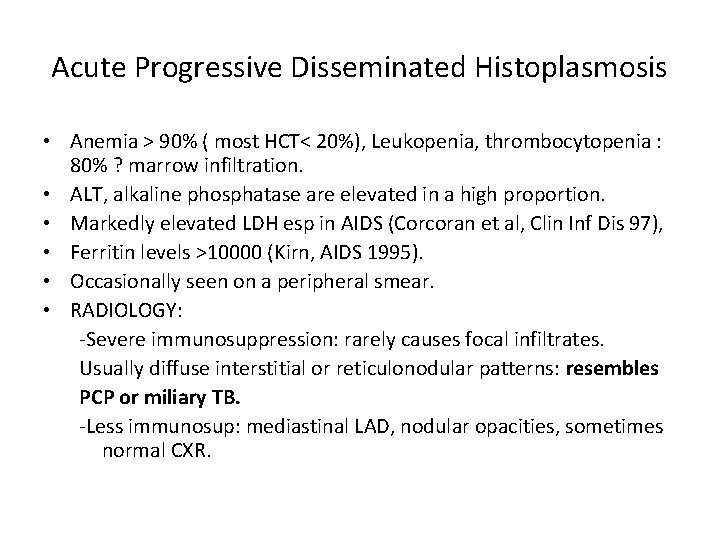
Acute Progressive Disseminated Histoplasmosis • Anemia > 90% ( most HCT< 20%), Leukopenia, thrombocytopenia : 80% ? marrow infiltration. • ALT, alkaline phosphatase are elevated in a high proportion. • Markedly elevated LDH esp in AIDS (Corcoran et al, Clin Inf Dis 97), • Ferritin levels >10000 (Kirn, AIDS 1995). • Occasionally seen on a peripheral smear. • RADIOLOGY: -Severe immunosuppression: rarely causes focal infiltrates. Usually diffuse interstitial or reticulonodular patterns: resembles PCP or miliary TB. -Less immunosup: mediastinal LAD, nodular opacities, sometimes normal CXR.
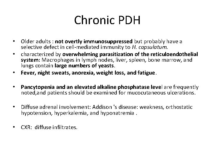
Chronic PDH • Older adults : not overtly immunosuppressed but probably have a selective defect in cell-mediated immunity to H. capsulatum. • characterized by overwhelming parasitization of the reticuloendothelial system: Macrophages in lymph nodes, liver, spleen, bone marrow, and lungs contain large numbers of yeasts. • Fever, night sweats, anorexia, weight loss, and fatigue. • Pancytopenia and an elevated alkaline phosphatase level are frequently noted, and patients should be examined for mucocutaneous ulcerations. • Diffuse adrenal involvement: Addison 's disease: weakness, orthostatic hypotension, hyperkalemia, and hyponatremia. • CXR: diffuse infiltrates.
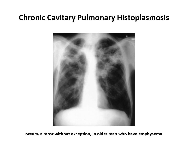
Chronic Cavitary Pulmonary Histoplasmosis occurs, almost without exception, in older men who have emphysema
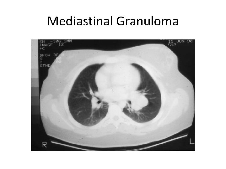
Mediastinal Granuloma
A definitive test • Culture : tissue or body fluids are plated onto Sabouraud’s dextrose agar and incubated at 25°C to allow for growth of the mycelial phase of H. capsulatum. • After several weeks up to 6 weeks, growth of a white to light tan mold occurs • Identification of the tuberculate macroconidia allows a presumptive diagnosis
A definitive test • Need to verify that the mold is H. capsulatum • A commercially available chemiluminescent DNA probe for H. capsulatum (Accu-Probe; Gen. Probe, Inc. , San Diego, CA) is the confirmatory test most often used for definitive identification. • Highly specific; false-positive tests are very rare but have been reported.
Culture The greatest yield of culture positivity: • Disseminated infection (samples can be taken from blood, bone marrow, liver, skin lesions, or any other site of infection) • Chronic cavitary pulmonary histoplasmosis, • Acute pulmonary histoplasmosis following exposure to a large inoculum of the organism. • • Low yield: mild to moderate acute pulmonary infection, granulomatous mediastinitis, mediastinal fibrosis, and Chronic meningitis. • Cultures usually yield no growth from CSF, and the diagnosis must be made in a different manner.
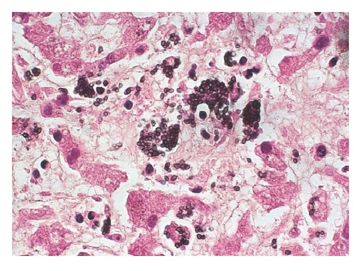
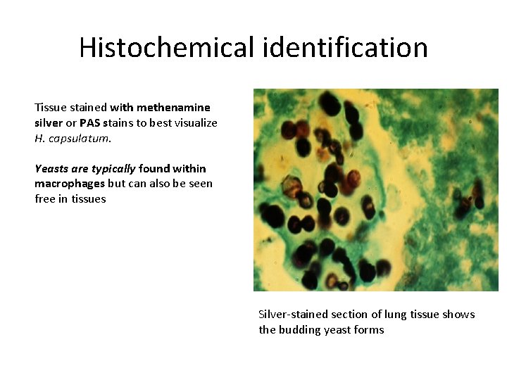
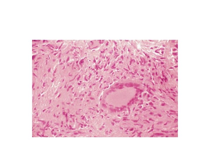
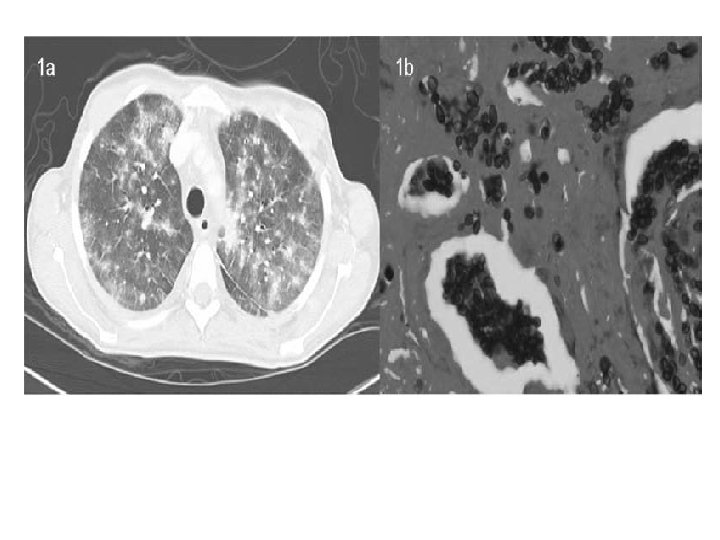
Histochemical identification Tissue stained with methenamine silver or PAS stains to best visualize H. capsulatum. Yeasts are typically found within macrophages but can also be seen free in tissues Silver-stained section of lung tissue shows the budding yeast forms
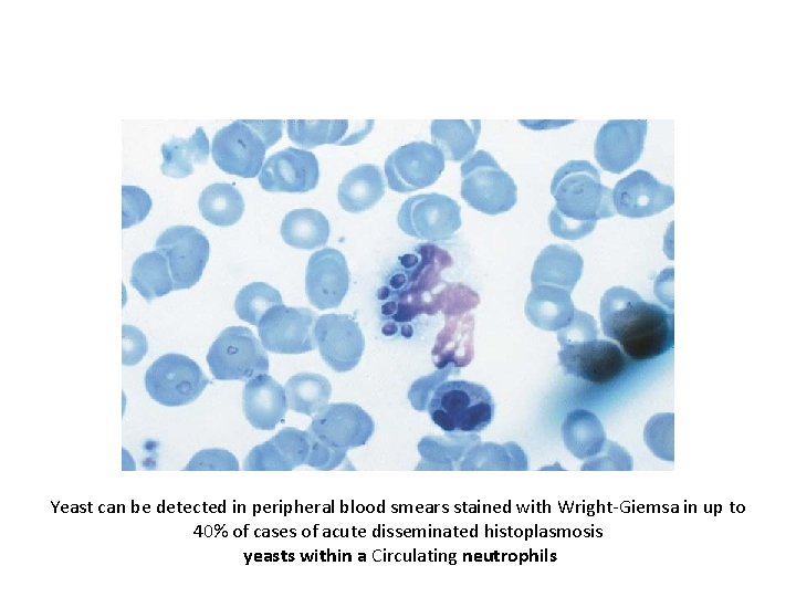
Yeast can be detected in peripheral blood smears stained with Wright-Giemsa in up to 40% of cases of acute disseminated histoplasmosis yeasts within a Circulating neutrophils
Histochemical identification • Severely ill with diffuse pulmonary infiltrates are likely to have organisms that are apparent on a lung biopsy specimen • Milder disease often have no organisms seen in biopsy samples. • Granulomatous mediastinitis: caseous material taken from necrotic nodes may contain a few yeast-like organisms typical of H. capsulatum. • Fibrosing mediastinitis typically reveals no organisms
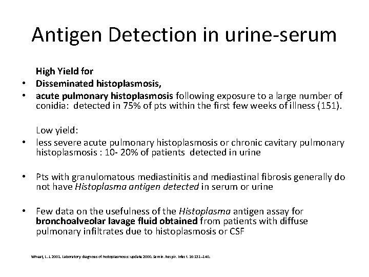
Antigen Detection in urine-serum • detects polysaccharide antigen in serum or urine by ELISA, is the mainstay of diagnosis, especially in those with disseminated disease. • The greatest experience was obtained in AIDS patients who had disseminated histoplasmosis and who had a large burden of organisms : Histoplasma antigen was detected in urine in 95% , in serum in 86% of patients Wheat, L. J. 2001. Laboratory diagnosis of histoplasmosis: update 2000. Semin. Respir. Infect. 16: 131– 140. • Fewer data are available for the use of the antigen assay in non-AIDS patients with disseminated histoplasmosis, but it appears to be a sensitive assay in this population also. • Published reports note that 92% of patients have antigenuria, but only approximately 50% have antigenemia
Antigen Detection in urine-serum High Yield for • Disseminated histoplasmosis, • acute pulmonary histoplasmosis following exposure to a large number of conidia: detected in 75% of pts within the first few weeks of illness (151). Low yield: • less severe acute pulmonary histoplasmosis or chronic cavitary pulmonary histoplasmosis : 10 - 20% of patients detected in urine • Pts with granulomatous mediastinitis and mediastinal fibrosis generally do not have Histoplasma antigen detected in serum or urine • Few data on the usefulness of the Histoplasma antigen assay for bronchoalveolar lavage fluid obtained from patients with diffuse pulmonary infiltrates due to histoplasmosis or CSF Wheat, L. J. 2001. Laboratory diagnosis of histoplasmosis: update 2000. Semin. Respir. Infect. 16: 131– 140.
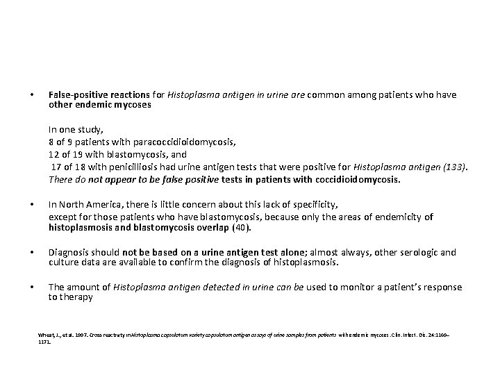
• False-positive reactions for Histoplasma antigen in urine are common among patients who have other endemic mycoses In one study, 8 of 9 patients with paracoccidioidomycosis, 12 of 19 with blastomycosis, and 17 of 18 with penicilliosis had urine antigen tests that were positive for Histoplasma antigen (133). There do not appear to be false positive tests in patients with coccidioidomycosis. • In North America, there is little concern about this lack of specificity, except for those patients who have blastomycosis, because only the areas of endemicity of histoplasmosis and blastomycosis overlap (40). • Diagnosis should not be based on a urine antigen test alone; almost always, other serologic and culture data are available to confirm the diagnosis of histoplasmosis. • The amount of Histoplasma antigen detected in urine can be used to monitor a patient’s response to therapy Wheat, J. , et al. 1997. Cross-reactivity in Histoplasma capsulatum variety capsulatum antigen assays of urine samples from patients with endemic mycoses. Clin. Infect. Dis. 24: 1169– 1171.
PCR assay • At this point, no PCR assay for routine use is commercially available.
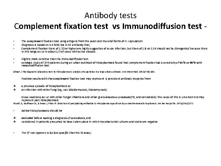
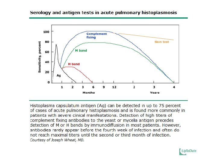
Antibody tests Complement fixation test vs Immunodiffusion test • • • The complement fixation test using antigens from the yeast and mycelial forms of H. capsulatum Diagnosis is based on a 4 -fold rise in CF antibody titer; Complement fixation titers of 1: 32 or higher are highly suggestive of acute infection, but titers of 1: 8 or 1: 16 should not be disregarded because titers in this range occur in about 1/3 of cases with active disease. • • Slightly more sensitive than the immunodiffusion test. serologic study of 276 patients during an urban outbreak of histoplasmosis found that complement fixation had a sensitivity of 95 % vs 90 % with immunodiffusion test. Wheat J. The diagnostic laboratory tests for histoplasmosis: analysis of experience in a large urban outbreak. Ann Intern Med. 1982; 97(5): 680. Positive results with the complement fixation test may represent a persistent antibody response from v v a previous episode of histoplasmosis or an infection with other fungi (eg, coccidioidomycosis, blastomycosis). Cross-reactions occur with other fungal infections and other granulomatous processes(TB, and sarcoidosis): the cause of this is uncertain but may represent past histoplasmosis. Picardi JL, Kauffman CA, Schwarz J, Phair JP. Detection of precipitating antibodies to Histoplasma capsulatum by counterimmunoelectrophoresis. Am Rev Respir Dis. 1976; 114(1): 171 • Active histoplasmosis should be v v excluded before making a diagnosis of sarcoidosis, and considered in patients presumed to have tuberculosis in which mycobacterial cultures and stains are negative • The CF test appears to be less specific than the ID assay;
immunodiffusion test Results are reported as M or H precipitins or bands. Most patients will develop an M band; M band becomes positive sooner than the H band persists longer H precipitin band is detectable in < 20% of cases often in • Disseminated infection, • Chronic cavitary pulmonary histoplasmosis, or • More severe acute pulmonary histoplasmosis ID test has greater specificity than the CF test.
Antibody tests • Tests for antibody are most useful in patients who have chronic forms of histoplasmosis that have allowed enough time for antibody to develop and in patients who have acute pulmonary histoplasmosis, in whom documentation of a fourfold rise in antibody titer to H. capsulatum can be diagnostic. • Because 2 to 6 weeks may be required for the appearance of antibodies, these assays are less useful for patients who have severe acute infection and in immunosuppressed patients, who mount a poor response. • For those patients who have mediastinal lymphadenopathy, antibody assays may or may not be positive and cannot be relied upon to make a definitive diagnosis. Tissue biopsy is always required in these instances. • False-positive CF tests occur in patients with lymphoma, tuberculosis, sarcoidosis, and other fungal infections, all of which may present as a mediastinal mass.
Antibody tests • One unique circumstance in which the presence of antibodies may be the only assay leading to a diagnosis of histoplas mosis is in a patient who has chronic meningitis due to H. capsulatum. • the presence of either CF or ID antibodies against H. capsulatum in the CSF allows one to make the diagnosis of Histoplasma meningitis even when the culture shows no growth
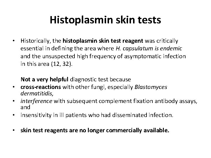
Histoplasmin skin tests • Historically, the histoplasmin skin test reagent was critically essential in defining the area where H. capsulatum is endemic and the unsuspected high frequency of asymptomatic infection in this area (12, 32). Not a very helpful diagnostic test because • cross-reactions with other fungi, especially Blastomyces dermatitidis, • interference with subsequent complement fixation antibody assays, and • insensitivity in ill patients who had disseminated infection. • skin test reagents are no longer commercially available.
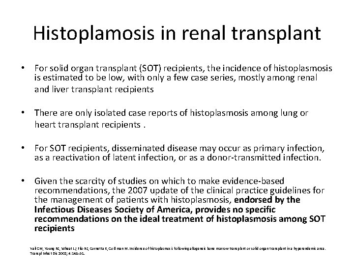
Histoplamosis in renal transplant • For solid organ transplant (SOT) recipients, the incidence of histoplasmosis is estimated to be low, with only a few case series, mostly among renal and liver transplant recipients • There are only isolated case reports of histoplasmosis among lung or heart transplant recipients. • For SOT recipients, disseminated disease may occur as primary infection, as a reactivation of latent infection, or as a donor-transmitted infection. • Given the scarcity of studies on which to make evidence-based recommendations, the 2007 update of the clinical practice guidelines for the management of patients with histoplasmosis, endorsed by the Infectious Diseases Society of America, provides no specific recommendations on the ideal treatment of histoplasmosis among SOT recipients Vail GM, Young RS, Wheat LJ, Filo RS, Cornetta K, Goldman M. Incidence of histoplasmosis following allogeneic bone marrow transplant or solid organ transplant in a hyperendemic area. Transpl Infect Dis 2002; 4: 148– 51.
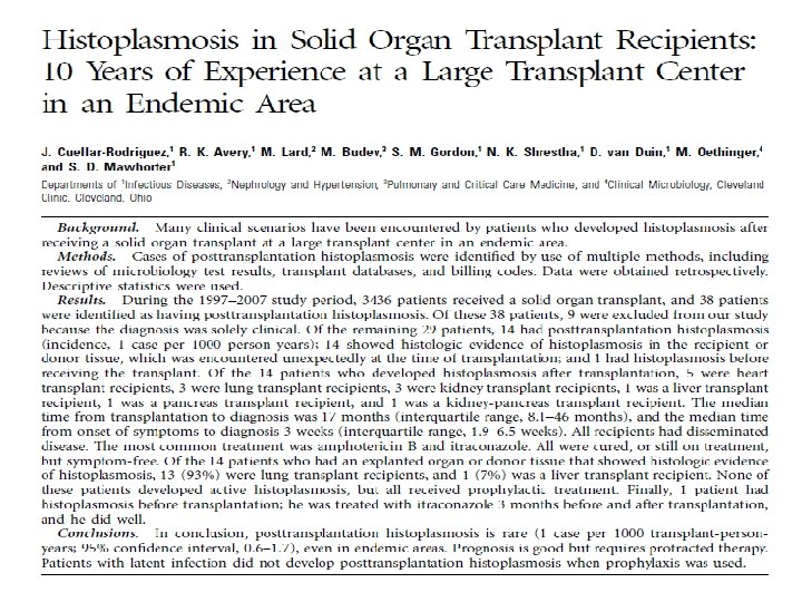
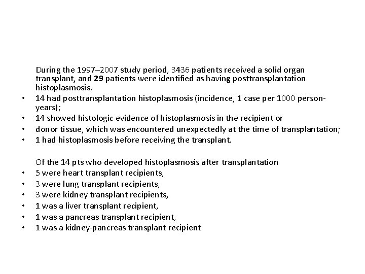
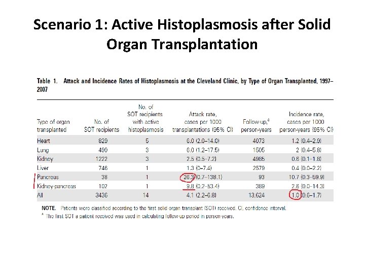
• • • During the 1997– 2007 study period, 3436 patients received a solid organ transplant, and 29 patients were identified as having posttransplantation histoplasmosis. 14 had posttransplantation histoplasmosis (incidence, 1 case per 1000 personyears); 14 showed histologic evidence of histoplasmosis in the recipient or donor tissue, which was encountered unexpectedly at the time of transplantation; 1 had histoplasmosis before receiving the transplant. • • • Of the 14 pts who developed histoplasmosis after transplantation 5 were heart transplant recipients, 3 were lung transplant recipients, 3 were kidney transplant recipients, 1 was a liver transplant recipient, 1 was a pancreas transplant recipient, 1 was a kidney-pancreas transplant recipient •
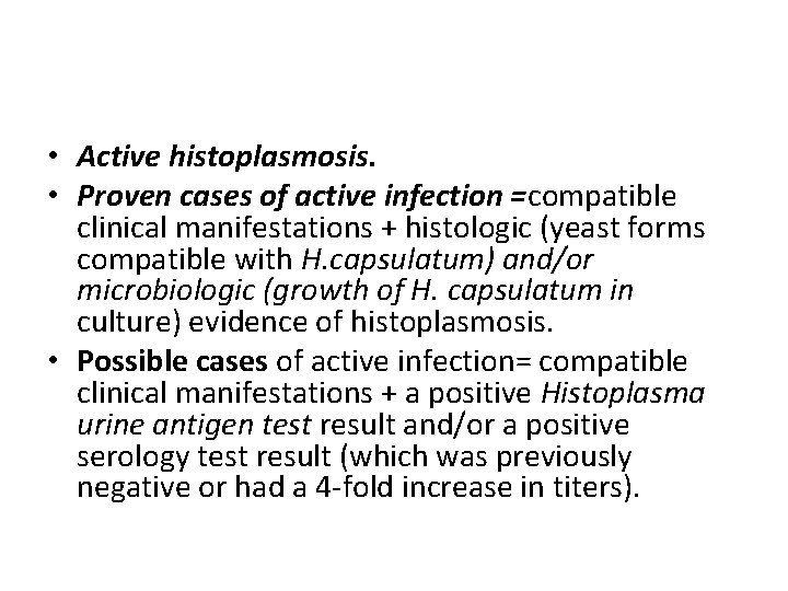
• Active histoplasmosis. • Proven cases of active infection =compatible clinical manifestations + histologic (yeast forms compatible with H. capsulatum) and/or microbiologic (growth of H. capsulatum in culture) evidence of histoplasmosis. • Possible cases of active infection= compatible clinical manifestations + a positive Histoplasma urine antigen test result and/or a positive serology test result (which was previously negative or had a 4 -fold increase in titers).
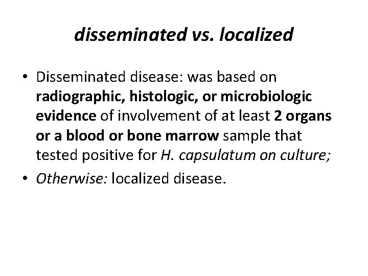
disseminated vs. localized • Disseminated disease: was based on radiographic, histologic, or microbiologic evidence of involvement of at least 2 organs or a blood or bone marrow sample that tested positive for H. capsulatum on culture; • Otherwise: localized disease.
Scenario 1: Active Histoplasmosis after Solid Organ Transplantation
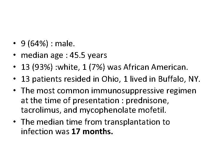
9 (64%) : male. median age : 45. 5 years 13 (93%) : white, 1 (7%) was African American. 13 patients resided in Ohio, 1 lived in Buffalo, NY. The most common immunosuppressive regimen at the time of presentation : prednisone, tacrolimus, and mycophenolate mofetil. • The median time from transplantation to infection was 17 months. • • •
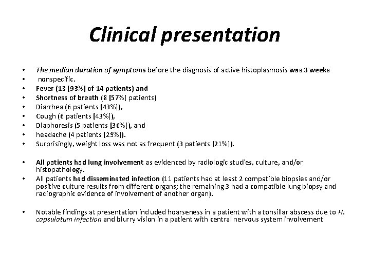
Clinical presentation • • • The median duration of symptoms before the diagnosis of active histoplasmosis was 3 weeks nonspecific. Fever (13 [93%] of 14 patients) and Shortness of breath (8 [57%] patients) Diarrhea (6 patients [43%]), Cough (6 patients [43%]), Diaphoresis (5 patients [36%]), and headache (4 patients [29%]). Surprisingly, weight loss was not as frequent (3 patients [21%]). • • All patients had lung involvement as evidenced by radiologic studies, culture, and/or histopathology. All patients had disseminated infection (11 patients had at least 2 compatible biopsies and/or positive culture results from different organs; the remaining 3 had a compatible lung biopsy and radiographic evidence of involvement of another organ). • Notable findings at presentation included hoarseness in a patient with a tonsillar abscess due to H. capsulatum infection and blurry vision in a patient with central nervous system involvement
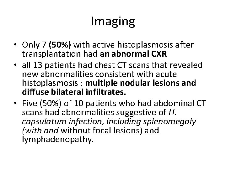
Imaging • Only 7 (50%) with active histoplasmosis after transplantation had an abnormal CXR • all 13 patients had chest CT scans that revealed new abnormalities consistent with acute histoplasmosis : multiple nodular lesions and diffuse bilateral infiltrates. • Five (50%) of 10 patients who had abdominal CT scans had abnormalities suggestive of H. capsulatum infection, including splenomegaly (with and without focal lesions) and lymphadenopathy.
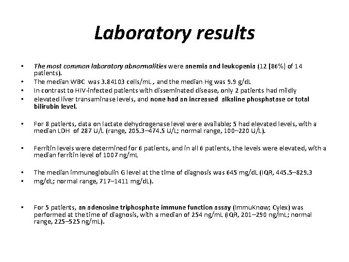
Laboratory results • • The most common laboratory abnormalities were anemia and leukopenia (12 [86%] of 14 patients). The median WBC was 3. 84103 cells/m. L , and the median Hg was 9. 9 g/d. L In contrast to HIV-infected patients with disseminated disease, only 2 patients had mildly elevated liver transaminase levels, and none had an increased alkaline phosphatase or total bilirubin level. • For 8 patients, data on lactate dehydrogenase level were available; 5 had elevated levels, with a median LDH of 287 U/L (range, 205. 3– 474. 5 U/L; normal range, 100– 220 U/L). • Ferritin levels were determined for 6 patients, and in all 6 patients, the levels were elevated, with a median ferritin level of 1007 ng/m. L • • The median immunoglobulin G level at the time of diagnosis was 645 mg/d. L (IQR, 445. 5– 829. 3 mg/d. L; normal range, 717– 1411 mg/d. L). • For 5 patients, an adenosine triphosphate immune function assay (Immu. Know; Cylex) was performed at the time of diagnosis, with a median of 254 ng/m. L (IQR, 201– 290 ng/m. L; normal range, 225– 525 ng/m. L).
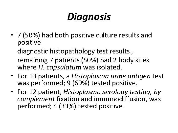
Diagnosis • 7 (50%) had both positive culture results and positive diagnostic histopathology test results , remaining 7 patients (50%) had 2 body sites where H. capsulatum was isolated. • For 13 patients, a Histoplasma urine antigen test was performed; 9 (69%) tested positive. • For 12 patient, Histoplasma serology testing, by complement fixation and immunodiffusion, was performed; 4 (33%) tested positive.
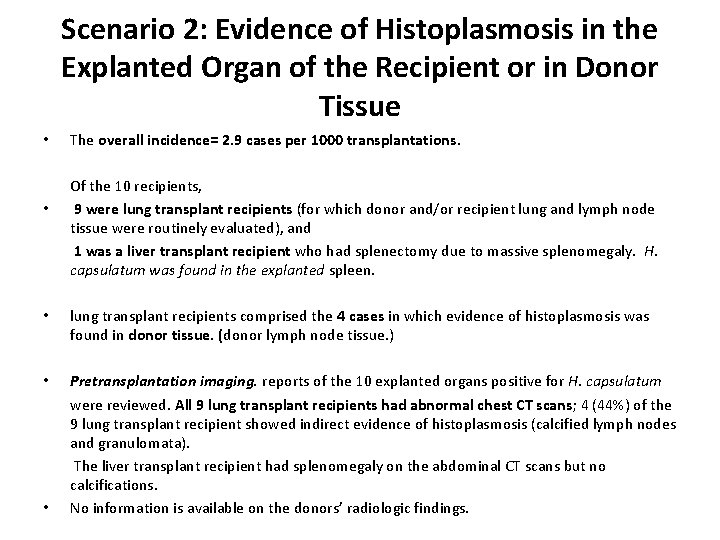
Scenario 2: Evidence of Histoplasmosis in the Explanted Organ of the Recipient or in Donor Tissue • • The overall incidence= 2. 9 cases per 1000 transplantations. Of the 10 recipients, 9 were lung transplant recipients (for which donor and/or recipient lung and lymph node tissue were routinely evaluated), and 1 was a liver transplant recipient who had splenectomy due to massive splenomegaly. H. capsulatum was found in the explanted spleen. • lung transplant recipients comprised the 4 cases in which evidence of histoplasmosis was found in donor tissue. (donor lymph node tissue. ) • Pretransplantation imaging. reports of the 10 explanted organs positive for H. capsulatum were reviewed. All 9 lung transplant recipients had abnormal chest CT scans; 4 (44%) of the 9 lung transplant recipient showed indirect evidence of histoplasmosis (calcified lymph nodes and granulomata). The liver transplant recipient had splenomegaly on the abdominal CT scans but no calcifications. No information is available on the donors’ radiologic findings. •
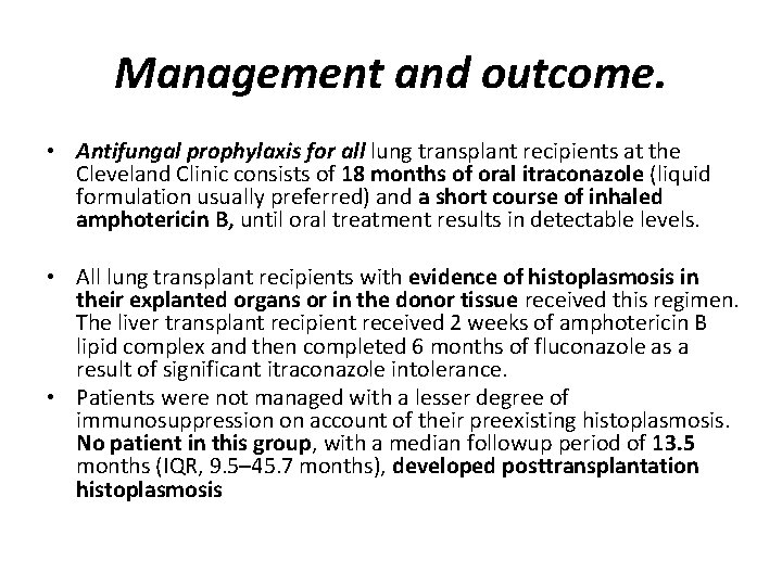
Management and outcome. • Antifungal prophylaxis for all lung transplant recipients at the Cleveland Clinic consists of 18 months of oral itraconazole (liquid formulation usually preferred) and a short course of inhaled amphotericin B, until oral treatment results in detectable levels. • All lung transplant recipients with evidence of histoplasmosis in their explanted organs or in the donor tissue received this regimen. The liver transplant recipient received 2 weeks of amphotericin B lipid complex and then completed 6 months of fluconazole as a result of significant itraconazole intolerance. • Patients were not managed with a lesser degree of immunosuppression on account of their preexisting histoplasmosis. No patient in this group, with a median followup period of 13. 5 months (IQR, 9. 5– 45. 7 months), developed posttransplantation histoplasmosis
Scenario 3: Pretransplantation Histoplasmosis • 1 pt had possible active histoplasmosis while on the waiting list for a kidney-liver transplant: had new onset of SOB ; a new lung nodule and a positive Histoplasma antibody (M band). Lung biopsy revealed loosely formed granulomata. • He received itraconazole for 3 months before the transplantation and continued treatment for 3 months after the transplantation. • Symptoms had subsided before he received the transplant. • The posttransplantation clinical course has been uneventful, with no evidence of recurrent histoplasmosis during 9 months of follow-up.
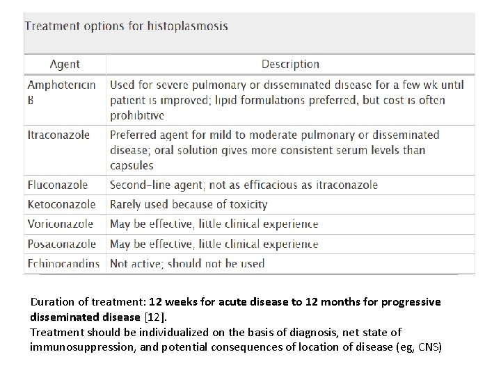
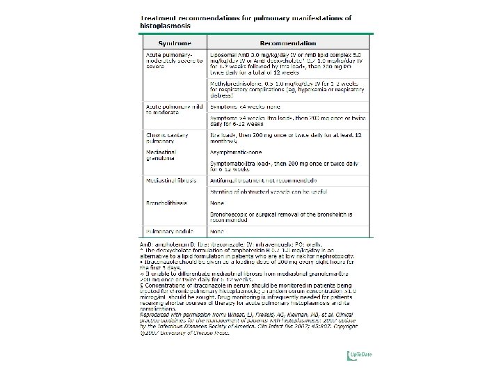
Duration of treatment: 12 weeks for acute disease to 12 months for progressive disseminated disease [12]. Treatment should be individualized on the basis of diagnosis, net state of immunosuppression, and potential consequences of location of disease (eg, CNS)