Anatomy FEMALE PELVIC THE PELVIS protective shield supports


- Slides: 42
Anatomy FEMALE PELVIC
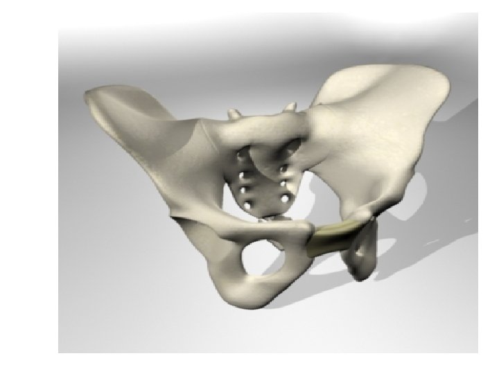
THE PELVIS • protective shield • supports the trunk • pelvis consists of the two innominate bones sacrum and coccyx. • There are four articulations • Sacroiliac Joints (x 2) – Between the ilium of the hip bones, and the sacrum • Sacrococcygeal symphysis – Between the sacrum and the coccyx. • Pubic symphysis – Between the pubis bodies of the two hip bones • It forms the ring like structure
Functions of the Pelvis The strong and rigid pelvis is adapted to serve a number of roles in the human body. The main functions being: Transfer of weight from the upper axial skeleton to the lower appendicular components of the skeleton, especially during movement. Provides attachment for a number of muscles and ligaments used in locomotion. Contains and protects the abdominopelvic and pelvic viscera
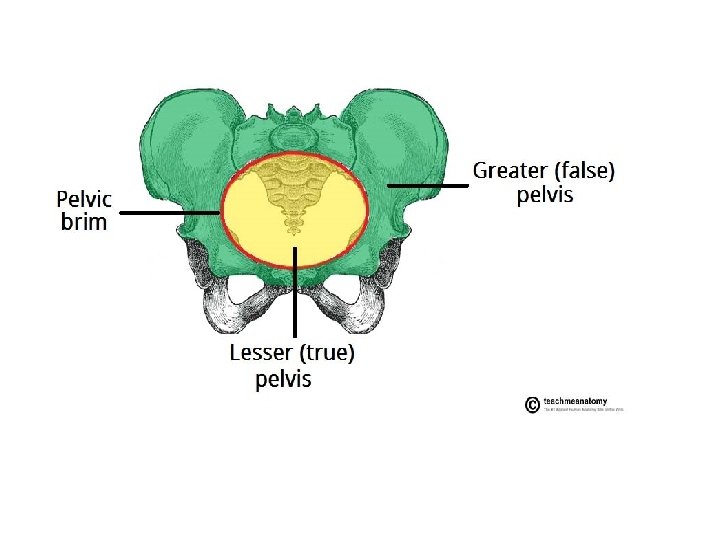
The Greater and Lesser Pelvis The osteology of the pelvic girdle allows the pelvic region to be divided into two: Greater pelvis(false pelvis) Lesser pelvis( true pelvis) • The superior portion of the pelvis is known as the greater pelvis(or false pelvis). It provides support for the lower abdominal viscera (ileum and sigmoid colon), and has no obstetric relevance. • The inferior portion of the pelvis is known as the lesser pelvis (or “true” pelvis). Within this reside the pelvic cavity and pelvic viscera. The junction between the greater and lesser pelvis is known as the pelvic inlet. The outer bony edges of the pelvic inlet are called the pelvic brim.
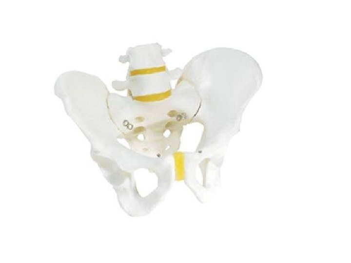
• The ring of bone is deeper posteriorly than anteriorly and forms a curved canal. • The inlet to this canal is at the level of the sacral promontory and superior aspect of the pubic bones
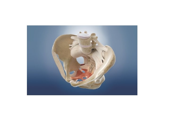
Iliolumber and saccrospinous lagaments
Saccrotuberous legament
• The outlet is formed by the pubic arch, ischial spines, sacrotuberous ligaments and the coccyx. • The enclosed space between the inlet and outlet is called the true pelvis, with the plane of the inlet being at right angles to the plane of the outlet.
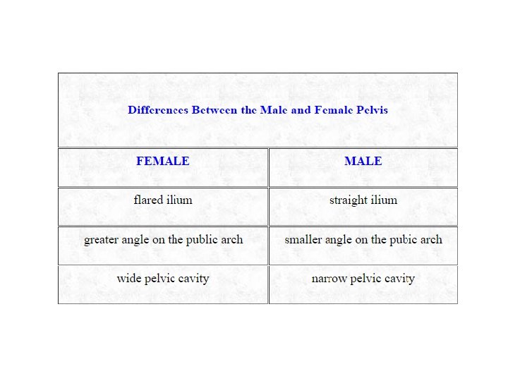
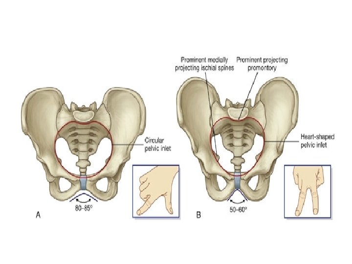
• The female true pelvis differs from the male in being shallower, having straighter sides, a wider angle between the pubic rami at the symphysis and a proportionately larger pelvic outlet.
THE PELVIC FLOOR AND MUSCLES OF THE PELVIS Gynae & Obs in PT
Pelvic Floor Musculature
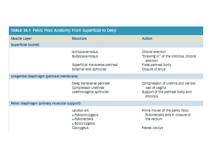
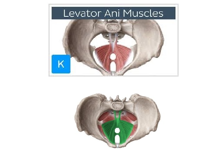
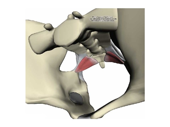
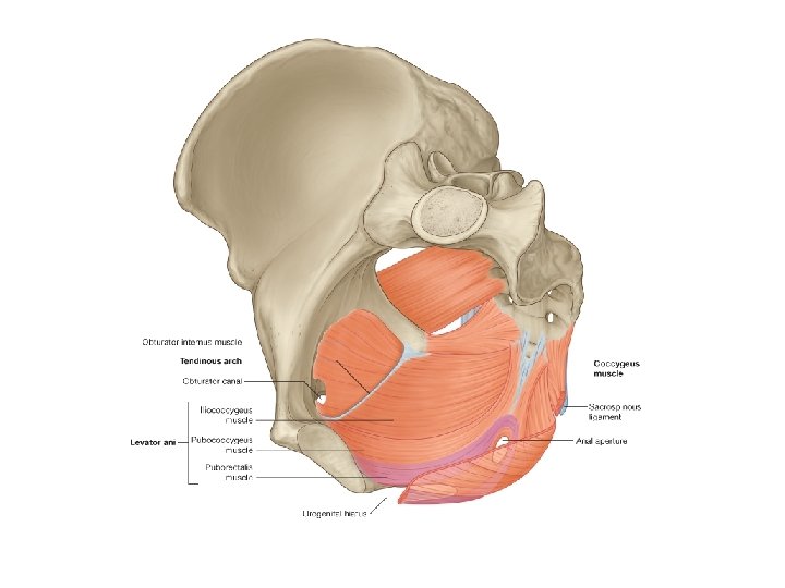
• The pelvic floor musculature is composed of three layers in a funnel-shaped orientation, with boney attachments to the pubic bone and the coccyx. • Laterally, the tissues blend into a fascial layer overlying the obturator internus. • The prime mover of the pelvic floor is the levator ani. • The levator ani, in combination with the coccygeus, forms the pelvic diaphragm.
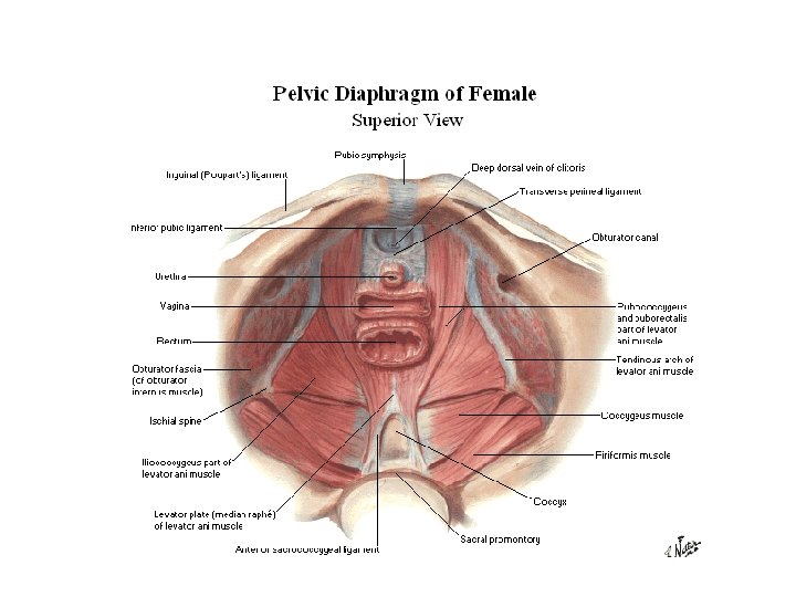
Female Pelvic Floor The female pelvic floor has three openings , This creates less inherent stability when compared to the male anatomy. Innervation The nerve supply to the perineal tissues includes the pudendal nerve (with its three terminal branches, the dorsal, perineal and rectal), the levator ani nerve, and direct branches from the sacral levels. This dual innervation provides a safeguard against direct damage during labor and delivery, that would be more likely with single nerve arrangement.
Function The pelvic floor musculature has the following essential roles: • Provide support for the pelvic organs and their contents • Withstand increases in intra-abdominal pressure • Contribute to stabilization of the spine / pelvis • Maintain continence (through sympathetic nerve fibers) to the urethral and anal sphincters • Reproductive function
Effects of childbirth on the pelvic floor Neurological Compromise Stretch and compression of the pudendal nerve occur during labor as the baby’s head travels through the birth canal. This stretch can be as much as 20% of the total length of the nerve. This compromise to the pudendal nerve is most intense during pushing (the second stage of labor), through the completion of delivery.
• Muscular impairment • Extreme stretching of pelvic floor tissue is inherent in the process of labor and delivery.
• An episiotomy • Is an incision made in the perineal body It is automatically considered a second-degree laceration • according to the following classification of perineal lacerations. • First degree—only skin • Second degree—includes underlying superficial muscle layer • Third degree—extends to anal sphincter • Fourth degree—tears through the sphincter and into the rectum, possibly into the deeper muscular layer of pelvic floor. • Although episiotomy is common, occurring in 33% to 51% of normal deliveries. Adverse effects are sphincter defects • Fecal incontinence • More worse with forceps delivery
Pelvic flour dysfunction
• Classification of Pelvic Floor Dysfunction • This is a very broad category that encompasses symptoms in a variety of combinations. Some patients with pelvic floor disuse atrophy, weakness, or nerve damage; others will have hypertonic pelvic floor musculature. • Pelvic pain is another far-reaching diagnosis; many of these patients will be seen by multiple doctors prior to physical therapy being considered. 1. Prolapse • A prolapse is a supportive impairment. It refers to the descent of any of the pelvic viscera out of their normal alignment because of muscular, fascial, and/or ligamentous deficits and because of increased abdominal pressure. • A prolapse often worsens over time and with subsequent pregnancies and can be aggravated by constipation and/or straining with elimination.
• • • Symptoms perineal pressure and heaviness, low back pain, abdominal pressure or pain, difficulties with defecation. Functional changes These symptoms can interfere with exercise, recreation, household responsibilities, including yard work, occasionally the ability to work outside the home.
• Treatment with biomechanical aspect, • Activation of the pelvic floor is necessary in coordination with deep segmental muscle activation and trunk strengthening activities to prevent excessive downward forces. Otherwise, trunk strengthening will likely increase a previously undetected prolapse or aggravate an existing condition.
2. Urinary or Fecal Incontinence • Involuntary loss of bladder or bowel contents, • Reason: • neuromuscular and musculoskeletal impairments, often occurs in combination with prolapse. • What we can do: • Strength training of the pelvic floor • (using principles of exercise physiology) improves structural support of the organs and connective tissue • Facilitation of more effective recruitment of motor units and more consistent, proficient contractions.
• Counterbracing of the pelvic floor musculature done intentionally and habitually prior to increases in intra-abdominal pressure becomes a form of behavioral modification with “trigger” activities.
3. Pain and Hypertonus Pain and hypertonus may be related to delayed healing of perineal lacerations, trauma to the soft tissues or the sacrococcygeal joint during delivery, pelvic obliquity, multiple gynecologic/visceral diagnoses, cauda equina involvement, scar tissue restrictions, as well as high incidence clinically of protective muscle spasm, guarding, and anxiety regarding movement in general. 4. One study described pelvic pain as being most commonly caused by endometriosis, adhesions, interstitial cystitis, or irritable bowel syndrome, occurring in as much as 20% of women aged 15 to 50 • 1. 2. 3.
• • • Functional impairments may include pain with ADLs, decreased sitting tolerance, difficulty with elimination of bladder and bowel contents. (In patients with pelvic pain impairments, often referred to as chronic pelvic pain (CPP), persistent tightness of the lumbar paraspinals and hip flexors is typically present.
• Risk Factors for Dysfunction 1. Childbirth • The most significant risk factor • stress incontinence during the first pregnancy doubled the risk of reoccurrence • Mothers older than 30 years of age, • multiple deliveries, • prolonged second stage of labor, • forced pushing, • use of forceps, • vacuumextraction or oxytocin, • third-degree perineal tears • birth weight greater than 8 lb
• Other Causes • Women who have never been pregnant may also present with pelvic floor dysfunction. • Excessive straining because of chronic constipation, smoking, chronic cough, obesity, and hysterectomy can contribute to these impairments in any woman. • High caffeine intake (more than 400 mg/day) is a specific risk factor for urge incontinence.
Interventions for Pelvic Floor Impairments • • Patient Education Neuromuscular Re education Biofeedback Manual treatments and modalities
• Patient Education • Teach the patient about pelvic floor anatomy and function. • All dimensions of muscle complex with visual aids for better understanding of anatomy and functions of pelvic floor muscle • Provide individual instruction in exercise performance. • Individualize the educational component along with exercise performance • Later confirmation of correct exercise
• Neuromuscular Re education • Facilitate pelvic floor muscular activation. Neuromuscular reeducation is required • As patient have significant disuse atrophy and proprioceptive deficits of the pelvic floor muscles. • For example, a proprioceptive neuromuscular facilitation technique to the levator ani can be an effective treatment option.
• Biofeedback • Use biofeedback with instrumentation. • Biofeed back is the technique of using monitoring devices to furnish information regarding ____ bodily function _____ in an attempt to get some voluntarily control over that function • Surface electromyography SEMG • Combine biofeedback with exercises. • Specific exercises with SEMG • Manual Treatment and Modalities • Manual treatment and modalities, including internal techniques, also play a role in the treatment of all pelvic floor disorders.