
Pelvic muscles and fascia Dr Stuti Tandon Assistant



- Slides: 27
Pelvic muscles and fascia Dr Stuti Tandon Assistant professor Department of anatomy Cims&h
Introduction
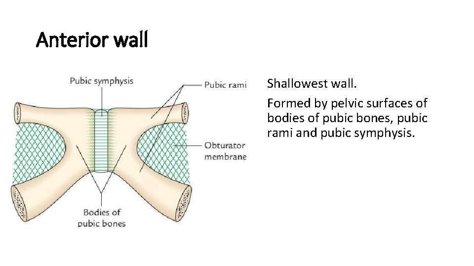
Anterior wall • Shallowest wall. • Formed by pelvic surfaces of bodies of pubic bones, pubic rami and pubic symphysis.
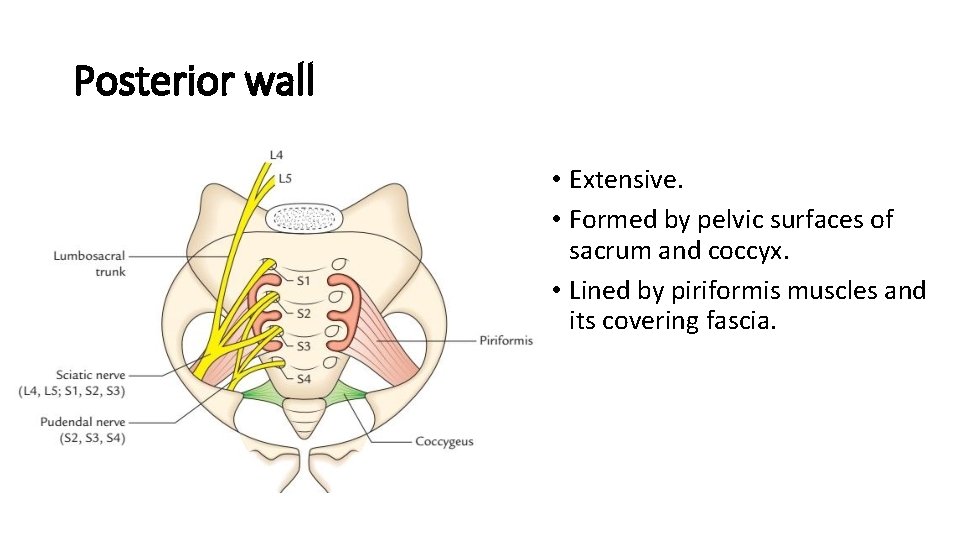
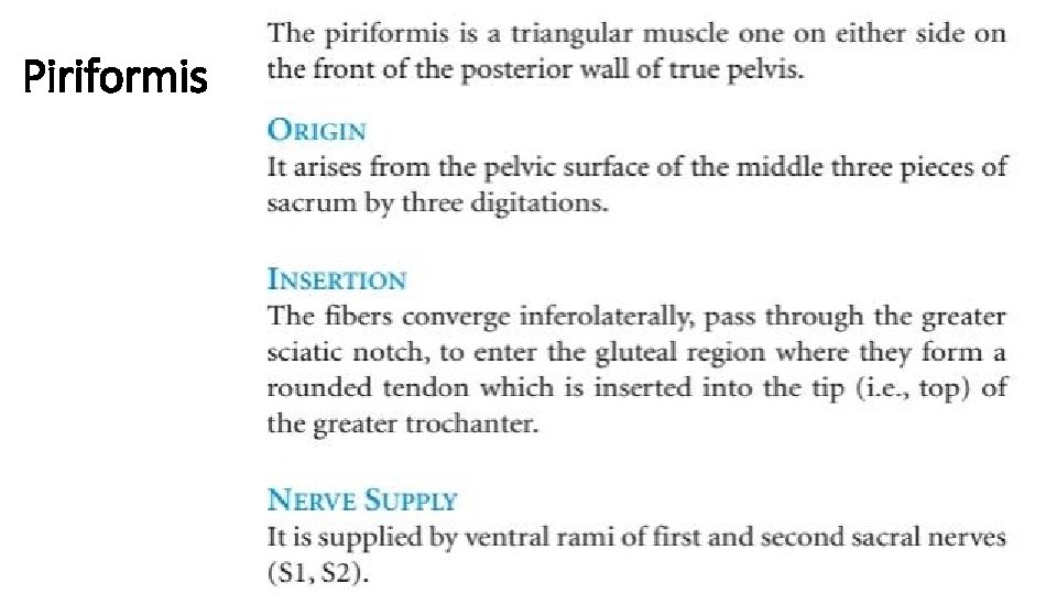
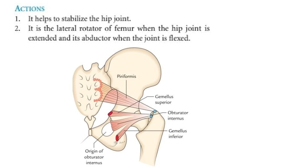
Posterior wall • Extensive. • Formed by pelvic surfaces of sacrum and coccyx. • Lined by piriformis muscles and its covering fascia.
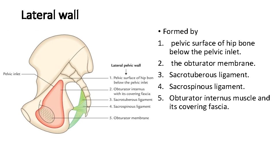
Lateral wall • Formed by 1. pelvic surface of hip bone below the pelvic inlet. 2. the obturator membrane. 3. Sacrotuberous ligament. 4. Sacrospinous ligament. 5. Obturator internus muscle and its covering fascia.
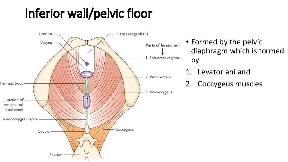
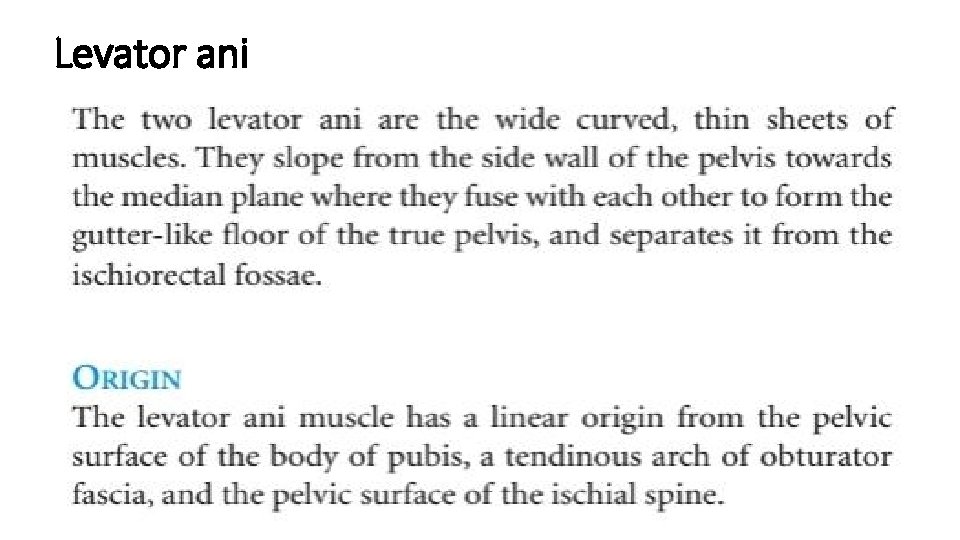
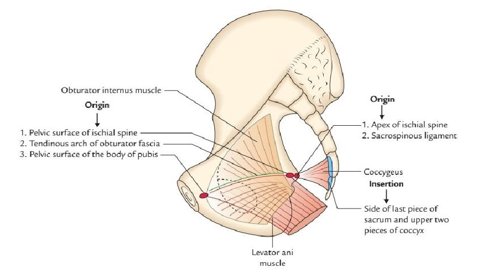
Inferior wall/pelvic floor • Formed by the pelvic diaphragm which is formed by 1. Levator ani and 2. Coccygeus muscles
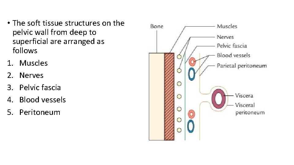
• The soft tissue structures on the pelvic wall from deep to superficial are arranged as follows 1. Muscles 2. Nerves 3. Pelvic fascia 4. Blood vessels 5. Peritoneum
Muscles of the pelvis 1. 2. 3. 4. Obturator internus Piriformis Levator ani and Coccygeus
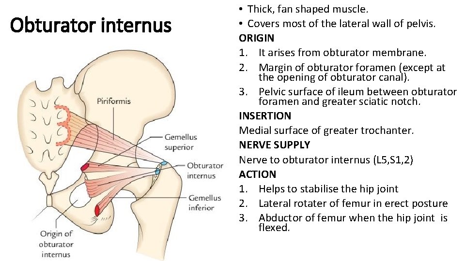
Obturator internus • Thick, fan shaped muscle. • Covers most of the lateral wall of pelvis. ORIGIN 1. It arises from obturator membrane. 2. Margin of obturator foramen (except at the opening of obturator canal). 3. Pelvic surface of ileum between obturator foramen and greater sciatic notch. INSERTION Medial surface of greater trochanter. NERVE SUPPLY Nerve to obturator internus (L 5, S 1, 2) ACTION 1. Helps to stabilise the hip joint 2. Lateral rotater of femur in erect posture 3. Abductor of femur when the hip joint is flexed.
Piriformis
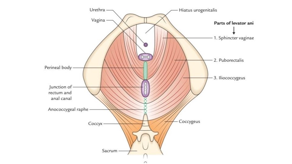
Levator ani
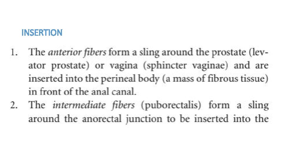
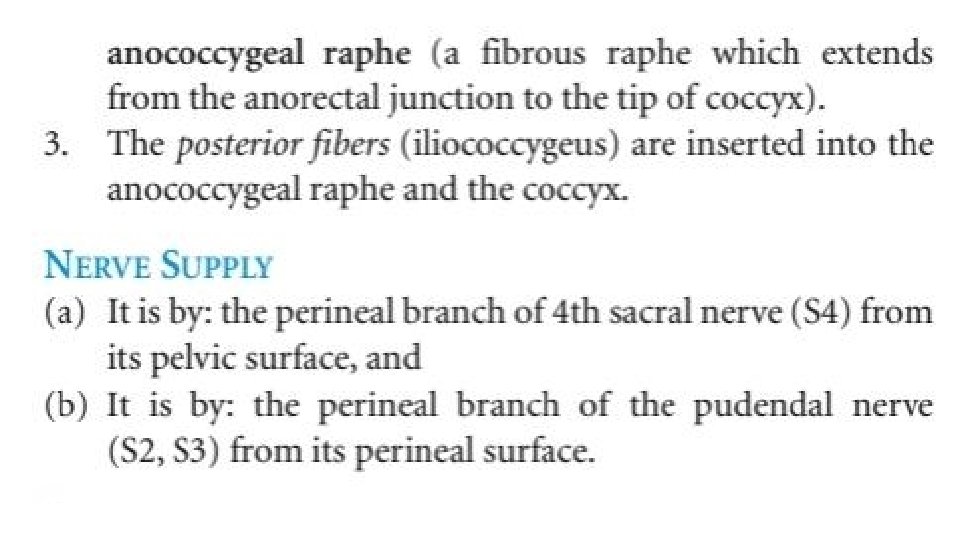
INSERTION
Coccygeus/ischiococcygeus • Small triangular muscle behind levator ani ORIGIN arises from its apex from pelvic surface of ischial spine and sacrospinous ligament
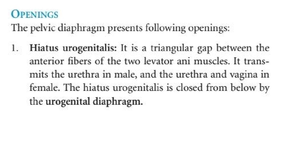
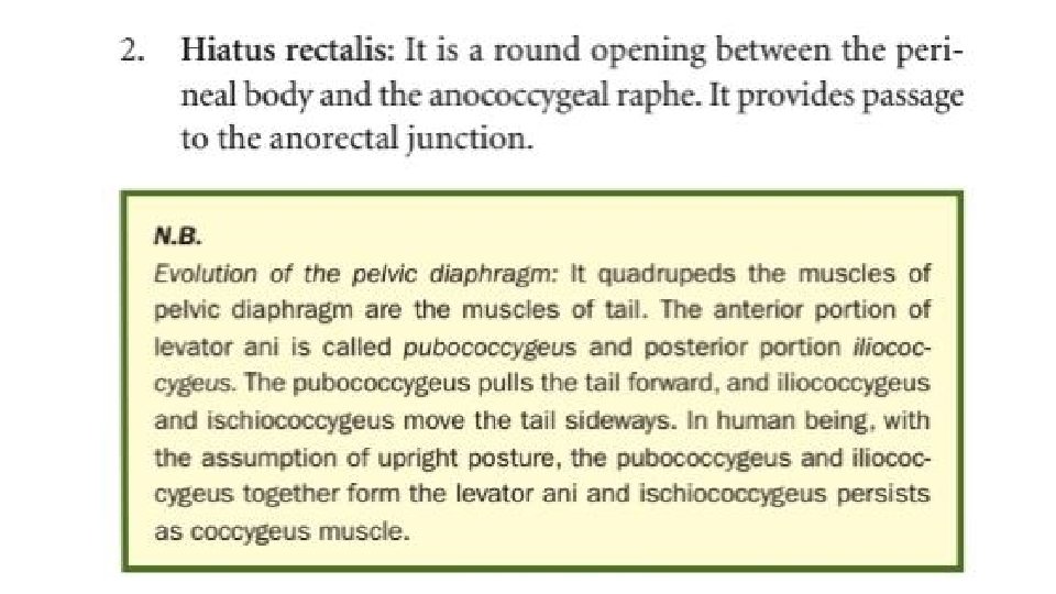
Pelvic diaphragm
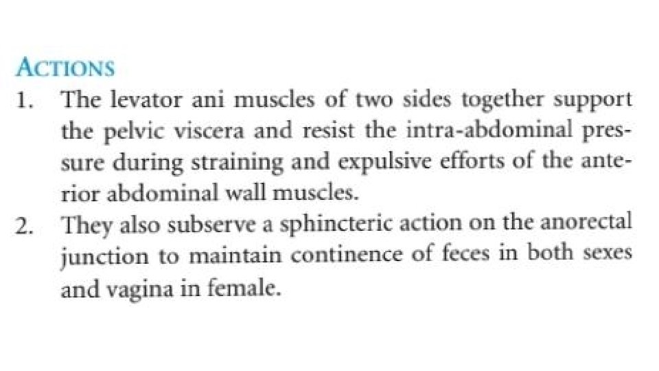
Functions • Provides principal support to pelvic viscera. • Has sphincteric action on rectum and vagina. • Also assists in increasing the intra abdominal pressure during defecation, micturition and parturition.
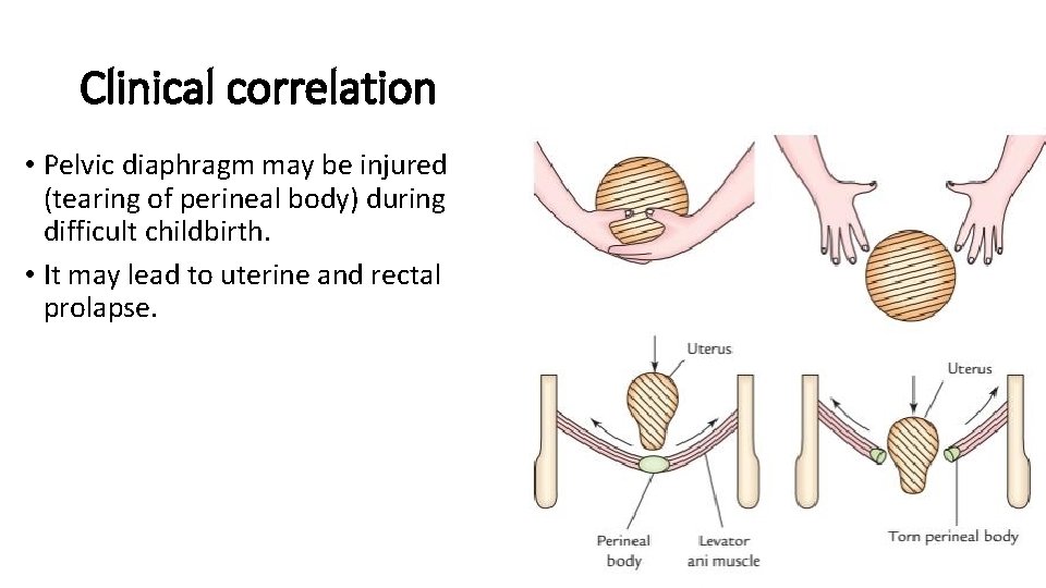
Clinical correlation • Pelvic diaphragm may be injured (tearing of perineal body) during difficult childbirth. • It may lead to uterine and rectal prolapse.
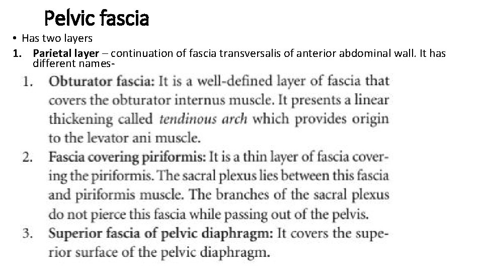
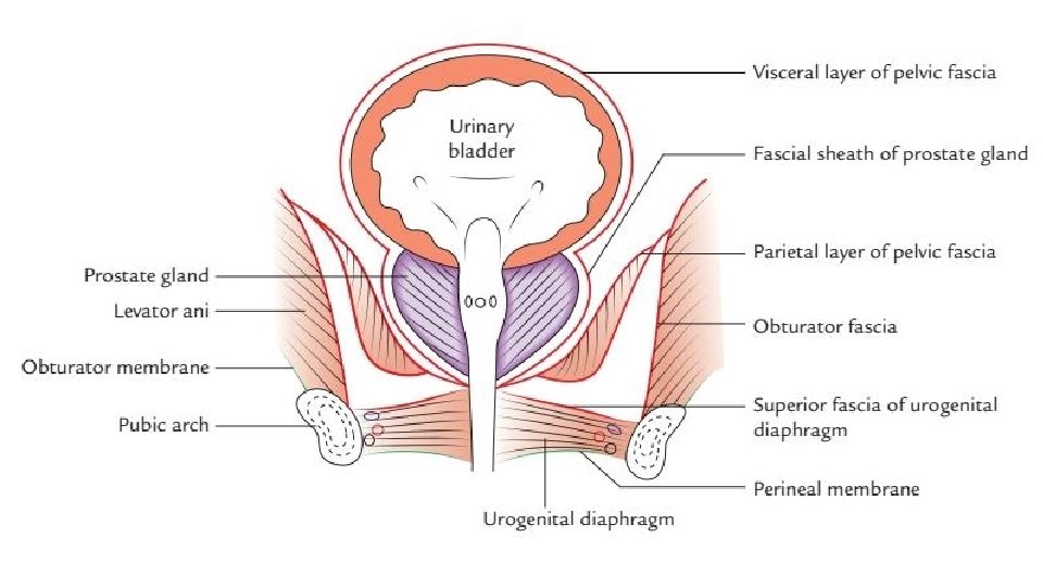
Pelvic fascia • Has two layers 1. Parietal layer – continuation of fascia transversalis of anterior abdominal wall. It has different names-
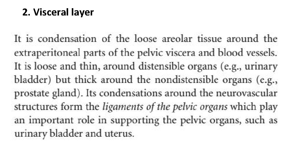
2. Visceral layer