Anatomy and Physiology Sixth Edition Rod R Seeley


- Slides: 45

Anatomy and Physiology, Sixth Edition Rod R. Seeley Idaho State University Trent D. Stephens Idaho State University Philip Tate Phoenix College Chapter 19 Lecture Outline* *See Power. Point Image Slides for all figures and tables pre-inserted into Power. Point without notes. 19 -1 Copyright © The Mc. Graw-Hill Companies, Inc. Permission required for reproduction or display.

Chapter 19 Cardiovascular System Blood 19 -2

Functions of Blood • Transport of: – Gases, nutrients, waste products – Processed molecules – Regulatory molecules • • Regulation of p. H and osmosis Maintenance of body temperature Protection against foreign substances Clot formation 19 -3

Composition of Blood 19 -4

Plasma • Liquid part of blood – Pale yellow made up of 91% water, 9% other • Colloid: Liquid containing suspended substances that don’t settle out – Albumin: Important in regulation of water movement between tissues and blood – Globulins: Immune system or transport molecules – Fibrinogen: Responsible formation of blood clots 19 -5

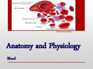
Formed Elements • Red blood cells (erythrocytes) • White blood cells (leukocytes) – Granulocytes • Neutrophils • Eosinophils • Basophils – Agranulocytes • Lymphocytes • Monocytes • Platelets (thrombocytes) 19 -6

Production of Formed Elements • Hematopoiesis or hemopoiesis: Process of blood cell production • Stem cells: All formed elements derived from single population – Proerythroblasts: Develop into red blood cells – Myeloblasts: Develop into basophils, neutrophils, eosinophils – Lymphoblasts: Develop into lymphocytes – Monoblasts: Develop into monocytes – Megakaryoblasts: Develop into platelets 19 -7

Hematopoiesis 19 -8

Erythrocytes • Structure – Biconcave, anucleate • Components – Hemoglobin – Lipids, ATP, carbonic anhydrase • Function – Transport oxygen from lungs to tissues and carbon dioxide from tissues to lungs 19 -9

Hemoglobin • Consists of: – 4 globin molecules: Transport carbon dioxide (carbonic anhydrase involved), nitric oxide – 4 heme molecules: Transport oxygen • Iron is required for oxygen transport 19 -10

Erythropoiesis • Production of red blood cells – Stem cells proerythroblasts early erythroblasts intermediate late reticulocytes • Erythropoietin: Hormone to stimulate RBC production 19 -11

Hemoglobin Breakdown 19 -12

Leukocytes • Types • Protect body against microorganisms and remove dead cells and debris • Movements – Ameboid – Diapedesis – Chemotaxis – Neutrophils: Small phagocytic cells – Eosinophils: Reduce inflammation – Basophils: Release histamine and increase inflammatory response – Lymphocytes: Immunity – Monocytes: Become macrophages 19 -13

Leukocytes

• Five Types of White Blood Cells and Their Functions – There are two different types of white blood cells and each looks different from one another under the microscope. – These include granulocytes and agranulocytes.

• Granulocytes have visible granules or grains inside the cells that have different cell functions. – Types of granulocytes include basophils, neutrophils, and eosinophils. • Agranulocytes are free of visible grains under the microscope and include lymphocytes and monocytes

• Together, they coordinate with one another to fight off things like cancer, cellular damage, and infectious diseases.

• Neutrophils – Neutrophils are the most common type of white blood cell in the body with levels of between 2000 to 7500 cells per mm 3 in the bloodstream. – Neutrophils are medium-sized white blood cells with irregular nuclei and many granules that perform various functions within the cell. • Function: Neutrophils function by attaching to the walls of the blood vessels, blocking the passageway of germs that try to gain access to the blood through a cut or infectious area

• Neutrophils are the first cells to reach an area where a breach in the body has been made. • They kill germs by means of a process known as phagocytosis or “cell-eating”. • Besides eating bacteria one-by-one, they also release a burst of super oxides that have the ability to kill many bacteria at the same time.

• Lymphocytes – Lymphocytes are small, round cells that have a large nucleus within a small amount of cytoplasm. – They have an important function in the immune system, being major players in the humoral immune system, which is the part of the immune system that relates to antibody production. – Lymphocytes tend to take up residence in lymphatic tissues, including the spleen, tonsils, and lymph nodes. – There about 1300 to 4000 lymphocytes per mm 3 of blood.

• Function: – B lymphocytes make antibodies, which is one of the final steps in disease resistance. – When B lymphocytes make antibodies, they prime pathogens for destruction and then make memory cells ready that can go into action at any time, remembering a previous infection with a specific pathogen. – T lymphocytes are another type of lymphocyte, differentiated in the thymus and important in cellmediated immunity.

• Monocytes – Monocytes are the largest of the types of white blood cells. – There are only about 200 -800 monocytes per mm 3 of blood. – Monocytes are agranulocytes, meaning they have few granules in the cytoplasm when seen under the microscope. – Monocytes turn into macrophages when they exit the bloodstream.

• Function: – As macrophages, monocytes do the job of phagocytosis (cell-eating) of any type of dead cell in the body, whether it is a somatic cell or a dead neutrophil. – Because of their large size, they have the ability to digest large foreign particles in a wound unlike other types of white blood cells.

• Eosinophils – There aren’t that many eosinophils in the bloodstream—only about 40 -400 cells per mm 3 of blood. – They have large granules that help in cellular functions. – Eosinophils are especially important when it comes to allergies and worm infestations.

• Function: – Eosinophils work by releasing toxins from their granules to kill pathogens. – The main pathogens eosinophils act against are parasites and worms. – High eosinophil counts are associated with allergic reactions.

• Basophils – Basophils are the least frequent type of white blood cell, with only 0 -100 cells per mm 3 of blood. – Basophils have large granules that perform functions that are not well known. – They are very colorful when stained and looked at under the microscope, making them easy to identify.

• Function: – Basophils have the ability to secrete anticoagulants and antibodies that have function against hypersensitivity reactions in the bloodstream. – They act immediately as part of the immune system’s action against foreign invaders. – Basophils contain histamine, which dilates the vessels to bring more immune cells to the area of injury.


• Macrophages: - are the main phagocytes of the body. • Neutrophils: - are the first responders and become phagocytic when they encounter infectious material.

• Eosinophils: - are weakly phagocytic but are important in defending the body against parasitic worms. • Mast cells: - have the ability to bind with, ingest, and kill a wide range of bacteria.

Natural killer cells • They are able to lyse and kill : - cancer cells - virally infected cells before the adaptive immune system has been activated

Leukocytes 19 -32

Thrombocytes • Cell fragments pinched off from megakaryocytes in red bone marrow • Important in preventing blood loss – Platelet plugs – Promoting formation and contraction of clots 19 -33

Hemostasis • Arrest of bleeding • Events preventing excessive blood loss – Vascular spasm: Vasoconstriction of damaged blood vessels – Platelet plug formation – Coagulation or blood clotting 19 -34

Platelet Plug Formation 19 -35

Coagulation • Stages – Activation of prothrombinase – Conversion of prothrombin to thrombin – Conversion of fibrinogen to fibrin • Pathways – Extrinsic – Intrinsic 19 -36

Clot Formation 19 -37

Fibrinolysis • Clot dissolved by activity of plasmin, an enzyme which hydrolyzes fibrin 19 -38

Blood Grouping • Determined by antigens (agglutinogens) on surface of RBCs • Antibodies (agglutinins) can bind to RBC antigens, resulting in agglutination (clumping) or hemolysis (rupture) of RBCs • Groups – ABO and Rh 19 -39

ABO Blood Groups 19 -40

Agglutination Reaction 19 -41

Rh Blood Group • First studied in rhesus monkeys • Types – Rh positive: Have these antigens present on surface of RBCs – Rh negative: Do not have these antigens present • Hemolytic disease of the newborn (HDN) – Mother produces anti-Rh antibodies that cross placenta and cause agglutination and hemolysis of fetal RBCs 19 -42

Erythroblastosis Fetalis 19 -43

Diagnostic Blood Tests • Type and crossmatch • Complete blood count – Red blood count – Hemoglobin measurement – Hematocrit measurement • White blood count • Differential white blood count • Clotting 19 -44

Blood Disorders • Erythrocytosis: RBC overabundance • Anemia: Deficiency of hemoglobin – – – Iron-deficiency Pernicious Hemorrhagic Hemolytic Sickle-cell • • • Hemophilia Thrombocytopenia Leukemia Septicemia Malaria Infectious mononucleosis • Hepatitis 19 -45
 Srednji rod rod imenica
 Endomysium
 Human anatomy & physiology edition 9
 Uterus perimetrium
 Peter piper picked
 The sixth sick sheik's sixth sheep's sick lyrics
 Biochemistry sixth edition
 Computer architecture a quantitative approach sixth edition
 Automotive technology principles diagnosis and service
 Automotive technology sixth edition
 Apa sixth edition
 Computer architecture a quantitative approach 6th
 Precalculus sixth edition
 Rational people think at the margin
 Computer architecture a quantitative approach sixth edition
 Physiology of sport and exercise 5th edition
 Upper respiratory tract
 Tattoo anatomy and physiology
 International anatomy olympiad
 Specialized stems examples
 Bone metabolism
 Anatomy of peptic ulcer
 Liver physiology and anatomy
 Difference between anatomy and physiology
 Epigastric region
 Cytoplasmic granules
 The central sulcus divides which two lobes? (figure 14-13)
 Http://anatomy and physiology
 Waistline
 Appendicitis anatomy and physiology
 Aohs foundations of anatomy and physiology 1
 Aohs foundations of anatomy and physiology 1
 Anatomical planes
 Anatomy and physiology chapter 8 special senses
 Chapter 13 anatomy and physiology of pregnancy
 Unit 26 agriscience
 Science olympiad forensics cheat sheet
 Anatomy and physiology chapter 2
 Stomach anatomy and physiology ppt
 Anatomy and physiology of pancreas
 Anatomy and physiology chapter 7
 Anatomy and physiology coloring workbook figure 14-1
 Chapter 10 blood anatomy and physiology
 Aohs foundations of anatomy and physiology 1
 Aohs foundations of anatomy and physiology 1
 What produces bile
