ANATOMY AND PHYSIOLOGY OF THE LARYNX Yard Doc


- Slides: 60

ANATOMY AND PHYSIOLOGY OF THE LARYNX Yard. Doc. Dr. Müzeyyen Doğan

Learning goal and objectives of the lesson n n Learning goal of the lesson: The learner should know the basic anatomic structures, embryology and physiology of the larynx Learning objectives of the lesson the learner will be able to identify the anatomic structures of the larynx identify the physiology of the larynx

LARYNX n n Adult: between 3 th and 6 th cervical vertebra İnfant: between 1 st and 4 th cervical vertebra Attaches to the hyoid bone and opens into the laryngopharynx superiorly Continuous with the trachea posteriorly

Embryology I n n Respiratory primordium n Third week– 26 days Respiratory primordium separated by tracheoesophageal folds n Fuse to form septum (4 -5 weeks)

Embryology II n n n Larynx from 4 th and 5 th arches Primitive larynx altered by hypobranchial eminence, epiglottis, arytenoids Laryngeal lumen obliterated and recanalized

Differences in adults and infants 1/3 size at birth n Narrow dimensions (subglottis vs. glottis) n Higher in neck and more pliable n Epiglottis narrower ●

Framework of the Larynx

Osseous Structure HYOID BONE n Greater cornu(cornu majus) n Lesser cornu (cornu minus) n Corpus hyoideum (body)

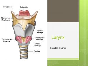
Cartilage -1 unpaired cartilages thyroid, cricoid, and epiglottis n paired cartilages arytenoids, corniculates, and cuneiforms n

Cartilage -2

Epıglottic Cartilage n n This leaf shaped cartilage is composed mainly of elastic cartilage that covers the laryngeal inlet during swallowing

Thyroid cartilage The thyroid cartilage is also known as the Adam’s apple. It is a large Vshaped cartilage that houses and protects the vocal cords and the opening to the trachea. The vocal cords attach to the thyroid cartilage at the front of the neck, just under the epiglottis.

Cricoid cartilage n n n Arcus cricoidea Lamina cricoidea The cricoid cartilage may be regarded as the base and support for the entire larynx.

Paired cartilages n arytenoids, corniculates, and cuneiforms

Arytenoid cartilage n n Pyramidal shape Joint with corniculate cartilage. Processus vocalis lig. vocale Processus muscularis m. cricoaritenoideus lateralis and posterior

Joints n CRICOTHYROID JOINT n CRICOARYTENOID JOINT

Cricoarytenoid Joint n Cricoid cartilage articulates with the arytenoids via the cricoarytenoid joints. (synovial joint).

Cricothyroid Joint n Cricoid cartilage articulates with the thyroid cartilage via the cricothyroid joints (synovial joint)

Ligaments and Membranes I extrinsic ligaments n thyrohyoid membrane n thyrohyoid ligaments n thyroepiglottic ligament, n hyoepiglottic ligament, n cricotracheal

Ligaments and Membranes II intrinsic ligaments n quadrangular membrane n vestibular ligament n conus elasticus n median cricothyroid ligament n vocal ligament

Thyrohyoid membran n This broad fibroelastic sheet is attached from the superior border and the superior horn of the thyroid cartilage to the posterior surface of the body and greater cornua of the hyoid bone )

Conus Elasticus This membrane arises from the inner surface of the cricoid arch

Quadrangular Membrane n It extends from the lateral margins of the epiglottis within the aryepiglottic fold and attaches to the arytenoid and corniculate cartilages. The inferior free edge is thickened to form the vestibular ligament (false vocal cord). The superior edge is also free and it is covered with aryepiglottic fold of mucosa.

Vocal cords Örtü-Gövde (Hirano) n Epitel n Reinke tabakası n Vokal ligaman n Vokalis kası

Larinks Kasları Intrinsic muscles: cricothyroids, posterior cricoarytenoids, lateral cricoarytenoids, transverse arytenoid, oblique arytenoids, and thyroarytenoids extrinsic muscles : strap muscles

Intrinsic Muscles n n n Cricoarytenoid Cricothyroid Interarytenoid Thyroepiglottic muscles

Extrinsic muscles Infrahyoid muscles n n Omohyoid Sternohyoid n Sternothyroid n Thyrohyoid

Extrinsic muscles Suprahyoid muscles n digastric n geniohyoid n mylohyoid n stylopharyngeus n thyrohyoid.

Suprahyoid muscles

Intrinsic Muscles n n n abduction of the vf. n Posteroir cricoarytenoid m adduction of the vf n thyroarytenoid / vocalis muscle n Lateral cricoarytenoid m n İnterarytenoid and obliq muscles tension of the vf. krikotiroid m

m. interarytenoideus • • Transvers Part: Adduction of the vocal folds by approximating the arytenoid cartilages Obliq Part: Sphincter of the inlet of the larynx during the act of swallowing Closes laryngeal inlet by approximating arytenoid cartilages.

m. cricoarytenoideus posterior abductor of the vocal cords n m. Cricoarytenoideus lateralis closes the glottis by adducting the vocal folds n

m. tyroaritenoideus n n n Thyroarytenoideus internus or vocalis muscle is the major tensor of the free edge of the vocal fold. Thyroarytenoideus externa contraction draws the arytenoid cartilages foreward toward the thyroid, thus shortening the vocal ligament. Thyroepiglotticus widens the inlet of the larynx

m. cricothyroideus n n The CT produces elongation and tension of the vocal fold ligament by elevating the arch of the cricoid cartilage upward toward the lowermost aspect of the thyroid ala. Contraction of the CT also rotates the arytenoids medially, adducting the vocal folds. Innervation external branch of the superior laryngeal nerve

Intrinsic Muscles

Vascular System n n Arterleri a. thyroidea superior ve inferiordan gelir Venleri (v. laringea superior ve inferior) v. thyroidea superior ve inferior v. jugularis interna

Arterial Drainage n a. carotis eksterna a. thyroidea superior a. laryngea superior a. cricothyroid eus Subclavian arter Turuncus thyroservicalis A. thyroidea inferior A. laryngea inferior n

Arterial Drainage n n n SUPERIOR LARYNGEAL A. INFERIOR LARYNGEAL A. CRICOTHYROID ARTER

Venous Drainage n n SUPERIOR LARYNGEAL V. INFERIOR LARYNGEAL V.

Lymphatic Drainage n n n Glottik bölgenin lenf drenajı zayıf Suprglottik bölgenin lenf drenajı derin servikal lenf nodları Subglottik bölgenin lenf drenajı alt derin servikal lenf nodları, pretrakeal lenf nodları, prelaringeal lenf nodları

Nerve -1 l Recurrent laryngeal nerve n Motor to all intrinsic muscles n Sensory to infra-glottic area n Frequency dependent Superior laryngeal nerve n Supra-glottic afferent (int. ) n Cricothyroid muscles

Nerve -2

Internal cavity of the larynx n n supraglottic space (also called the vestibule which is surrounded by the piriform fossa) preepiglottic space paraglottic space (which contains the ventricles) subglottic space (which is the area below the true vocal folds).

supraglottic space n n Superior border : free margin of the epiglottis and aryepiglottic folds Inferior border: lower margin of the ventricular or false vocal folds

preepiglottic space n n n Superior border : hyoepiglottic ligament Anterior border: thyrohyoid membrane and ligament Posterior border: anterior surface of the epiglottis and thyroepiglottic ligament

supraglottic space n n Superior border : free margin of the epiglottis and aryepiglottic folds Inferior border: lower margin of the ventricular or false vocal folds

paraglottic space n n Superior border : quadrangular membrane Inferior border: conus elasticus Lateral border: inner surface of the thyroid cartilage Medial border: ventricle

Laryngeal Histology n n n It is lined mainly by a pseudostratified, ciliated, columnar epithelium. It also contains a mucosa with laryngeal glands and a few taste buds. The true vocal folds have a specialized histology different from the rest of the larynx. Virtually all the laryngeal membranes and ligaments consist of elastic and collagenous fibers. All the laryngeal muscle is cross-striated muscle.

Laryngeal Physiology n n 1. protection 2. respiration 3. phonation 4. effort closure

Speech Production n n begins in cerebral cortex precentral gyrus to motor nuclei then coordinated activity

Phonation

Myoelastic - aerodynamic theory n n n 1. glottis closed 2. subglottal pressure increases 3. vocal folds are blown open in a zipper-like fashion (from lower towards the upper lip) 4. glottis open 5. subglottal pressure decreases 6. recoiling forces and the Bernoulli effect adduct the vocal folds back together (also in a zipperlike fashion with the edges of the lower lip closing first)

Phonatory cycle n n vocal folds approximated infraglottic pressure builds up pressure opens folds from bottom up upper portion with strong elastic properties

Movements of Vocal Cords

PHONATION

Physıcal Examination I n n n Inspection Palpation Indirect Laryngoscopy Direct Laryngoscopy Radiography n n n Neck films, chest films Barium swallow CT/MRI

Physical Examination Iı n n n Videolaryngostroboscopy Glottography Laryngeal EMG

CADAVER I

CADAVER II

CADAVER III
 Respiratory
 Tattoo anatomy and physiology
 International anatomy olympiad
 Structure anatomy and physiology in agriculture
 Anatomy and physiology of bone
 Gastric ulcer anatomy
 Liver anatomy and physiology
 Epigastric region
 Wpigastric region
 Blood in anatomy and physiology
 Chapter 14 anatomy and physiology
 3 layers of muscle
 Http://anatomy and physiology
 Chapter 1 introduction to human anatomy and physiology
 Appendectomy anatomy and physiology
 Aohs foundations of anatomy and physiology 1
 Aohs foundations of anatomy and physiology 1
 Anatomical planes
 Anatomy and physiology chapter 8 special senses
 Chapter 13 anatomy and physiology of pregnancy
 Unit 26 animal anatomy physiology and nutrition
 Science olympiad anatomy and physiology 2020 cheat sheet
 Anatomy and physiology chapter 2
 Physiology of stomach ppt
 Anatomy and physiology of diabetes
 Chapter 7 anatomy and physiology
 Chapter 14 the digestive system and body metabolism
 Chapter 10 blood anatomy and physiology
 Aohs foundations of anatomy and physiology 1
 Aohs foundations of anatomy and physiology 1
 What produces bile
 Anatomy and physiology chapter 15
 Cornell notes for anatomy and physiology
 Human anatomy & physiology edition 9
 Anatomy and physiology chapter 1
 Holes anatomy and physiology chapter 1
 Holes essential of human anatomy and physiology
 Anatomy and physiology unit 7 cardiovascular system
 Anatomy and physiology chapter 15
 Anatomy and physiology
 Chapter 1 introduction to anatomy and physiology
 The speed at which the body consumes energy
 Aohs foundations of anatomy and physiology 1
 Cranial cephalic
 Anatomy and physiology exam 1
 Welcome to anatomy and physiology
 Physiology of the foot and ankle
 Integumentary system psoriasis
 Pancreas anatomy histology
 Anatomy and physiology vocabulary
 Anatomy and physiology
 Biceps muscle names
 Anatomy and physiology
 Anatomy and physiology
 Anatomy and physiology
 Anatomy and physiology
 Thyroid anatomy
 Anatomy and physiology
 Anatomy and physiology
 Anatomy and physiology
 Anatomy and physiology
