ACUTE KIDNEY INJURY Prof Dr Glin Kantarc MD

- Slides: 59
ACUTE KIDNEY INJURY Prof. Dr. Gülçin Kantarcı, MD Internal Medicine and Nephrology
REFERENCE &SUGGESTED READING ü http: //accessmedicine. com Current Medical Diagnosis and Treatment, Maxine A. Papadakis, Stephen J. Mc. Phee, Eds. Michael W. Rabow, Associate Ed. Chapter 22. Kidney Disease (ACUTE RENAL FAILURE: GENERAL) Current Diagnosis & Treatment: Nephrology & Hypertension Edgar V. Lerma, Jeffrey S. Berns, Allen R. Nissenson Chapter 9 -11 -14 ACUTE RENAL FAILURE ü http: //www. uptodate. com (Overview of the management of acute kidney injury, Diagnostic approach to the patient with acute kidney injury (acute renal failure) or chronic kidney disease, Definition of acute kidney injury, Etiology and diagnosis of prerenal disease and acute tubular necrosis in acute kidney injury, Crush-related acute kidney injury )
CONTENTS • Definitions of Acute Kidney Injury (AKI). • Staging of AKI • Diagnostic Work-Up for AKI • Management of AKI • Therapeutic approach to AKI • Case presentations
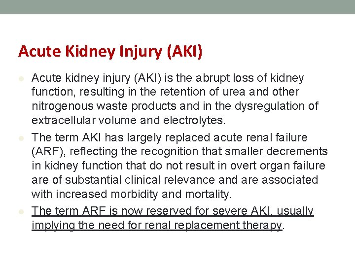
Acute Kidney Injury (AKI) l l l Acute kidney injury (AKI) is the abrupt loss of kidney function, resulting in the retention of urea and other nitrogenous waste products and in the dysregulation of extracellular volume and electrolytes. The term AKI has largely replaced acute renal failure (ARF), reflecting the recognition that smaller decrements in kidney function that do not result in overt organ failure are of substantial clinical relevance and are associated with increased morbidity and mortality. The term ARF is now reserved for severe AKI, usually implying the need for renal replacement therapy.
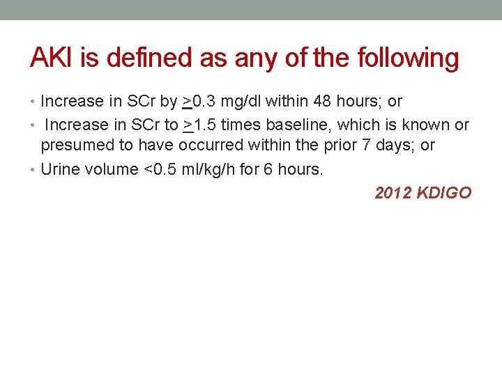
AKI is defined as any of the following • Increase in SCr by >0. 3 mg/dl within 48 hours; or • Increase in SCr to >1. 5 times baseline, which is known or presumed to have occurred within the prior 7 days; or • Urine volume <0. 5 ml/kg/h for 6 hours. 2012 KDIGO
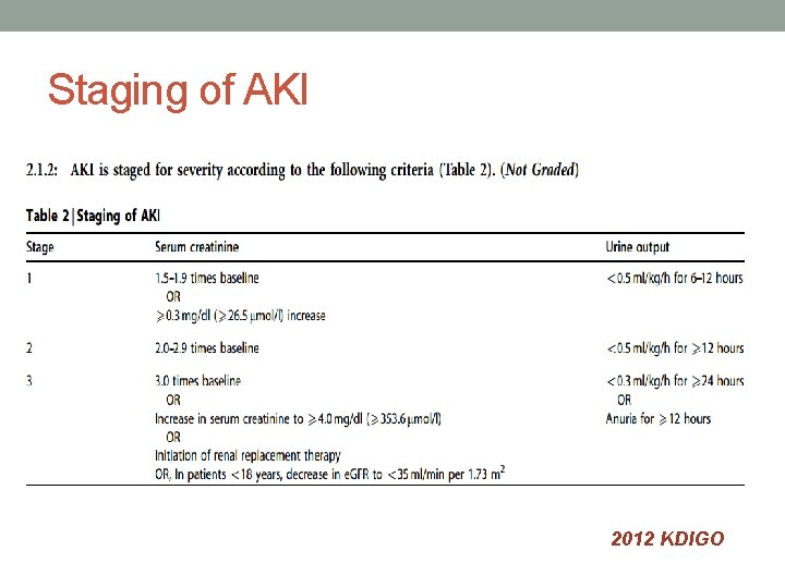
Staging of AKI 2012 KDIGO
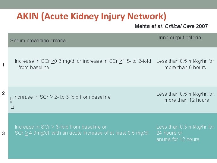
AKIN (Acute Kidney Injury Network) Mehta et al. Critical Care 2007 Serum creatinine criteria Increase in SCr >0. 3 mg/dl or increase in SCr >1. 5 - to 2 -fold from baseline 1 2 b Increase in SCr > 2 - to 3 fold from baseline b Urine output criteria Less than 0. 5 ml/kg/hr for more than 6 hours Less than 0. 5 ml/kg/hr for more than 12 hours � 3 Increase in SCr > 3 -fold from baseline or SCr > 4. 0 mg/dl with an acute increase of at least 0. 5 mg/dl Less than 0. 3 ml/kg/hr for 24 hours or anuria for 12 hours
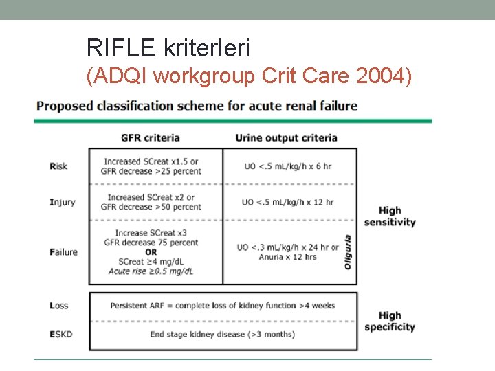
RIFLE kriterleri (ADQI workgroup Crit Care 2004)
• The change in serum creatinine was specified as occurring over not more than seven days. Subsequent to publication of RIFLE, it was noted that the change in serum creatinine concentrations do not correlate with the percent decrease in GFR that is cited in the RIFLE classification; a 1. 5 -fold increase in serum creatinine corresponds to a 33 rather than 25 percent decrease in GFR. • However, given the absence of readily available methods for measurement of GFR when serum creatinine is not in steady state, as is the case during AKI, changes in GFR are not included in the Acute Kidney Injury Network (AKIN) and KDIGO 2012 classification system.
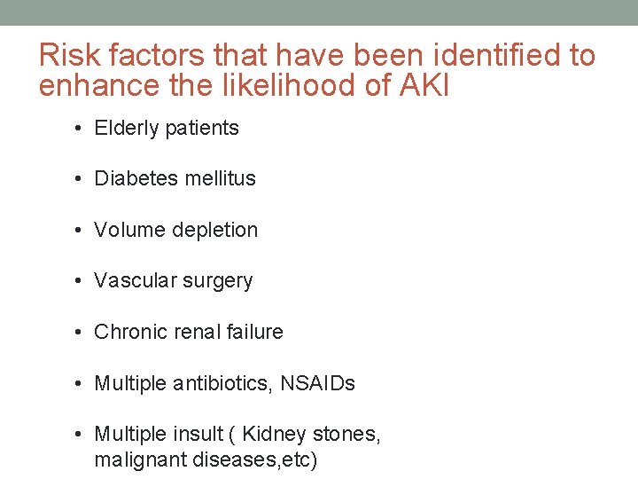
Risk factors that have been identified to enhance the likelihood of AKI • Elderly patients • Diabetes mellitus • Volume depletion • Vascular surgery • Chronic renal failure • Multiple antibiotics, NSAIDs • Multiple insult ( Kidney stones, malignant diseases, etc)
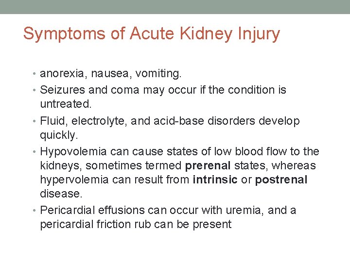
Symptoms of Acute Kidney Injury • anorexia, nausea, vomiting. • Seizures and coma may occur if the condition is untreated. • Fluid, electrolyte, and acid-base disorders develop quickly. • Hypovolemia can cause states of low blood flow to the kidneys, sometimes termed prerenal states, whereas hypervolemia can result from intrinsic or postrenal disease. • Pericardial effusions can occur with uremia, and a pericardial friction rub can be present
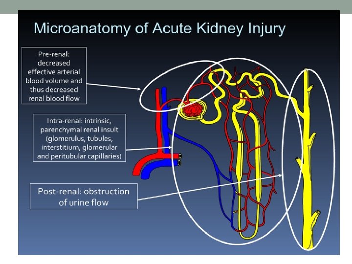
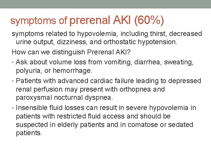
symptoms of prerenal AKI (60%) symptoms related to hypovolemia, including thirst, decreased urine output, dizziness, and orthostatic hypotension. How can we distinguish Prerenal AKI? • Ask about volume loss from vomiting, diarrhea, sweating, polyuria, or hemorrhage. • Patients with advanced cardiac failure leading to depressed renal perfusion may present with orthopnea and paroxysmal nocturnal dyspnea. • Insensible fluid losses can result in severe hypovolemia in patients with restricted fluid access and should be suspected in elderly patients and in comatose or sedated patients.
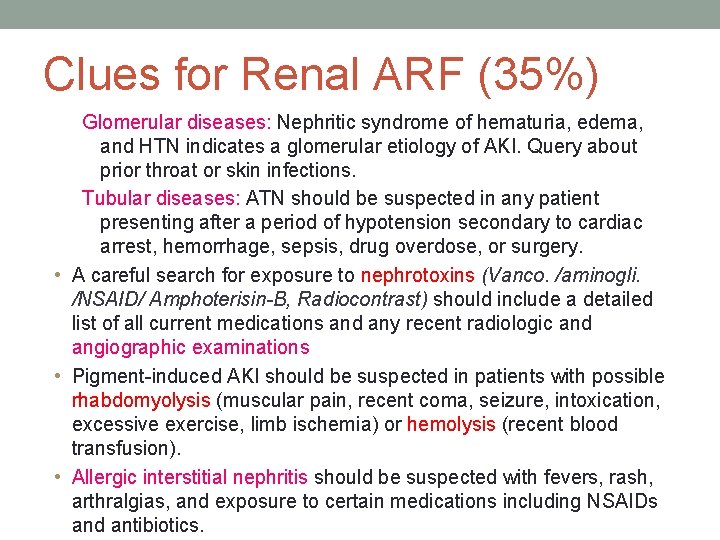
Clues for Renal ARF (35%) Glomerular diseases: Nephritic syndrome of hematuria, edema, and HTN indicates a glomerular etiology of AKI. Query about prior throat or skin infections. Tubular diseases: ATN should be suspected in any patient presenting after a period of hypotension secondary to cardiac arrest, hemorrhage, sepsis, drug overdose, or surgery. • A careful search for exposure to nephrotoxins (Vanco. /aminogli. /NSAID/ Amphoterisin-B, Radiocontrast) should include a detailed list of all current medications and any recent radiologic and angiographic examinations • Pigment-induced AKI should be suspected in patients with possible rhabdomyolysis (muscular pain, recent coma, seizure, intoxication, excessive exercise, limb ischemia) or hemolysis (recent blood transfusion). • Allergic interstitial nephritis should be suspected with fevers, rash, arthralgias, and exposure to certain medications including NSAIDs and antibiotics.
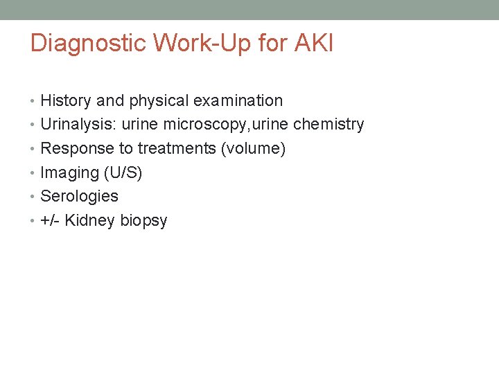
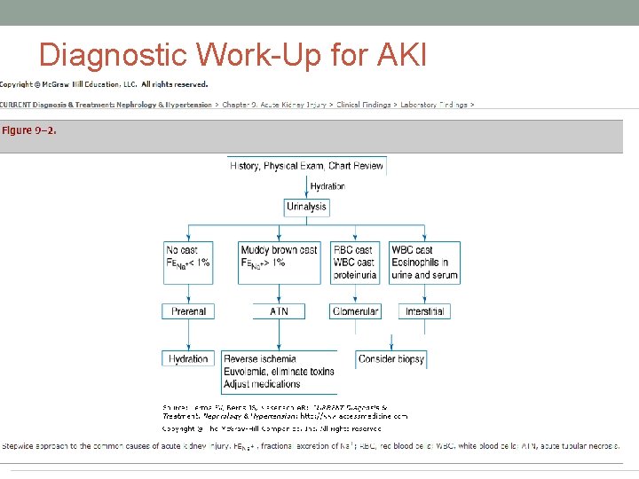
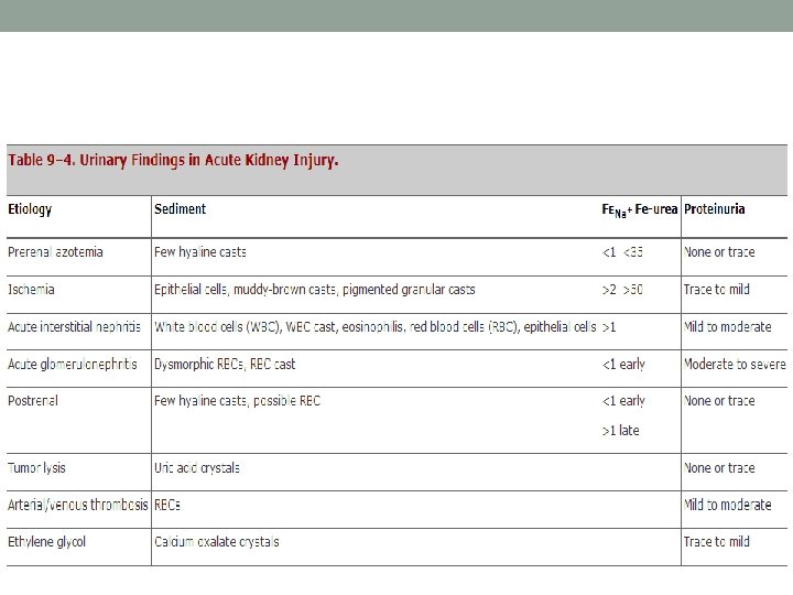
Diagnostic Work-Up for AKI • History and physical examination • Urinalysis: urine microscopy, urine chemistry • Response to treatments (volume) • Imaging (U/S) • Serologies • +/- Kidney biopsy
Diagnostic Work-Up for AKI
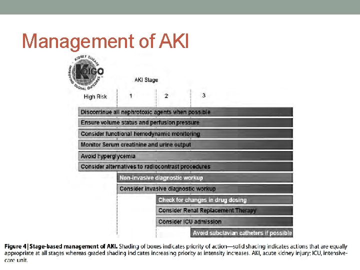
Management of AKI
• most cases of community-acquired AKI’s are secondary to volume depletion, as many as 90% of cases are estimated to have a potentially reversible cause. • Hospital-acquired AKI often occurs in an ICU setting and is commonly part of multiorgan failure.
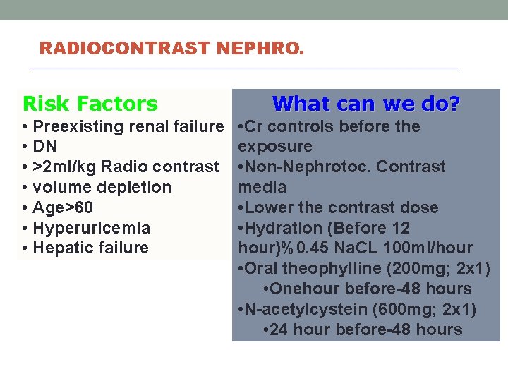
RADIOCONTRAST NEPHRO. Risk Factors • Preexisting renal failure • DN • >2 ml/kg Radio contrast • volume depletion • Age>60 • Hyperuricemia • Hepatic failure What can we do? • Cr controls before the exposure • Non-Nephrotoc. Contrast media • Lower the contrast dose • Hydration (Before 12 hour)%0. 45 Na. CL 100 ml/hour • Oral theophylline (200 mg; 2 x 1) • Onehour before-48 hours • N-acetylcystein (600 mg; 2 x 1) • 24 hour before-48 hours
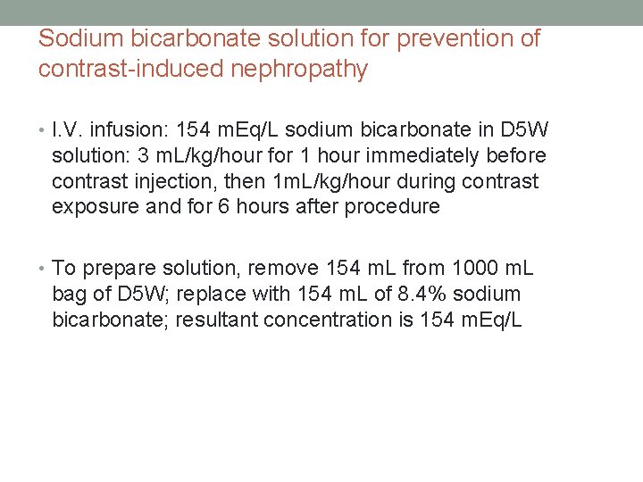
Sodium bicarbonate solution for prevention of contrast-induced nephropathy • I. V. infusion: 154 m. Eq/L sodium bicarbonate in D 5 W solution: 3 m. L/kg/hour for 1 hour immediately before contrast injection, then 1 m. L/kg/hour during contrast exposure and for 6 hours after procedure • To prepare solution, remove 154 m. L from 1000 m. L bag of D 5 W; replace with 154 m. L of 8. 4% sodium bicarbonate; resultant concentration is 154 m. Eq/L
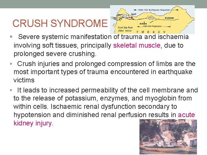
CRUSH SYNDROME § Severe systemic manifestation of trauma and ischaemia involving soft tissues, principally skeletal muscle, due to prolonged severe crushing. § Crush injuries and prolonged compression of limbs are the most important types of trauma encountered in earthquake victims § It leads to increased permeability of the cell membrane and to the release of potassium, enzymes, and myoglobin from within cells. Ischaemic renal dysfunction secondary to hypotension and diminished renal perfusion results in acute kidney injury.
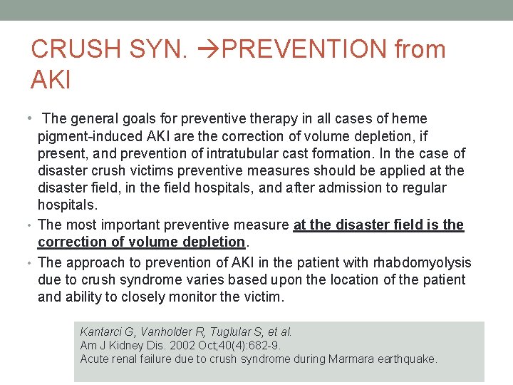
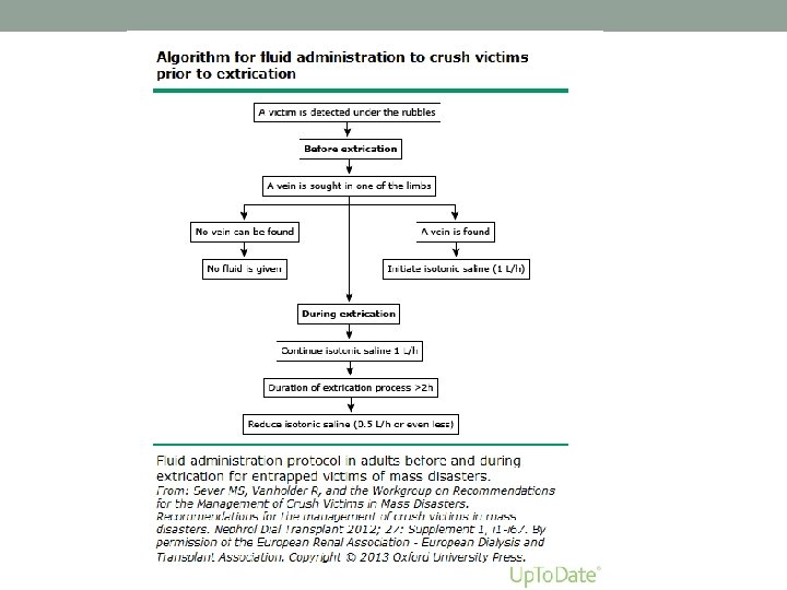
CRUSH SYN. PREVENTION from AKI • The general goals for preventive therapy in all cases of heme pigment-induced AKI are the correction of volume depletion, if present, and prevention of intratubular cast formation. In the case of disaster crush victims preventive measures should be applied at the disaster field, in the field hospitals, and after admission to regular hospitals. • The most important preventive measure at the disaster field is the correction of volume depletion. • The approach to prevention of AKI in the patient with rhabdomyolysis due to crush syndrome varies based upon the location of the patient and ability to closely monitor the victim. Kantarci G, Vanholder R, Tuglular S, et al. Am J Kidney Dis. 2002 Oct; 40(4): 682 -9. Acute renal failure due to crush syndrome during Marmara earthquake.
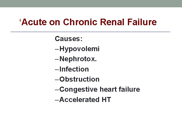
‘Acute on Chronic Renal Failure Causes: – Hypovolemi – Nephrotox. – Infection – Obstruction – Congestive heart failure – Accelerated HT
Prevention Early detection
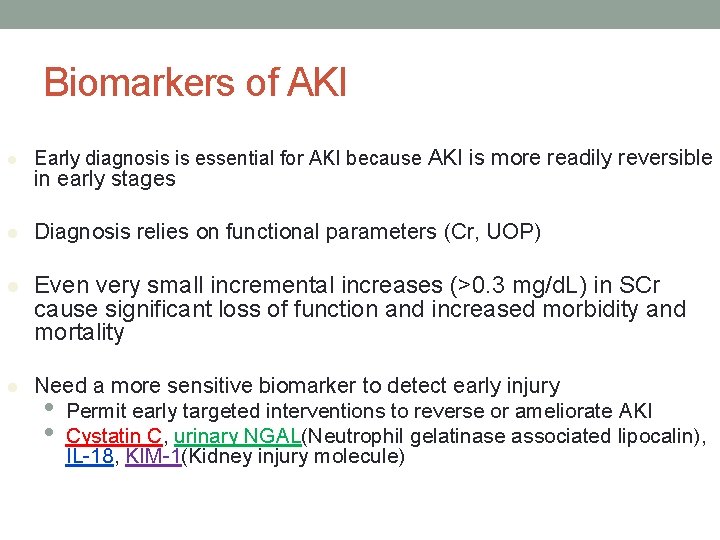
Biomarkers of AKI l Early diagnosis is essential for AKI because AKI is more readily reversible l Diagnosis relies on functional parameters (Cr, UOP) l Even very small incremental increases (>0. 3 mg/d. L) in SCr cause significant loss of function and increased morbidity and mortality l Need a more sensitive biomarker to detect early injury • Permit early targeted interventions to reverse or ameliorate AKI • Cystatin C, urinary NGAL(Neutrophil gelatinase associated lipocalin), IL-18, KIM-1(Kidney injury molecule) in early stages
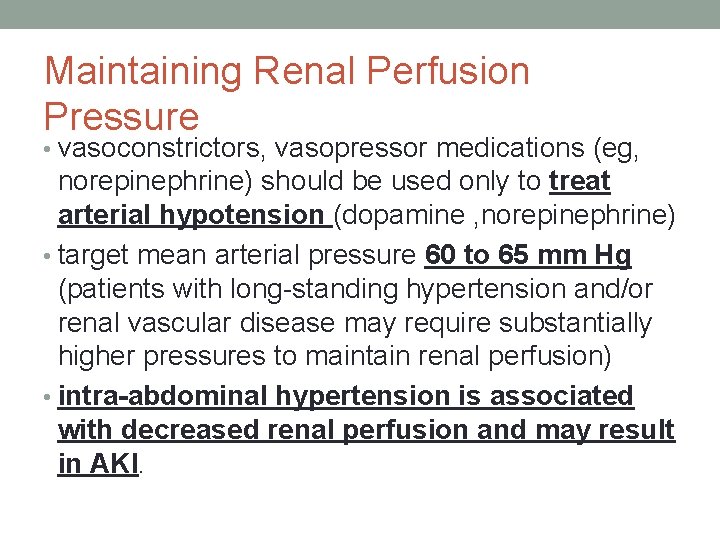
Maintaining Renal Perfusion Pressure • vasoconstrictors, vasopressor medications (eg, norepinephrine) should be used only to treat arterial hypotension (dopamine , norepinephrine) • target mean arterial pressure 60 to 65 mm Hg (patients with long-standing hypertension and/or renal vascular disease may require substantially higher pressures to maintain renal perfusion) • intra-abdominal hypertension is associated with decreased renal perfusion and may result in AKI.
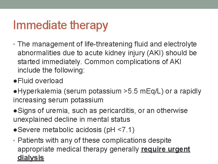
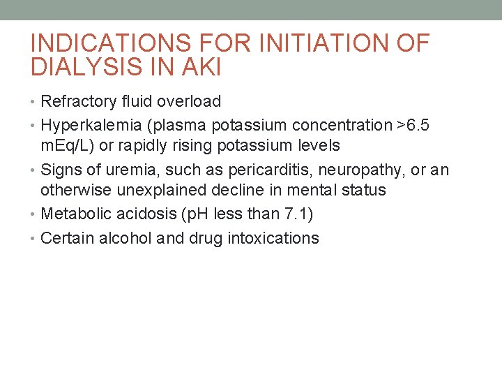
Immediate therapy • The management of life-threatening fluid and electrolyte abnormalities due to acute kidney injury (AKI) should be started immediately. Common complications of AKI include the following: ●Fluid overload ●Hyperkalemia (serum potassium >5. 5 m. Eq/L) or a rapidly increasing serum potassium ●Signs of uremia, such as pericarditis, or an otherwise unexplained decline in mental status ●Severe metabolic acidosis (p. H <7. 1) • Patients with any of these complications despite appropriate medical therapy generally require urgent dialysis
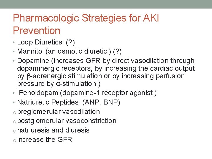
Pharmacologic Strategies for AKI Prevention • Loop Diuretics (? ) • Mannitol (an osmotic diuretic ) (? ) • Dopamine (increases GFR by direct vasodilation through dopaminergic receptors, by increasing the cardiac output by β-adrenergic stimulation or by increasing perfusion pressure by α-stimulation ) • Fenoldopam (dopamine-1 receptor agonist ) • Natriuretic Peptides (ANP, BNP) o preglomerular vasodilation o postglomerular vasoconstriction o natriuresis and diuresis o increase the GFR
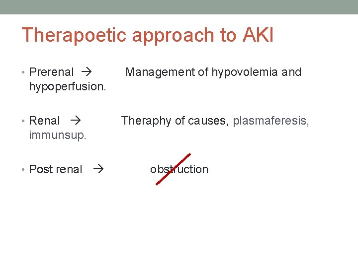
Therapoetic approach to AKI • Prerenal Management of hypovolemia and hypoperfusion. • Renal Theraphy of causes, plasmaferesis, immunsup. • Post renal obstruction
INDICATIONS FOR INITIATION OF DIALYSIS IN AKI • Refractory fluid overload • Hyperkalemia (plasma potassium concentration >6. 5 m. Eq/L) or rapidly rising potassium levels • Signs of uremia, such as pericarditis, neuropathy, or an otherwise unexplained decline in mental status • Metabolic acidosis (p. H less than 7. 1) • Certain alcohol and drug intoxications
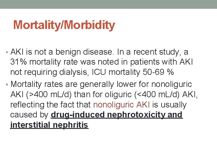
Mortality/Morbidity • AKI is not a benign disease. In a recent study, a 31% mortality rate was noted in patients with AKI not requiring dialysis, ICU mortality 50 -69 % • Mortality rates are generally lower for nonoliguric AKI (>400 m. L/d) than for oliguric (<400 m. L/d) AKI, reflecting the fact that nonoliguric AKI is usually caused by drug-induced nephrotoxicity and interstitial nephritis
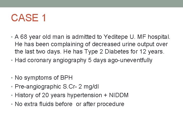
CASE 1 • A 68 year old man is admitted to Yeditepe U. MF hospital. He has been complaining of decreased urine output over the last two days. He has Type 2 Diabetes for 12 years. • Had coronary angiography 5 days ago-uneventfully • No symptoms of BPH • Pre-angiographic S. Cr- 2 mg/dl • History of 20 years hypertension + NIDDM • No extra fluids before or after procedure
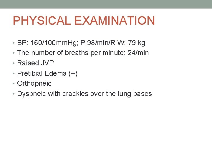
PHYSICAL EXAMINATION • BP: 160/100 mm. Hg; P: 98/min/R W: 79 kg • The number of breaths per minute: 24/min • Raised JVP • Pretibial Edema (+) • Orthopneic • Dyspneic with crackles over the lung bases
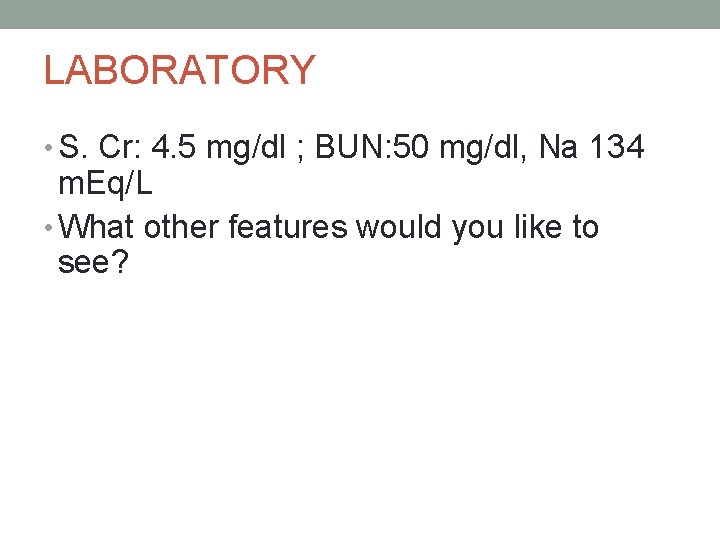
LABORATORY • S. Cr: 4. 5 mg/dl ; BUN: 50 mg/dl, Na 134 m. Eq/L • What other features would you like to see?
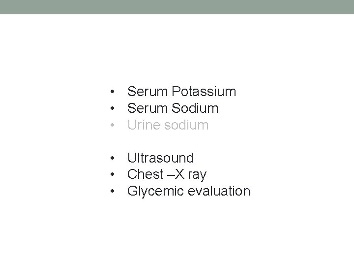
• Serum Potassium • Serum Sodium • Urine sodium • Ultrasound • Chest –X ray • Glycemic evaluation
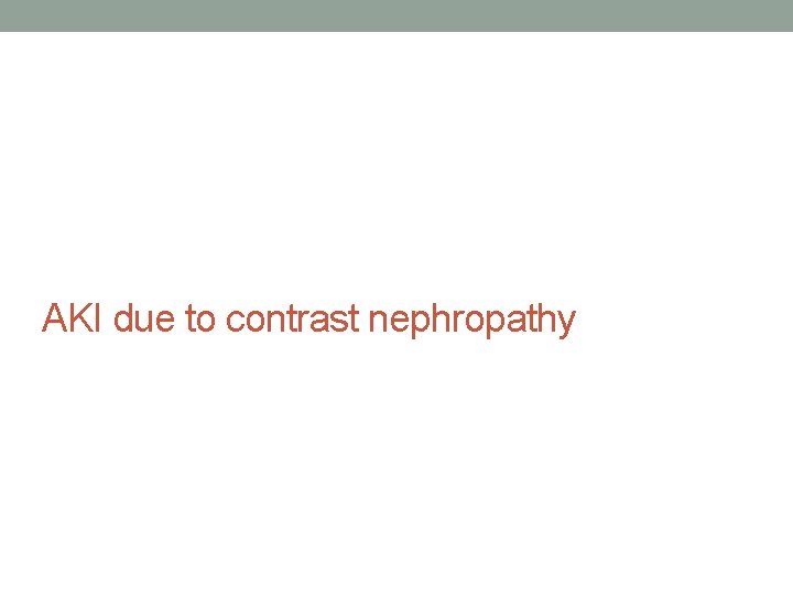
AKI due to contrast nephropathy
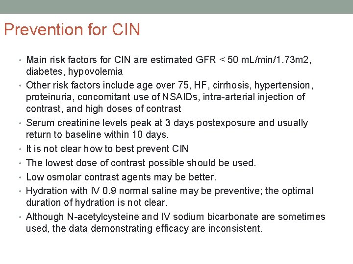
Prevention for CIN • Main risk factors for CIN are estimated GFR < 50 m. L/min/1. 73 m 2, • • diabetes, hypovolemia Other risk factors include age over 75, HF, cirrhosis, hypertension, proteinuria, concomitant use of NSAIDs, intra-arterial injection of contrast, and high doses of contrast Serum creatinine levels peak at 3 days postexposure and usually return to baseline within 10 days. It is not clear how to best prevent CIN The lowest dose of contrast possible should be used. Low osmolar contrast agents may be better. Hydration with IV 0. 9 normal saline may be preventive; the optimal duration of hydration is not clear. Although N-acetylcysteine and IV sodium bicarbonate are sometimes used, the data demonstrating efficacy are inconsistent.
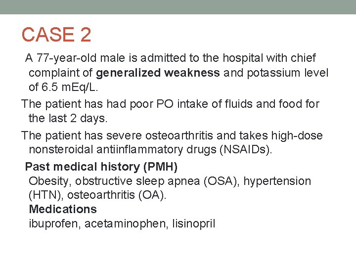
CASE 2 A 77 -year-old male is admitted to the hospital with chief complaint of generalized weakness and potassium level of 6. 5 m. Eq/L. The patient has had poor PO intake of fluids and food for the last 2 days. The patient has severe osteoarthritis and takes high-dose nonsteroidal antiinflammatory drugs (NSAIDs). Past medical history (PMH) Obesity, obstructive sleep apnea (OSA), hypertension (HTN), osteoarthritis (OA). Medications ibuprofen, acetaminophen, lisinopril
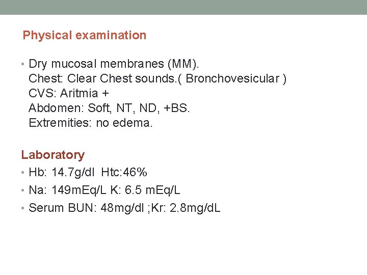
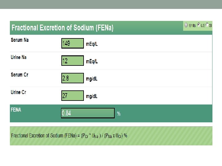
Physical examination • Dry mucosal membranes (MM). Chest: Clear Chest sounds. ( Bronchovesicular ) CVS: Aritmia + Abdomen: Soft, NT, ND, +BS. Extremities: no edema. Laboratory • Hb: 14. 7 g/dl Htc: 46% • Na: 149 m. Eq/L K: 6. 5 m. Eq/L • Serum BUN: 48 mg/dl ; Kr: 2. 8 mg/d. L
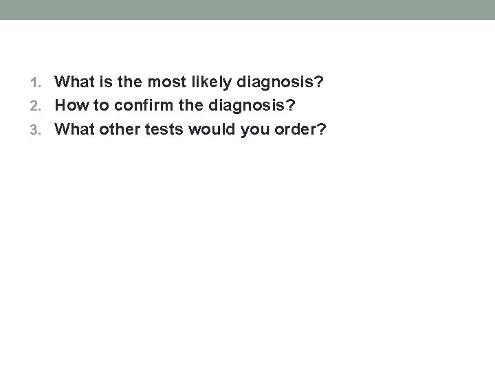
1. What is the most likely diagnosis? 2. How to confirm the diagnosis? 3. What other tests would you order?
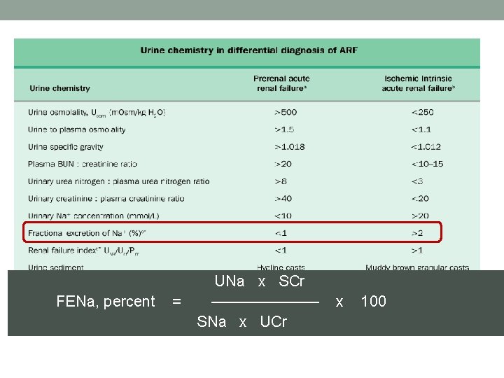
FENa, percent = UNa x SCr ——————— SNa x UCr x 100
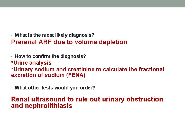
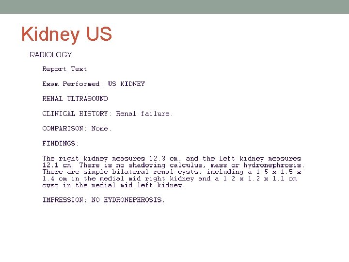
• What is the most likely diagnosis? Prerenal ARF due to volume depletion • How to confirm the diagnosis? *Urine analysis *Urinary sodium and creatinine to calculate the fractional excretion of sodium (FENA) • What other tests would you order? Renal ultrasound to rule out urinary obstruction and nephrolithiasis
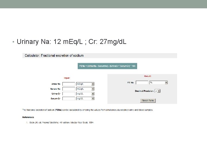
• Urinary Na: 12 m. Eq/L ; Cr: 27 mg/d. L
Kidney US
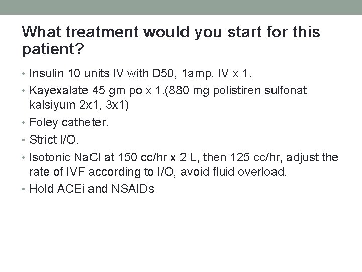
What treatment would you start for this patient? • Insulin 10 units IV with D 50, 1 amp. IV x 1. • Kayexalate 45 gm po x 1. (880 mg polistiren sulfonat kalsiyum 2 x 1, 3 x 1) • Foley catheter. • Strict I/O. • Isotonic Na. Cl at 150 cc/hr x 2 L, then 125 cc/hr, adjust the rate of IVF according to I/O, avoid fluid overload. • Hold ACEi and NSAIDs
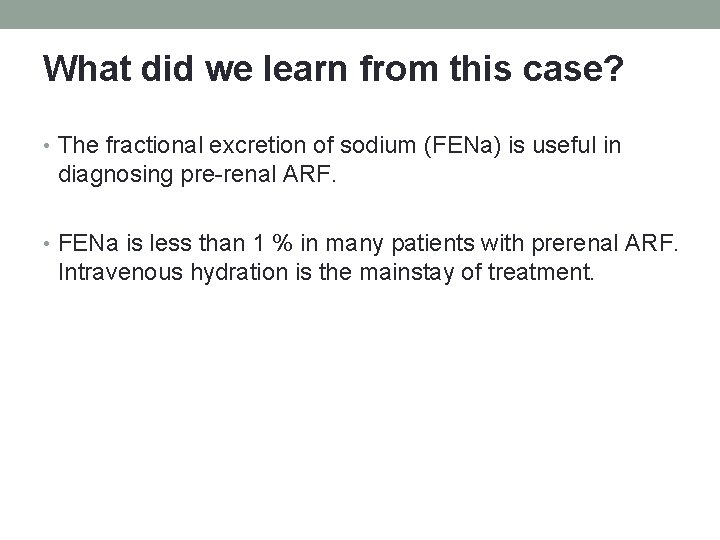
What did we learn from this case? • The fractional excretion of sodium (FENa) is useful in diagnosing pre-renal ARF. • FENa is less than 1 % in many patients with prerenal ARF. Intravenous hydration is the mainstay of treatment.
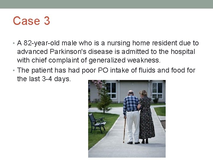
Case 3 • A 82 -year-old male who is a nursing home resident due to advanced Parkinson's disease is admitted to the hospital with chief complaint of generalized weakness. • The patient has had poor PO intake of fluids and food for the last 3 -4 days.
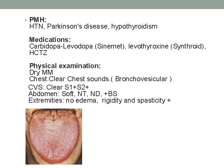
• PMH: HTN, Parkinson's disease, hypothyroidism Medications: Carbidopa-Levodopa (Sinemet), levothyroxine (Synthroid), HCTZ Physical examination: Dry MM Chest: Clear Chest sounds. ( Bronchovesicular ) CVS: Clear S 1+S 2+ Abdomen: Soft, NT, ND, +BS Extremities: no edema, rigidity and spasticity +
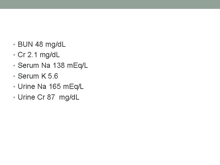
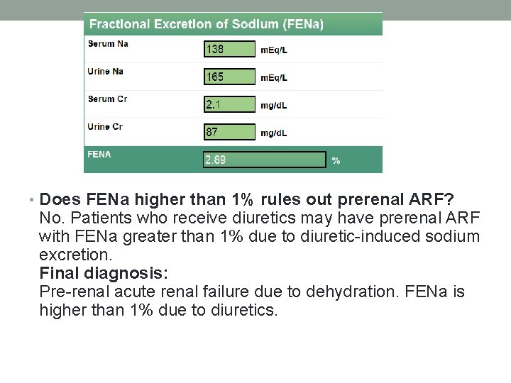
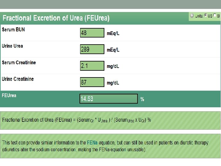
• BUN 48 mg/d. L • Cr 2. 1 mg/d. L • Serum Na 138 m. Eq/L • Serum K 5. 6 • Urine Na 165 m. Eq/L • Urine Cr 87 mg/d. L
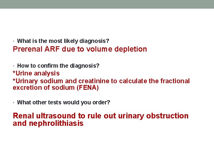
• What is the most likely diagnosis? Prerenal ARF due to volume depletion • How to confirm the diagnosis? *Urine analysis *Urinary sodium and creatinine to calculate the fractional excretion of sodium (FENA) • What other tests would you order? Renal ultrasound to rule out urinary obstruction and nephrolithiasis
• Does FENa higher than 1% rules out prerenal ARF? No. Patients who receive diuretics may have prerenal ARF with FENa greater than 1% due to diuretic-induced sodium excretion. Final diagnosis: Pre-renal acute renal failure due to dehydration. FENa is higher than 1% due to diuretics.
• In patients who are receiving diuretics and suppose to have prerenal AKI • what else do we need for to the proper diagnosis ?
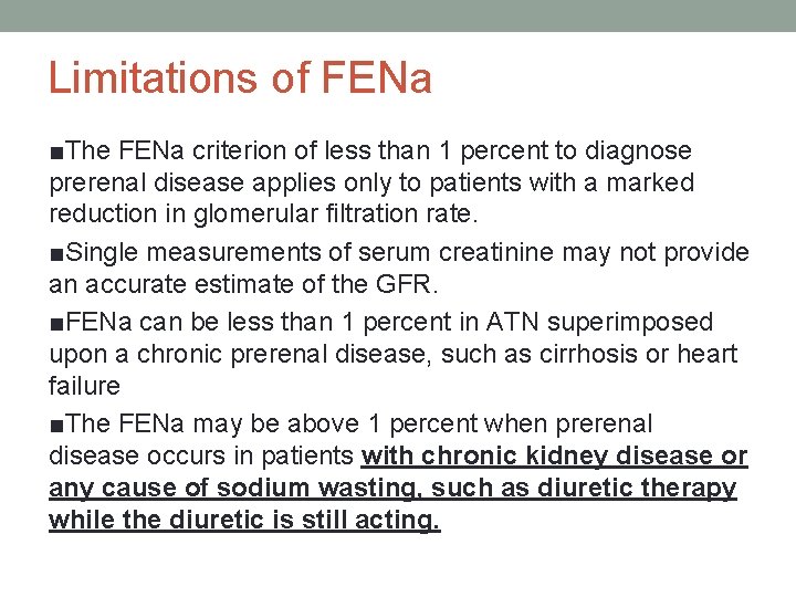
Limitations of FENa ■The FENa criterion of less than 1 percent to diagnose prerenal disease applies only to patients with a marked reduction in glomerular filtration rate. ■Single measurements of serum creatinine may not provide an accurate estimate of the GFR. ■FENa can be less than 1 percent in ATN superimposed upon a chronic prerenal disease, such as cirrhosis or heart failure ■The FENa may be above 1 percent when prerenal disease occurs in patients with chronic kidney disease or any cause of sodium wasting, such as diuretic therapy while the diuretic is still acting.
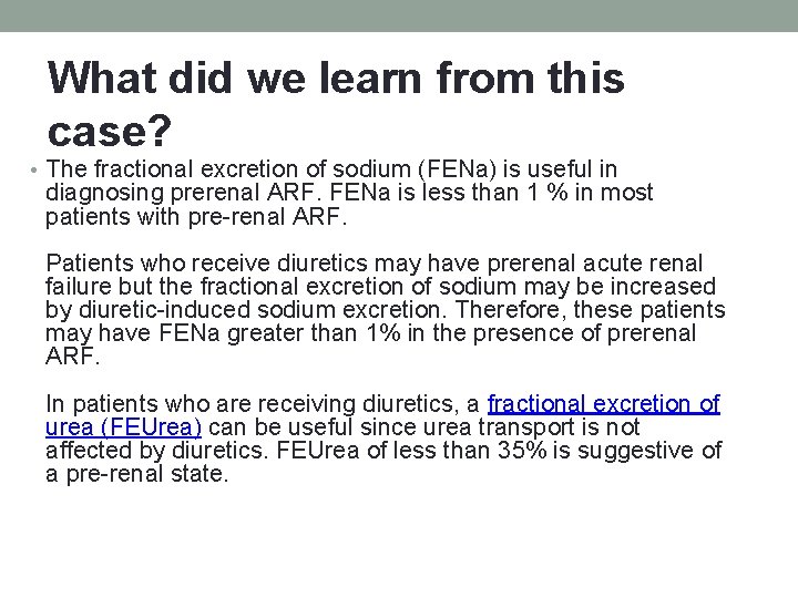
What did we learn from this case? • The fractional excretion of sodium (FENa) is useful in diagnosing prerenal ARF. FENa is less than 1 % in most patients with pre-renal ARF. Patients who receive diuretics may have prerenal acute renal failure but the fractional excretion of sodium may be increased by diuretic-induced sodium excretion. Therefore, these patients may have FENa greater than 1% in the presence of prerenal ARF. In patients who are receiving diuretics, a fractional excretion of urea (FEUrea) can be useful since urea transport is not affected by diuretics. FEUrea of less than 35% is suggestive of a pre-renal state.