https www yenepoya edu in 11272020 12 00



- Slides: 80
https: //www. yenepoya. edu. in 11/27/2020 12: 00 AM Yenepoya Nursing College (NAME) 1
Unit VI URINARY CALCULI/ UROLITHIASIS/ RENAL CALCULI PRESENTED BY: Ms. Pavithra K Lecturer, MSN dept REVIEWED BY: Mr. Gireesh G R Assoc. Professor, MSN dept
At the end of the class students will be able to • Define Renal Calculi • Enlist the causes of Renal Calculi • Explain the patho- physiology of Renal Calculi • Enlist the clinical features of Renal Calculi • Enumerate the management of Renal Calculi 11/27/2020 12: 00 AM (NAME) 3
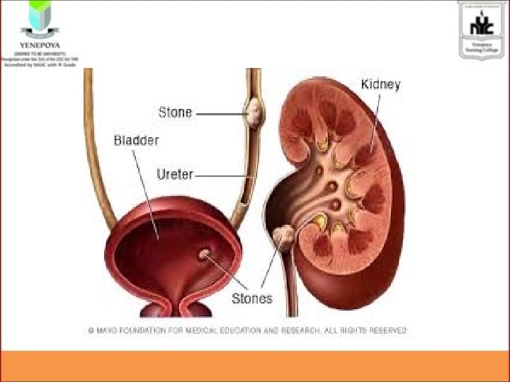
• Kidney stones, or renal calculi, are solid masses made of crystals. • Stone formation anywhere in urinary system • are small, hard deposits that form in the urine • made up of mineral and acid salts. • kidney stones form in the kidneys, they can be transported throughout the urinary tract, which includes the kidneys, ureters, bladder, and urethra.
Incidence • Kidney stones are most commonly found in people between the ages of 20 -40, • about one million people are treated for them every year in the United States. • Males
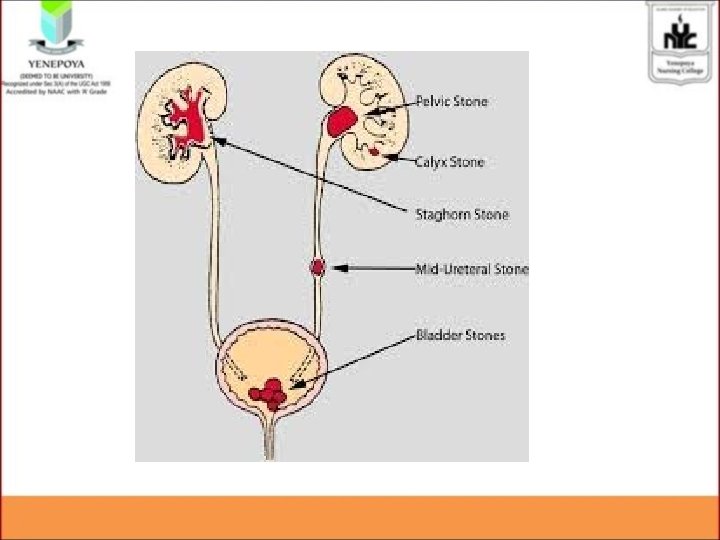
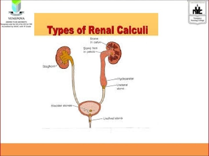
• Sites Pelvis Ureters Bladder
Risk factors • • • Immobiliy Dehydration Reduced fluid intake Metabolic disturbances-hypercalcemia Diet rich in purine, oxalate, calcium
• • • Previous renal calculi Urinary catheterization Neurogenic bladder Acute urinary retension Overactive parathyroid gland UTI
Etiology • Urinary stasis – Infection – Foreign body – Dehyration – Obstruction in the urinary tract – Bladder neck obstruction – Failure to empty the bladder – Urinary diversion – Immobilization
• Supersaturation of urine – Highly concentrated – Crystallization/ precipitation • Inhibitor substances – Citrate – magnisium
• Medications- drugs Crystallize – magnesium trisilicate – ciprofloxacin – indinavir – Ephedrine – Calcium carbonate, Calcium gluconate
TYPES • • • Calcium stones Oxalate stones/ mulberry Struvite stones Uric acid stones Xanthine stones Cystine stones
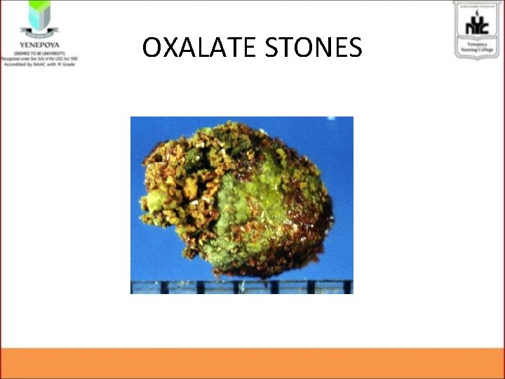
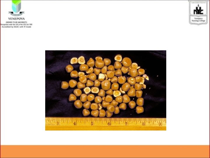
OXALATE STONES • Insoluble in urine • Occur in persons who take large number of cereals • Known as mulberry stones • Has sharp projections, uneven surfaces and appears as single ones
OXALATE STONES
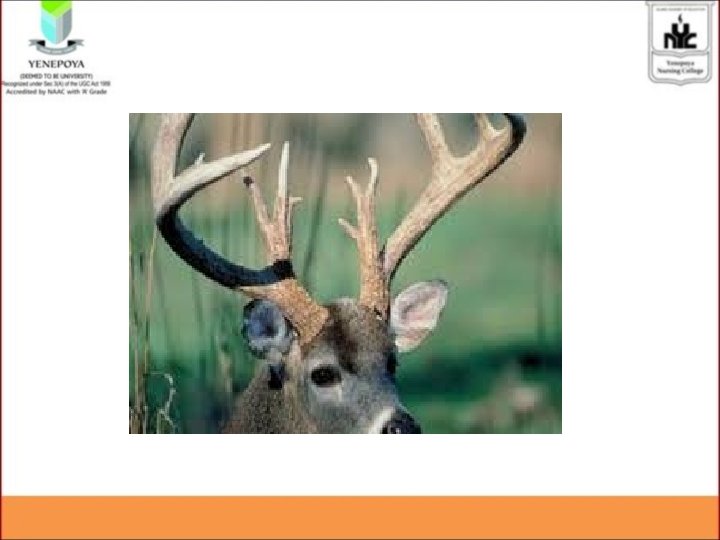
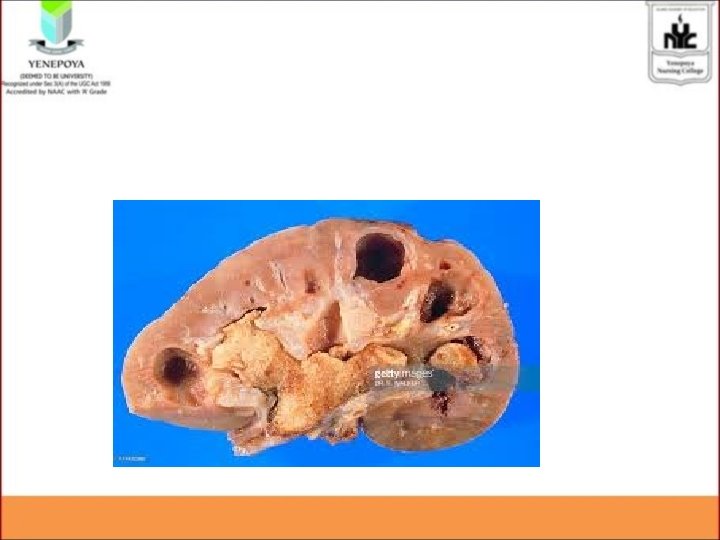
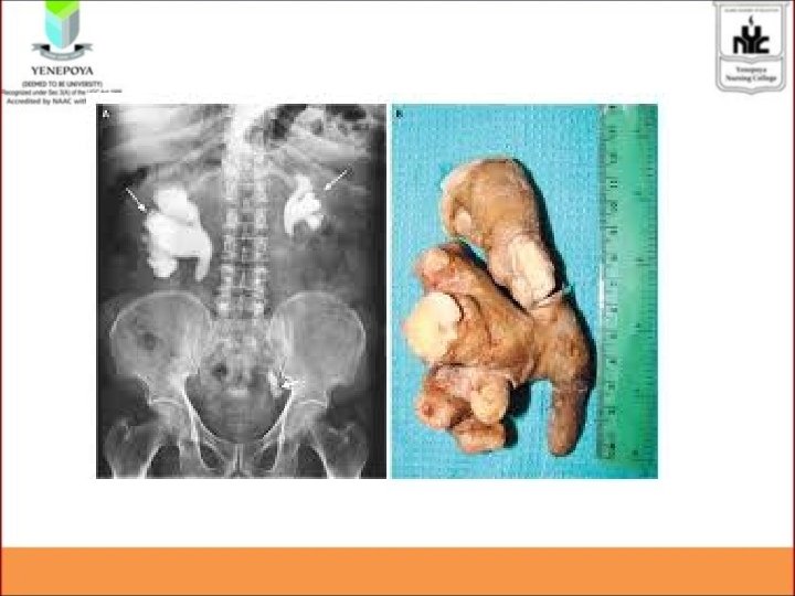
CALCIUM STONES • • • In persons with increase calcium level Exist as calcium phosphate, calcium oxalate Vary from small to large size Has stang horn appearance Common in females of 20 -30 yrs
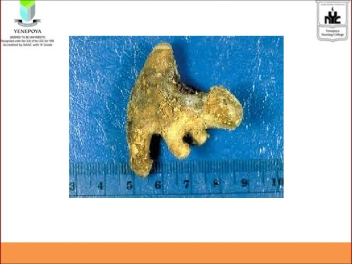
STRUVITE STONES • Are triple phosphate stones • Contains phosphate of magnesium, calcium, ammonium • Stones will be hard
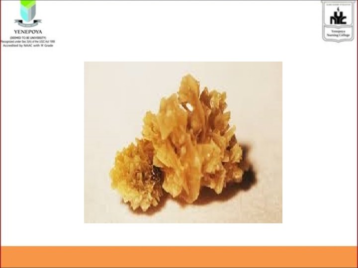
URIC ACID STONES • Seen in patients with gout • Those who take high purine foods like shell fish, mushroom, asparagus etc
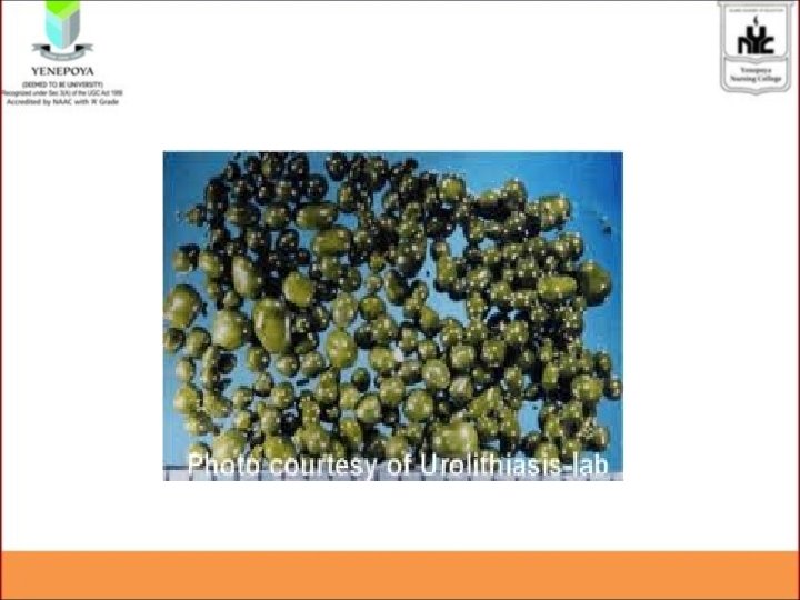
XANTHINE STONES • Hereditary disorders • seen in children and adolescents
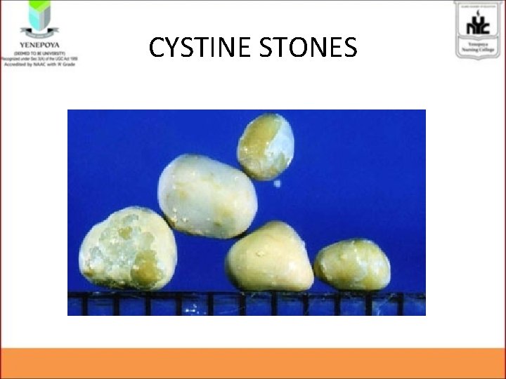
CYSTINE STONES • heriditaryhey occur in both men and women who have the genetic disorder cystinuria. • cystine — an acid that occurs naturally in the body — leaks from the kidneys into the urine.
CYSTINE STONES
Pathophysiology • • • Etiology Crystal growth Crystal aggregation Stone formation Randalls plaque- Back pain
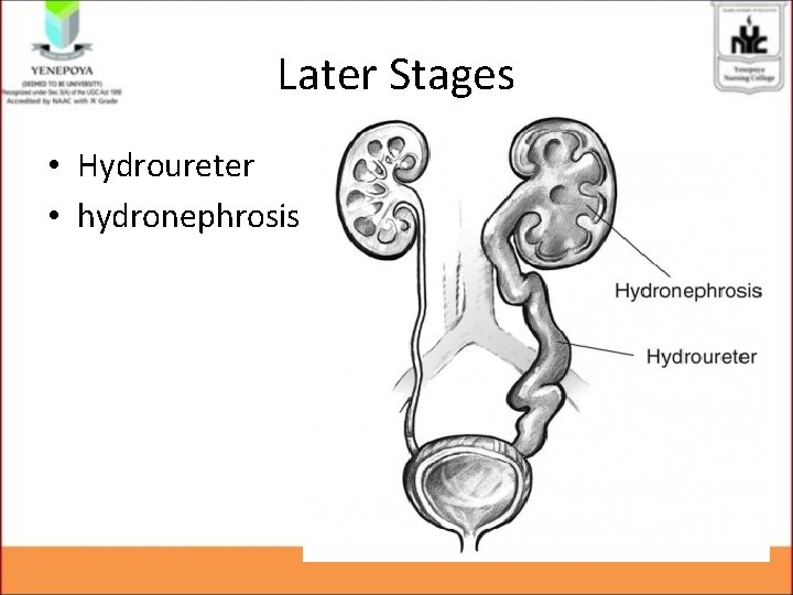
• Progression of stone • Surface erosion of the urinary tract— Hematuria • Increase in the size • Moves down • Obstruct • Complictions – Hydroureter, Hydronephrosis
CLINICAL FEATURES • Depends on – size of calculi – location
1. Pain • Renal colic • Radiates medially, • downward towards bladder in females and testis in males • Renal colic is associated with fever, nausea and vomiting
URETERIC CALCULI • Sudden quality pain • pain radiating to thigh and regions of kidney • Patient have frequent desire to void • Patient have hematuria due to sharp projections of stones
• • High grade fever Anorexia Nausea Vomiting Fullness in the bladder Decreased bladder capacity Intermittent micturation diaphoresis
• • • Dysuria Urgency Frequency Hematuria Pyuria Urinary retension
Later Stages • Hydroureter • hydronephrosis
DIAGNOSTIC EVALUATION • History collection – Previous history or family history – Episodes of dehydration – Prolonged immobility – UTI – medications • Physical examination – Location, severity and radiation of pain – Clinical features of renal calculi
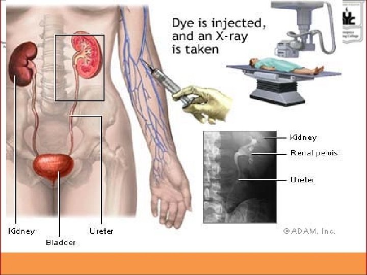
• • Ultrasonography Urine analysis Intravenous pyelogram X-ray KUB
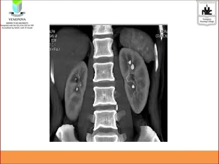
• CT • MRI • Blood investigations – calcium, phosphorus, uric acid, • KFT/ RFT – Serum urea (15 -45 mg/dl) – Serum creatinine ( 0. 6 -1. 2 mg/dl)
Management Goals: • To identify the cause • To relieve the symptoms • To stop the progression of the disease • To control infection • To relieve obstruction • To prevent the complications
Small stones Drinking water. • 2 to 3 litres per day • Increase fluid intake leads to forced diuresis • Helps to flush out stones from the urinary system
Pain relievers. • To relieve mild pain, • pain relievers such as ibuprofen, acetaminophen or naproxen sodium Alpha blockers • relaxes the muscles of ureter, helping the kidney stone to move quickly and with less pain. • Doxazosin, Prazosin, Terazosin.
URINARY ANALGESICS • Pyridium • Relieves irritation of barrier mucosa • So it should be combined with antibiotics ANTIBIOTICS • Amoxicillin • cefotaxim
Medications • Uric acid stones – allopurinol (Zyloprim, Aloprim) – to reduce uric acid levels in your blood and urine
Calcium oxalate stones • Increase hydration. • Reduce dietary oxalate. • administer thiazide diuretics. – Thiazide diuretics. Eg: chlorthalidone – Increases calcium excretion • administer cellulose phosphate to prevent GI absorption. • Administer potassium citrate to maintain alkaline urine.
Cystine • Increase hydration. • Administer alpha-penicillamine and tiopronin to prevent cystin crystallization. • Give potassium citrate to maintain alkaline urine
Other management Ensure adequate output Dietary regulations Decrease calcium diet in hypercalcemia Restrict high oxalate food- tomato, instant coffee, cola drinks, chocolates, citrus fruits etc • Restrict purine containing foods like shellfish, mushrooms, asparagus and organ meats • •
• URETEROSCOPY • involves first visualizing the stone and then destroying it. • Access to the stone is accomplished by inserting a ureteroscope into the ureter and then inserting a electrohydraulic lithotriptor, • to fragment and remove the stones. • A stent may be inserted and left in place for one month to maintain theureter patent.
LASER LITHOTRIPSY. • A newer treatment for calculi is laser lithotripsy. • Lasers are used together with a uretero-scope to remove or loosen impacted stones.
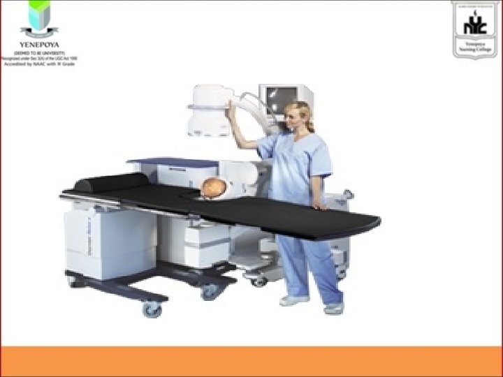
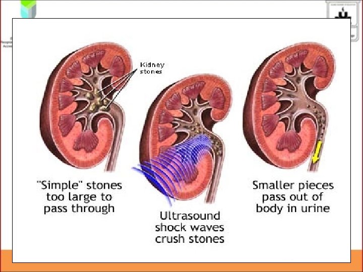
• EXTRACORPOREAL SHOCK WAVE LITHOTRIPSY (ESWL) • ESWL is a non-invasive procedure used to break up stones. • In ESWL, a high-energy amplitude of pressure, or shock wave, is generated are focussed over the area of the renal stone. • Shock waves pass through the skin and fragment the stone.
• Repeated shock waves focused on the stone eventually reduce it to many small pieces. • These small pieces are excreted in the urine, usually without difficulty. • the fragments may be passed upto 3 months after the procedure
Surgical management Indication • Size • Location • Failure of ESWL • Obstruction – Hydroureter – hydronephrosis
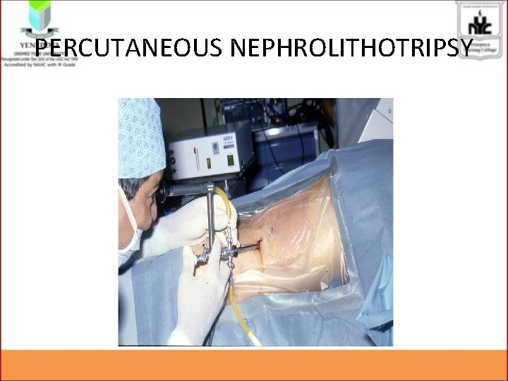
• PERCUTANEOUS LITHOTRIPSY • It is used to treat larger stones. • Percutaneous tract is formed and a nephroscope is inserted. • To extract or fragment the stones.
PERCUTANEOUS NEPHROLITHOTRIPSY
Open surgical management • Ureterolithotomy • is the removal of a stone from the ureter through a flank incision or an abdominal incision. • A Penrose drain are usually placed postoperatively.
• Cystolithotomy, • removal of bladder calculi through a suprapubic incision • is used only when stones cannot be crushed and removed transurethrally.
• Nephrolithotomy- incision is made over kidney to remove the stones • Pyelolithotomy- opening in pelvis of kidney and remove stones • Nephrectomy- removal of kidney – Partial – complete
• • • Post operatively…. . Monitor urine output Check for hematuria Observe for urinary retention Check for the stone fragments in the urine Check for the signs of infection
complications • • Obstruction Infection Impaired Renal function Peri-renal hematoma- bleeding arround the kidney caused by trauma of shock waves
Nursing Management Nursing assessment: • Assess for pain and discomfort, including severity, location, and radiation of pain. • Assess for associated symptoms, including nausea, vomiting, diarrhea, and abdominal distention. • Observe for signs of urinary tract infection(chills, fever, frequency, and hesitancy) and obstruction (oliguria, or anuria).
• Observe urine for hematuria; strain for stones • Focus on Previous history of urinary tract stones - that may have precipitated current episode of renal or ureteral colic. • Assess patient’s knowledge about renal stones and measures to prevent recurrence
Nursing diagnosis • Acute pain r/t obstructing urinary calculus • Altered urinary elimination related to presence of urinary calculi • Risk tor infection r/t obstructing urinary calculus • Altered renal peripheral tissue perfusion r/t postrenal obstruction
Acute pain r/t obstructing urinary calculus • Monitor pain closely and report promptly increases in severity. • Administer opioid analgesics (IV or intramuscular) with IV NSAID as prescribed. • Comfortable position. • Assist patient to ambulate to obtain some pain relief. • Comfort devices • Hot application
Impaired urinary elimination • Record I&O and characteristics of urine • Determine patient’s normal voiding pattern and note variations. • Promote sufficient intake of fluids. • Strain all urine. Document any stones expelled and send to laboratory for analysis. • catheterization
Risk tor infection r/t obstructing urinary calculus • Administer antibiotics- IV or oral • Assess the urine for colour, cloudiness and odour. • Monitor for the signs and symptoms of infection like fever, tachycardia.
Other nursing interventions • Encourage increased fluid intake and ambulation. • Begin IV fluids if patient cannot take adequate oral fluids. • Monitor total urine output
• Instruct patient to report decreased urine volume, bloody or cloudy urine, fever, and pain. • Instruct patient to report any sudden increase in pain. • Monitor vital signs • monitor for early indications of infection
Prevention • Increase fluid intake to maintain urine output at 2 -3 litres per day. • Reduce salt intake. • Reduce the amount of meat and animal protein. • Reduce oxalate intake (foods rich in oxalate include chocolate, nuts) and urate-rich foods (eg, fish).
• take regular cranberry juice- citrate , oxalate and phosphate stones can be prevented. • Maintain calcium intake at normal levels (lowering intake increases excretion of calcium oxalate).
EVALUATION • • • What is Renal calculi? What are the causes of Renal calculi? Explain the Patho-physiology of Renal calculi? List down the clinical features of Renal calculi Enumerate the Management of Renal calculi
REFERENCE • Smeltzer – Brunner & Suddharth Textbook of Medical Surgical Nursing, Wolters kluwer publishers, 12 th edition 2009. • Black – Medical Surgical Nursing, Elsevier publishers, 8 th edition 2009. • Nettina – Lippincott manual of Nursing Practice, Wolters kluwer publishers, 7 th edition 2014. • Lewis – Medical Surgical Nursing, Elsevier publishers, 10 th edition, 2017