
LIFE SCIENCES GRADE 11 CAPS STRUCTURED CLEAR PRACTICAL




- Slides: 68
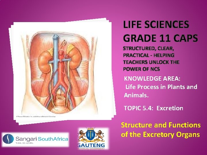
LIFE SCIENCES GRADE 11 CAPS STRUCTURED, CLEAR, PRACTICAL - HELPING TEACHERS UNLOCK THE POWER OF NCS KNOWLEDGE AREA: Life Process in Plants and Animals. TOPIC 5. 4: Excretion Structure and Functions of the Excretory Organs
SUMMARY OF THE PRESENTATION: Introduction Gut Lungs Skin Liver Kidneys Structure of the Urinary System
INTRODUCTION: �Excretion is the removal of metabolic wastes from the living organism. �Metabolic wastes are waste substances that are produced during metabolic reactions. � Some examples of the metabolic wastes are urea, uric acid and carbon dioxide. � These substances can be very dangerous if allowed to accumulate in the body. � For example if carbon dioxide is allowed to collect in the body it combines with water to form carbonic acid. � This would change the p. H of the tissue fluid and affect the functioning of enzymes.
TERMINOLOGY: TERM: Excretion DEFINITION: USE IN SENTENCE: Excretion is the removal of metabolic waste from the body Carbon dioxide leaves the body through excretion
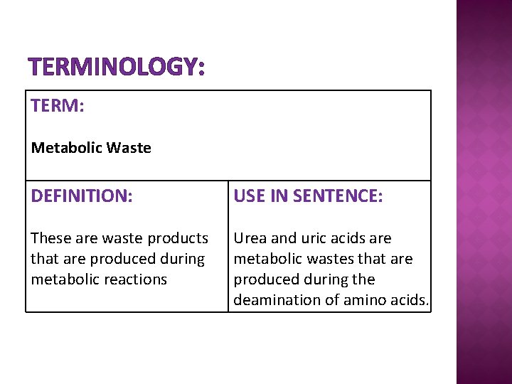
TERMINOLOGY: TERM: Metabolic Waste DEFINITION: USE IN SENTENCE: These are waste products that are produced during metabolic reactions Urea and uric acids are metabolic wastes that are produced during the deamination of amino acids.
EXCRETORY ORGANS AND THEIR FUNCTIONS Gut: � At the end of the digestion the undigested and indigestible food needs to leave the body. � This occurs through the process of defaecation or egestion. �But indigestible food is not an example of excretory waste. � This is so because they are not produced as a result of any metabolic activities. � Therefore defaecation or egestion are not examples of excretory processes. � Therefore the gut is not an example of an excretory organ.
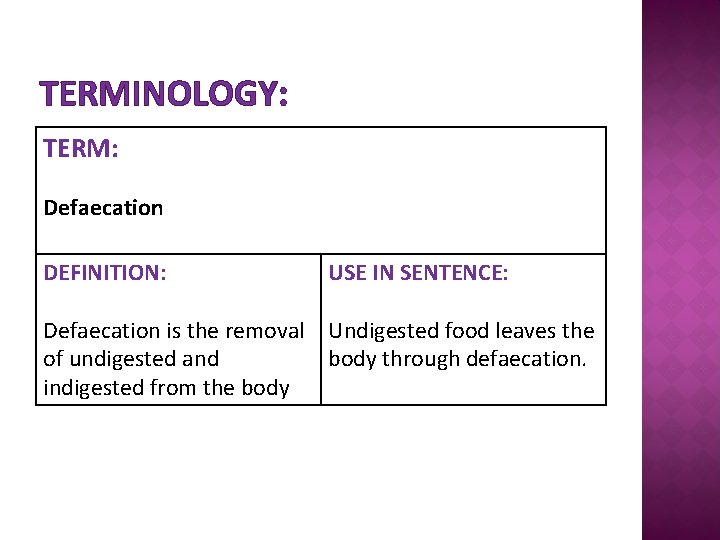
TERMINOLOGY: TERM: Defaecation DEFINITION: USE IN SENTENCE: Defaecation is the removal Undigested food leaves the of undigested and body through defaecation. indigested from the body
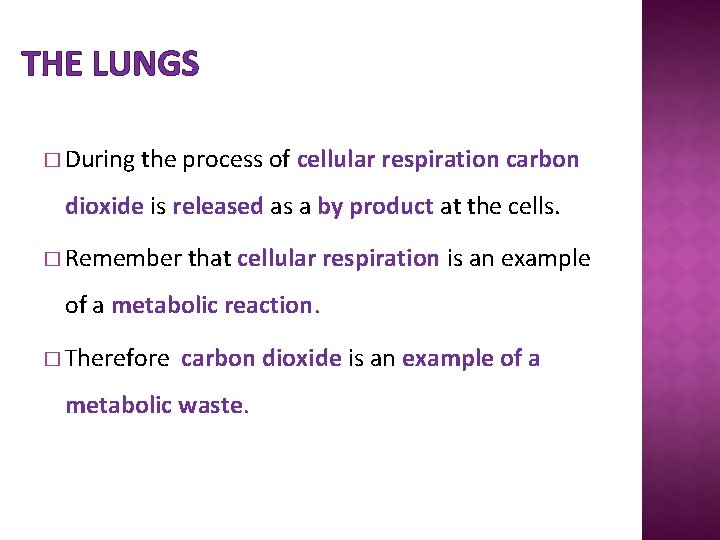
THE LUNGS � During the process of cellular respiration carbon dioxide is released as a by product at the cells. � Remember that cellular respiration is an example of a metabolic reaction. � Therefore carbon dioxide is an example of a metabolic waste.
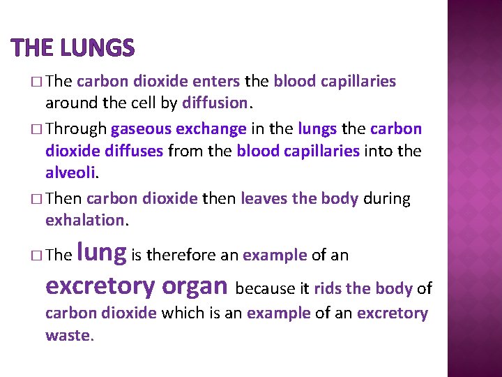
THE LUNGS � The carbon dioxide enters the blood capillaries around the cell by diffusion. � Through gaseous exchange in the lungs the carbon dioxide diffuses from the blood capillaries into the alveoli. � Then carbon dioxide then leaves the body during exhalation. lung is therefore an example of an excretory organ because it rids the body of � The carbon dioxide which is an example of an excretory waste.
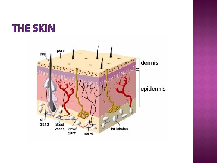
THE SKIN � When the body temperature increases we perspire in order to lower our body temperature. � The sweat or perspiration reaches the surface of the skin through pores on the skin. � These pores carry the sweat form the sweat glands. �Sweat is made up of water, salt and urea. � Since these substances are examples of metabolic waste, the organ. skin is also an example of an excretory
THE SKIN
THEIR LIVER � The liver is an excretory organ because it is responsible for the deamination of the amino acids. � This means that it breaks down the excess amino acids. � The excess amino acids are broken down into urea. �Urea is an example of a nitrogenous waste. � The urea is then carried by the blood to the kidneys where it forms a part of urine.
TERMINOLOGY: TERM: Deamination DEFINITION: USE IN SENTENCE: Deamination refers to the break down of the excess amino acids. The excess amino acids undergo deamination.
THE KIDNEYS � The kidneys are an example of an excretory organ because they are involved in the production of urine. � Metabolic wastes from all over the body come to the kidneys. � In the kidneys this blood is filtered and all the metabolic waste forms urine. � Therefore the metabolic waste leaves the body as urine.
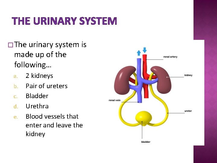
THE URINARY SYSTEM � The urinary system is made up of the following… a. b. c. d. e. 2 kidneys Pair of ureters Bladder Urethra Blood vessels that enter and leave the kidney
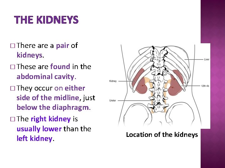
THE KIDNEYS � There a pair of kidneys. � These are found in the abdominal cavity. � They occur on either side of the midline, just below the diaphragm. � The right kidney is usually lower than the left kidney. Location of the kidneys
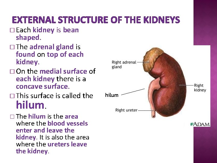
EXTERNAL STRUCTURE OF THE KIDNEYS � Each kidney is bean shaped. � The adrenal gland is found on top of each kidney. � On the medial surface of each kidney there is a concave surface. � This surface is called the hilum. � The hilum is the area where the blood vessels enter and leave the kidney. It is also the area where the ureters leave the kidney. hilum
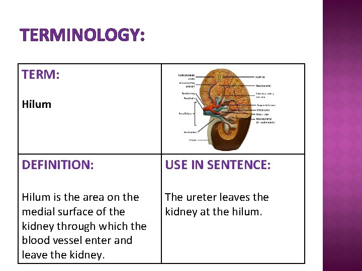
TERMINOLOGY: TERM: Hilum DEFINITION: USE IN SENTENCE: Hilum is the area on the medial surface of the kidney through which the blood vessel enter and leave the kidney. The ureter leaves the kidney at the hilum.
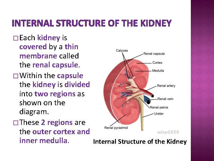
INTERNAL STRUCTURE OF THE KIDNEY � Each kidney is covered by a thin membrane called the renal capsule. � Within the capsule the kidney is divided into two regions as shown on the diagram. � These 2 regions are the outer cortex and inner medulla. Internal Structure of the Kidney
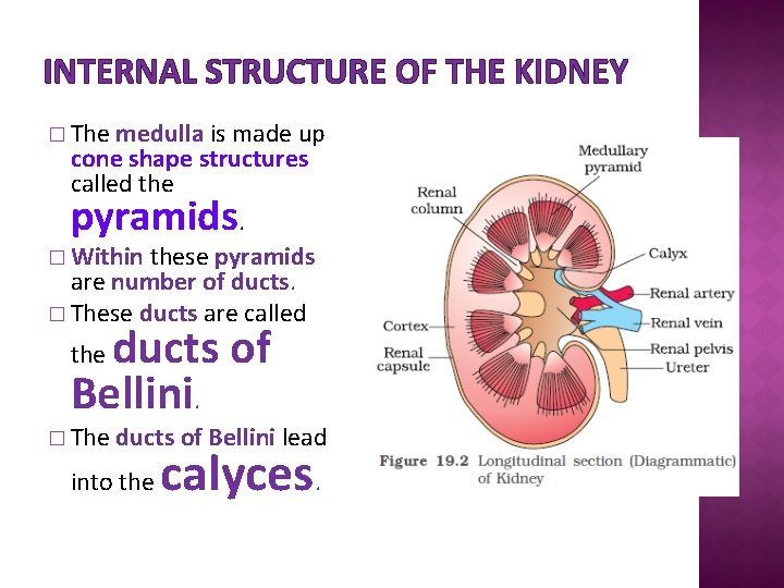
INTERNAL STRUCTURE OF THE KIDNEY � The medulla is made up cone shape structures called the. � Within these pyramids are number of ducts. � These ducts are called pyramids ducts of Bellini. the � The ducts of Bellini lead into the calyces.
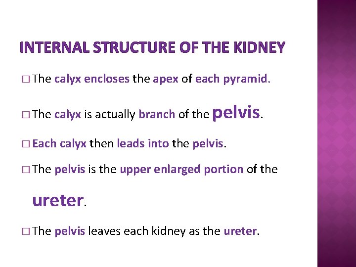
INTERNAL STRUCTURE OF THE KIDNEY � The calyx encloses the apex of each pyramid. � The calyx is actually branch of the pelvis. � Each � The calyx then leads into the pelvis is the upper enlarged portion of the ureter. � The pelvis leaves each kidney as the ureter.
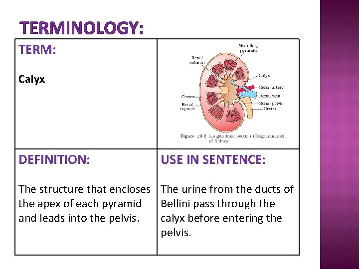
TERMINOLOGY: TERM: Calyx DEFINITION: USE IN SENTENCE: The structure that encloses The urine from the ducts of the apex of each pyramid Bellini pass through the and leads into the pelvis. calyx before entering the pelvis.
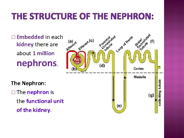
THE STRUCTURE OF THE NEPHRON: � Embedded in each kidney there about 1 million nephrons. The Nephron: � The nephron is the functional unit of the kidney.
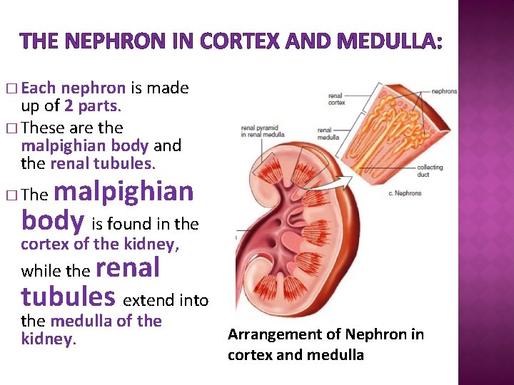
THE NEPHRON IN CORTEX AND MEDULLA: � Each nephron is made up of 2 parts. � These are the malpighian body and the renal tubules. malpighian body is found in the � The cortex of the kidney, while the renal tubules extend into the medulla of the kidney. Arrangement of Nephron in cortex and medulla
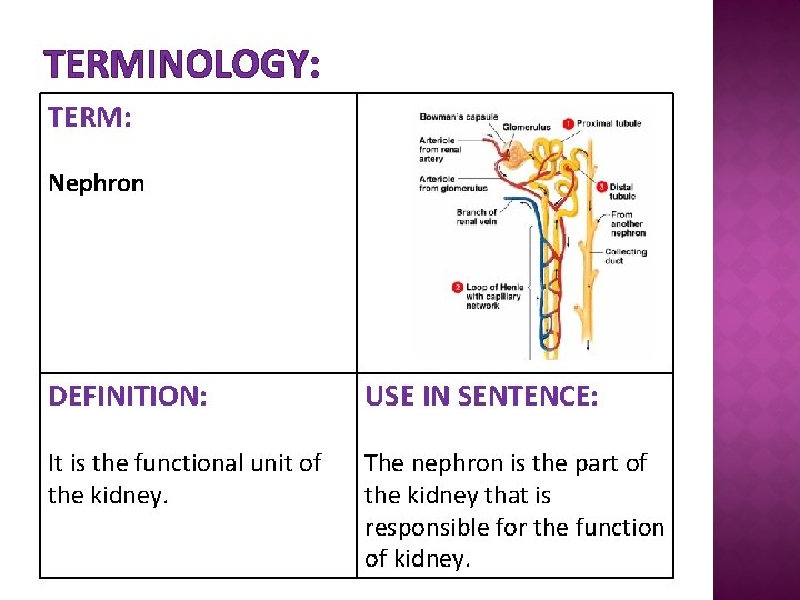
TERMINOLOGY: TERM: Nephron DEFINITION: USE IN SENTENCE: It is the functional unit of the kidney. The nephron is the part of the kidney that is responsible for the function of kidney.
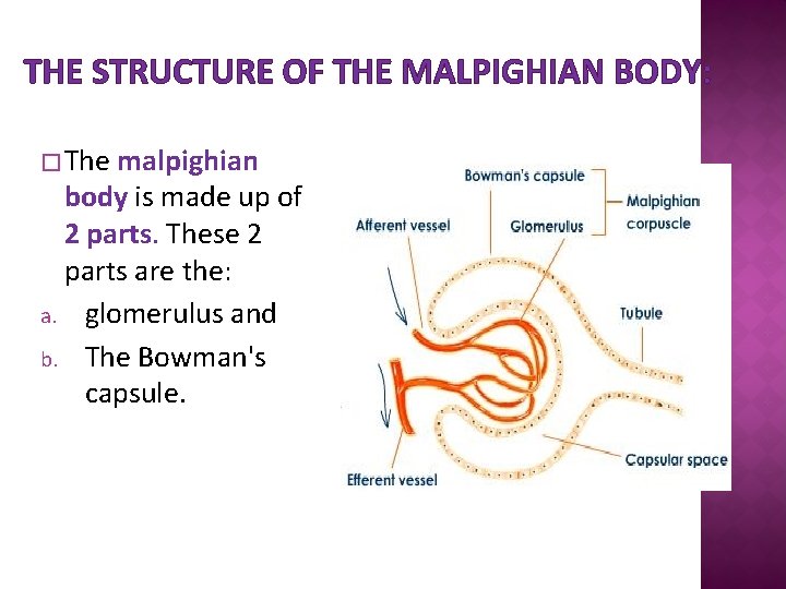
THE STRUCTURE OF THE MALPIGHIAN BODY: � The malpighian body is made up of 2 parts. These 2 parts are the: a. glomerulus and b. The Bowman's capsule.
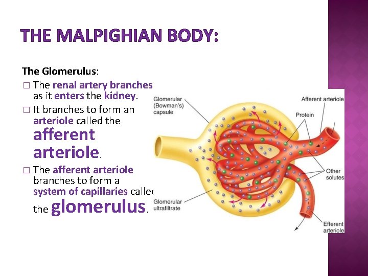
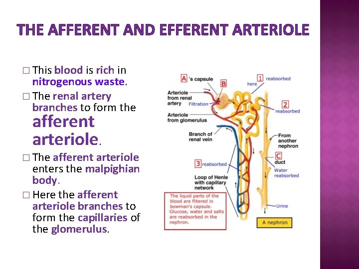
THE MALPIGHIAN BODY: The Glomerulus: � The renal artery branches as it enters the kidney. � It branches to form an arteriole called the afferent arteriole. � The afferent arteriole branches to form a system of capillaries called the glomerulus.
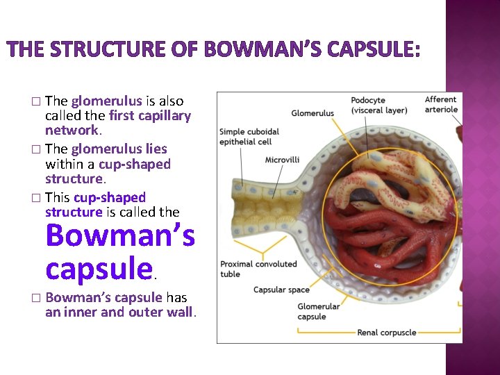
THE STRUCTURE OF BOWMAN’S CAPSULE: The glomerulus is also called the first capillary network. � The glomerulus lies within a cup-shaped structure. � This cup-shaped structure is called the � Bowman’s capsule has an inner and outer wall.
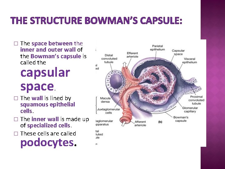
THE STRUCTURE BOWMAN’S CAPSULE: � The space between the inner and outer wall of the Bowman’s capsule is called the capsular space. The wall is lined by squamous epithelial cells. � The inner wall is made up of specialized cells. � These cells are called � podocytes.
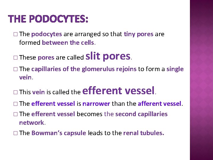
THE PODOCYTES: � The podocytes are arranged so that tiny pores are formed between the cells. � These pores are called slit pores. � The capillaries of the glomerulus rejoins to form a single vein. � This � The vein is called the efferent vessel is narrower than the afferent vessel. � The efferent vessel becomes the second capillaries network. � The Bowman’s capsule leads to the renal tubules.
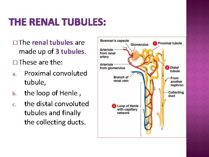
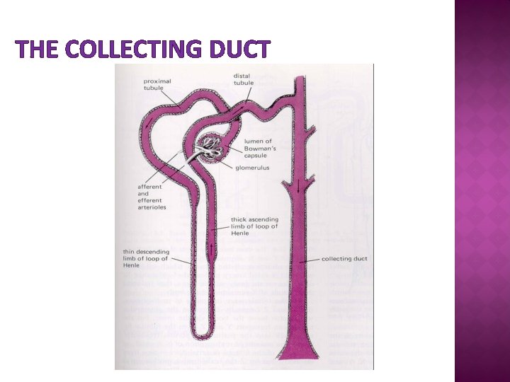
THE RENAL TUBULES: � The renal tubules are made up of 3 tubules. � These are the: a. Proximal convoluted tubule, b. the loop of Henle , c. the distal convoluted tubules and finally the collecting ducts.
THE RENAL TUBULES: � The proximal convoluted tubule leads form the Bowman’s capsule. � The word convoluted means twisted. � It is long and twisted, this creates a large surface area. � These tubules are lined on the inside by cuboidal epithelial cells with brush borders. � The proximal convoluted tubule leads to the loop of Henle.
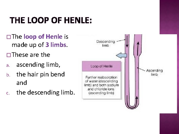
THE LOOP OF HENLE: � The loop of Henle is made up of 3 limbs. � These are the a. ascending limb, b. the hair pin bend and c. the descending limb.
THE LOOP OF HENLE: � The descending limb of the loop of Henle opens into the distal � This convoluted tubule. is the last part of the renal tubules. � It is also long and convoluted. � It is also lined by the cubiodal epithelium with a brush border.
THE COLLECTING DUCT
THE COLLECTING DUCT: � The distal convoluted tubule leads into the ducts. collecting � The collecting ducts start in the renal cortex and continues into the medulla. � These collecting tubules join to form the Bellini � The ducts of Bellini are found in the pyramids. � The ducts open into the calyces of the pyramid.
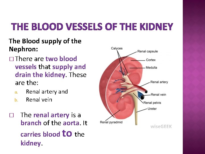
THE BLOOD VESSELS OF THE KIDNEY The Blood supply of the Nephron: � There are two blood vessels that supply and drain the kidney. These are the: a. b. � Renal artery and Renal vein The renal artery is a branch of the aorta. It carries blood to the kidney.
TERMINOLOGY: TERM: Afferent arteriole DEFINITION: USE IN SENTENCE: A branch of the renal artery that enters the malpighian body. The afferent arteriole is a branch of the renal artery.
THE AFFERENT AND EFFERENT ARTERIOLE � This blood is rich in nitrogenous waste. � The renal artery branches to form the afferent arteriole. � The afferent arteriole enters the malpighian body. � Here the afferent arteriole branches to form the capillaries of the glomerulus.
TERMINOLOGY: TERM: Renal artery DEFINITION: USE IN SENTENCE: The blood vessel that carries blood rich in nitrogenous waste to the kidney. The renal artery branches to form the afferent arteriole.
THE BLOOD VESSELS OF THE URINARY SYSTEM: � These capillaries then rejoin to form the efferent vessel. � The efferent vessel then leaves the Bowman’s capsule. � The efferent vessel then divides to form the capillaries of the second capillary network. � These capillaries rejoin to form small veins called venules. � These � The venules unite to form the renal vein leaves the kidney.
TERMINOLOGY TERM: Efferent vessel DEFINITION: USE IN SENTENCE: The blood vessel that leaves the malpighian body and then divides to form the second capillary network. The efferent vessel is narrower than afferent vessel therefore creating pressure in the glomerulus.
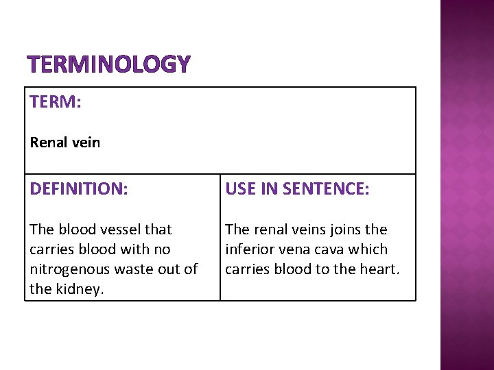
TERMINOLOGY TERM: Renal vein DEFINITION: USE IN SENTENCE: The blood vessel that carries blood with no nitrogenous waste out of the kidney. The renal veins joins the inferior vena cava which carries blood to the heart.
THE BLOOD VESSELS OF THE URINARY SYSTEM � The renal vein carries blood that has no nitrogenous waste. � The renal vein then joins the inferior vena cava, which carries this blood to the heart.
TERMINOLOGY: � Excretion: is the removal of metabolic waste from the body. � Metabolic Waste: these are waste products that are produced during metabolic reactions. � Defaecation: is the removal of undigested and indigested from the body. � Deamination: refers to the break down of the excess amino acids. � Hilum: is the area on the medial surface of the kidney through which the blood vessel enter and leave the kidney. � Calyx: is the structure that encloses the apex of each pyramid and leads into the pelvis.
TERMINOLOGY: � Nephron: is the functional unit of the kidney. � Malpighian body: part of the nephron, made up of the Bowman’s capsule and glomerulus. � Bowman’s capsule: cup shaped structure that contains the glomerulus. � Glomerulus: also known as the first capillary network, a bunch of capillaries formed when the afferent arteriole branches. � Capsular Space: the space found between the inner and outer wall of the Bowman’s capsule. � Podocytes: these are specialized cells found on the inner wall of the Bowman’s capsules. � Slit pores: tiny pores found between the podocytes.
TERMINOLOGY: � Renal artery: a blood vessel that carries blood rich in nitrogenous waste to the kidney. � Renal Vein: the blood vessel that carries blood with no nitrogenous waste out of the kidney. � Afferent arteriole: a branch of the renal artery that enters the malpighian body. � Efferent arteriole: the narrow blood vessel that leaves the malpighian body and then divides to form the second capillary network.
QUESTION 1 The pair of blood vessels that is directly responsible for glomerular filtration… A. B. C. D. Afferent vessel and renal artery Efferent vessel and renal vein Afferent and efferent arteriole Renal artery and renal vein
QUESTION 2 The … is not an example of an excretory organ. A. B. C. D. Lung Skin Gut Liver
QUESTION 3 Largest excretory organ in the body… A. B. C. D. Lungs Skin Gut Liver
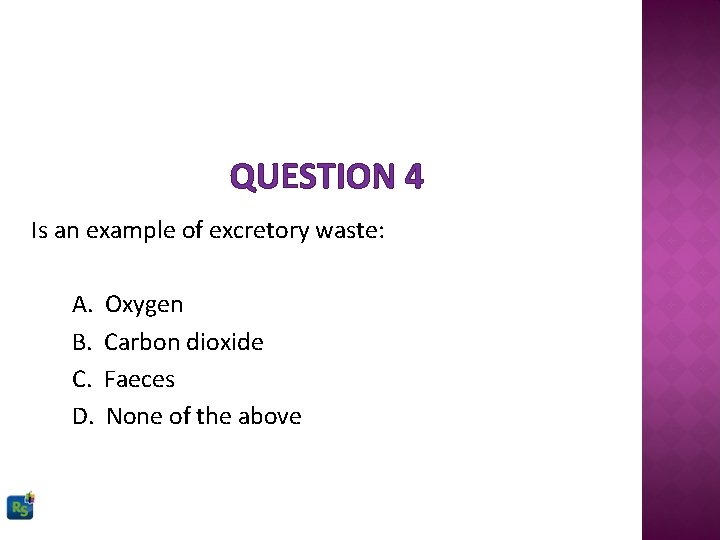
QUESTION 4 Is an example of excretory waste: A. B. C. D. Oxygen Carbon dioxide Faeces None of the above
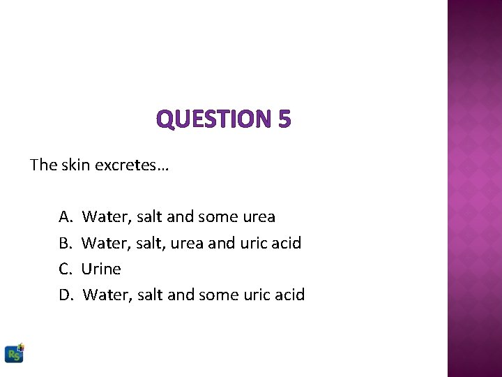
QUESTION 5 The skin excretes… A. B. C. D. Water, salt and some urea Water, salt, urea and uric acid Urine Water, salt and some uric acid
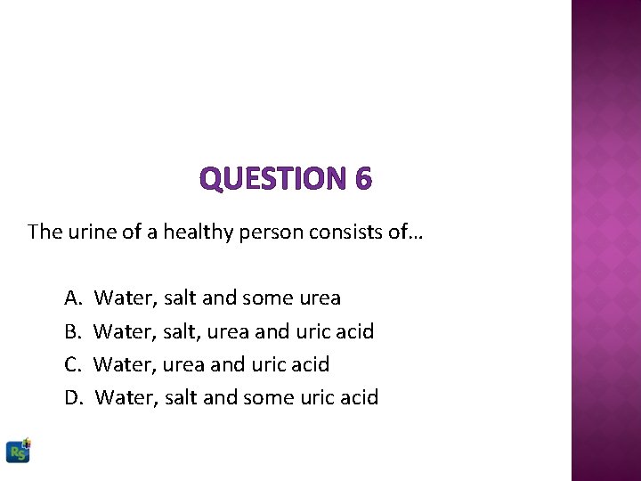
QUESTION 6 The urine of a healthy person consists of… A. B. C. D. Water, salt and some urea Water, salt, urea and uric acid Water, salt and some uric acid
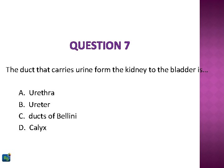
QUESTION 7 The duct that carries urine form the kidney to the bladder is… A. B. C. D. Urethra Ureter ducts of Bellini Calyx
QUESTION 8 Common tube for both urine and semen in males is the… A. B. C. D. Urethra Ureter ducts of Bellini Calyx
QUESTION 9 Encloses the apex of the pyramid… A. B. C. D. Urethra Ureter ducts of Bellini Calyx
QUESTION 10 Tubes found in the pyramids are called… A. B. C. D. Urethra Ureter ducts of Bellini Calyx
QUESTION 11 The thin membrane that surrounds the kidney is called the… A. B. C. D. Vertical septum Plural membrane Pericardium Renal capsule
QUESTION 12 Found in the cortex of the kidney… A. B. C. D. Malpighian body Renal tubules Collecting tubules None of the above
QUESTION 13 The proximal convoluted tubule… A. B. C. D. Leaves the malpighian body. Enters the distal convoluted tubules Leads to the collecting duct Is a straight tube
QUESTION 14 The collecting duct… A. B. C. D. Leaves the malpighian body. Enters the distal convoluted tubules Leads to the collecting duct Is a straight tube
QUESTION 15 The loop of Henle… A. B. C. D. Leaves the malpighian body. Leads to the distal convoluted tubules Leads to the collecting duct Is a straight tube
QUESTION 16 The distal convoluted tubule… A. Leaves the malpighian body. B. Enters the distal convoluted tubules C. Leads to the collecting duct D. Is a straight tube
QUESTION 17 The functional unit of the kidney is the… A. B. C. D. Cortex Medulla Nephron Alveoli
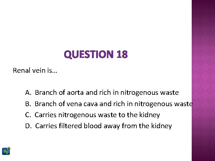
QUESTION 18 Renal vein is… A. B. C. D. Branch of aorta and rich in nitrogenous waste Branch of vena cava and rich in nitrogenous waste Carries nitrogenous waste to the kidney Carries filtered blood away from the kidney
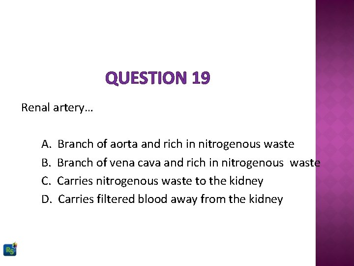
QUESTION 19 Renal artery… A. B. C. D. Branch of aorta and rich in nitrogenous waste Branch of vena cava and rich in nitrogenous waste Carries nitrogenous waste to the kidney Carries filtered blood away from the kidney
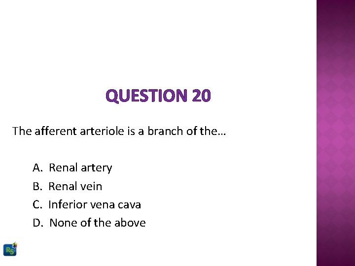
QUESTION 20 The afferent arteriole is a branch of the… A. B. C. D. Renal artery Renal vein Inferior vena cava None of the above
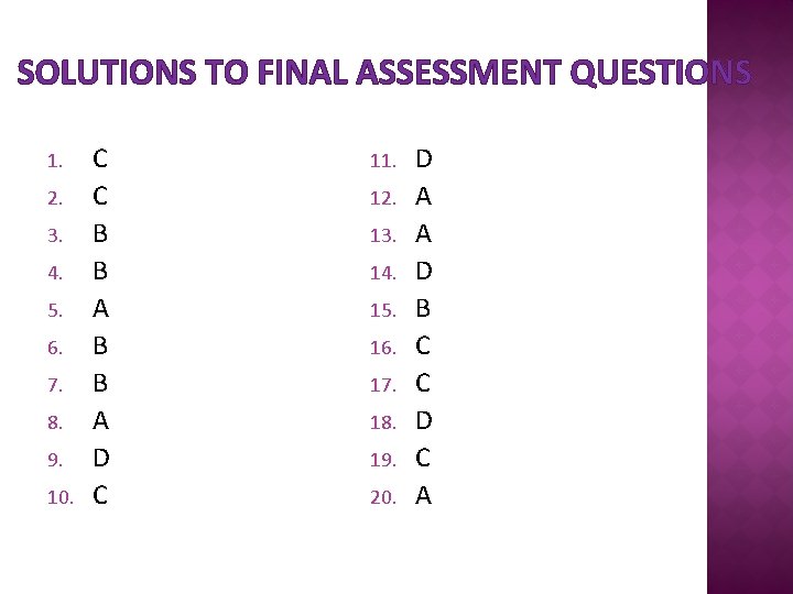
SOLUTIONS TO FINAL ASSESSMENT QUESTIONS 1. 2. 3. 4. 5. 6. 7. 8. 9. 10. C C B B A D C 11. 12. 13. 14. 15. 16. 17. 18. 19. 20. D A A D B C C D C A