23 8 The Small Intestine Gross Anatomy Small


- Slides: 50

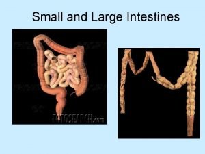
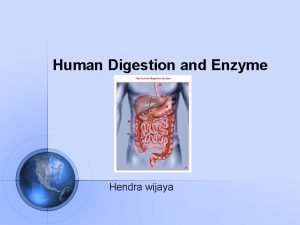
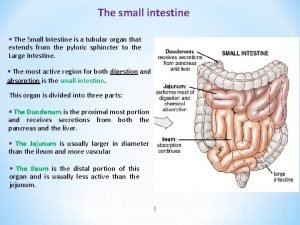
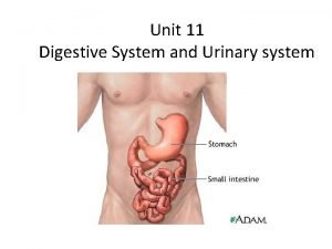
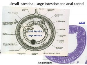
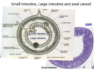
23. 8 The Small Intestine Gross Anatomy • Small intestine is the major organ of digestion and absorption • 2– 4 m long (7– 13 ft) from pyloric sphincter to ileocecal valve, point at which it joins large intestine • Small diameter of 2. 5– 4 cm (1. 0– 1. 6 inches) © 2016 Pearson Education, Inc.

Gross Anatomy (cont. ) • Subdivisions – Duodenum: mostly retroperitoneal; ~25. 0 cm (10. 0 in) long; curves around head of pancreas • Has most features – Jejunum: ~2. 5 m (8 ft) long; attached posteriorly by mesentery – Ileum: ~3. 6 m (12 ft) long; attached posteriorly by mesentery; joins large intestine at ileocecal valve © 2016 Pearson Education, Inc.

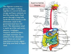
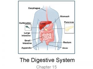
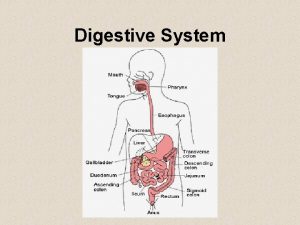
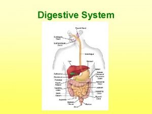
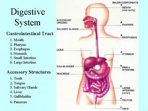
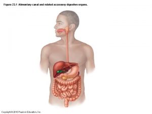
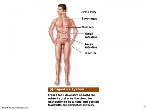
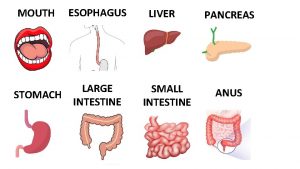
Figure 23. 1 Alimentary canal and related accessory digestive organs. Mouth (oral cavity) Tongue* Parotid gland Sublingual gland Submandibular gland Salivary glands* Pharynx Esophagus Stomach Pancreas* (Spleen) Liver* Gallbladder* Transverse colon Duodenum Small intestine Jejunum Ileum Descending colon Ascending colon Cecum Sigmoid colon Rectum Appendix Anus © 2016 Pearson Education, Inc. Anal canal Large intestine

Figure 23. 27 Relationship of the liver, gallbladder and pancreas to the duodenum. Liver • Right and left hepatic ducts • Common hepatic duct • Bile duct and sphincter Accessory pancreatic duct Gallbladder • Mucosa with folds • Cystic duct Duodenum • Major duodenal papilla © 2016 Pearson Education, Inc. Pancreas • Tail of pancreas • Main pancreatic duct and sphincter • Head of pancreas Jejunum Hepatopancreatic ampulla and sphincter

Microscopic Anatomy • Modifications of small intestine for absorption – Small intestine’s length and other structural modifications provide huge surface area for nutrient absorption • Surface area is increased 600 to ~200 m 2 (size of a tennis court) – Modifications include: • Circular folds • Villi • Microvilli © 2016 Pearson Education, Inc.

Microscopic Anatomy (cont. ) – Circular folds • Permanent folds (~1 cm deep) that force chyme to slowly spiral through lumen, allowing more time for nutrient absorption – Villi • Fingerlike projections of mucosa (~1 mm high) with a core that contains dense capillary bed and lymphatic capillary called a lacteal for absorption – Microvilli • Cytoplasmic extensions of mucosal cell that give fuzzy appearance called the brush border that contains membrane -bound enzymes brush border enzymes, used for final carbohydrate and protein digestion © 2016 Pearson Education, Inc.

Figure 23. 29 b Structural modifications of the small intestine that increase its surface area for digestion and absorption. Microvilli (brush border) Enterocytes (absorptive cells) Lacteal Goblet cell Blood capillaries Mucosaassociated lymphoid tissue Intestinal crypt Villus Enteroendocrine cells Paneth cells Venule Muscularis mucosae Lymphatic vessel Duodenal gland Submucosa © 2016 Pearson Education, Inc.

Figure 23. 29 c Structural modifications of the small intestine that increase its surface area for digestion and absorption. Enterocytes (absorptive cells) Goblet cells Villi Intestinal crypt © 2016 Pearson Education, Inc.

Figure 23. 30 Microvilli of the small intestine. Mucus granules Microvilli forming the brush border Enterocyte (absorptive cell) © 2016 Pearson Education, Inc.

Microscopic Anatomy (cont. ) • Histology of the small intestine wall – Modifications of mucosa and submucosa of small intestine reflect its function in digestion – Intestinal crypts: tubular glands scattered between villi – Five main types of cells found in villi and crypts 1. Enterocytes: make up bulk of epithelium – Simple columnar absorptive cells bound by tight junctions and contain many microvilli – Function » Villi: absorb nutrients and electrolytes » Crypts: produce intestinal juice, watery mixture of mucus that acts as carrier fluid for chyme © 2016 Pearson Education, Inc.

Microscopic Anatomy (cont. ) • Histology of the small intestine wall (cont. ) 2. Goblet cells: mucus-secreting cells found in epithelia of villi and crypts 3. Enteroendocrine cells: source of enterogastrones (examples: CCK and secretin) – Found scattered in villi but some in crypts 4. Paneth cells: found deep in crypts, specialized secretory cells that fortify small intestine’s defenses – Secrete antimicrobial agents (defensins and lysozyme) that can destroy bacteria 5. Stem cells that continuously divide to produce other cell types – Villus epithelium renewed every 2– 4 days © 2016 Pearson Education, Inc.

Intestinal Juice • 1– 2 L secreted daily in response to distension or irritation of mucosa • Major stimulus for production is hypertonic or acidic chyme • Slightly alkaline and isotonic with blood plasma • Consists largely of water but also contains mucus – Mucus is secreted by duodenal glands and goblet cells of mucosa © 2016 Pearson Education, Inc.

Digestive Processes in the Small Intestine • Chyme from stomach contains partially digested carbohydrates and proteins and undigested fats • Takes 3– 6 hours in small intestine to absorb all nutrients and most water • Sources of enzymes for digestion – Substances such as bile, bicarbonate, digestive enzymes are imported from liver and pancreas – Brush border enzymes bound to plasma membrane perform final digestion of chyme © 2016 Pearson Education, Inc.

Digestive Processes in the Small Intestine (cont. ) • Motility of the small intestine – After a meal • Segmentation is most common motion of small intestine – Initiated by intrinsic pacemaker cells – Mixes/moves contents toward ileocecal valve – Intensity is altered by long and short reflexes and hormones • Peristalsis increases, initiated by rise in hormone motilin in late intestinal phase (every 90– 120 minutes) – Meal remnants, bacteria, and debris are moved toward large intestine – Complete trip from duodenum to ileum takes ~2 hours © 2016 Pearson Education, Inc.

Digestive Processes in the Small Intestine (cont. ) • Ileocecal valve control – Ileocecal sphincter relaxes and admits chyme into large intestine when: • Gastroileal reflex enhances force of segmentation in ileum • Gastrin increases motility of ileum – Ileocecal valve flaps close when chyme exerts backward pressure • Prevents regurgitation into ileum © 2016 Pearson Education, Inc.

Table 23. 3 Control of Small Intestinal Motility © 2016 Pearson Education, Inc.

Table 23. 2 -1 Overview of the Functions of the Gastrointestinal Organs © 2016 Pearson Education, Inc.

Table 23. 2 -2 Overview of the Functions of the Gastrointestinal Organs (continued) © 2016 Pearson Education, Inc.

23. 9 The Large Intestine Gross Anatomy • Large intestine has three unique features not seen elsewhere: – Teniae coli: three bands of longitudinal smooth muscle in muscularis – Haustra: pocketlike sacs – Epiploic appendages: fat-filled pouches of visceral peritoneum © 2016 Pearson Education, Inc.

Gross Anatomy (cont. ) • Subdivisions of large intestine 1. Cecum: first part of large intestine 2. Appendix: masses of lymphoid tissue • Part of MALT of immune system • Bacterial storehouse capable of recolonizing gut when necessary © 2016 Pearson Education, Inc.

Gross Anatomy (cont. ) • Subdivisions of large intestine (cont. ) 3. Colon: has several regions, most which are retroperitoneal (except for transverse and sigmoid regions) • Ascending colon: travels up right side of abdominal cavity to level of right kidney • Transverse colon: travels across abdominal cavity • Descending colon: travels down left side of abdominal cavity • Sigmoid colon: S-shaped portion that travels through pelvis 4. Rectum: three rectal valves stop feces from being passed with gas (flatus) 5. Anal canal: last segment of large intestine that opens to body exterior at anus © 2016 Pearson Education, Inc.

Figure 23. 32 a Mesenteries of the abdominal digestive organs. Falciform ligament Liver Gallbladder Spleen Stomach Round ligament Greater omentum Small intestine Cecum © 2016 Pearson Education, Inc.

Clinical – Homeostatic Imbalance 23. 11 • Appendicitis: acute inflammation of appendix; usually results from a blockage by feces that traps infectious bacteria • Venous drainage can be impaired, leading to ischemia and necrosis (tissue death) • Ruptured appendix can cause peritonitis • Symptoms: pain in umbilical region, moving to lower right abdominal quadrant – loss of appetite, nausea, and vomiting are also seen • Treatment: surgical removal (appendectomy) © 2016 Pearson Education, Inc.

Bacterial Flora • Bacterial flora: consist of 1000+ different types of bacteria – Outnumber our own cells 10 to 1 • Enter from small intestine or anus to colonize colon • Metabolic functions – Fermentation • Ferment indigestible carbohydrates and mucin • Release irritating acids and gases (~500 ml/day) © 2016 Pearson Education, Inc.

Bacterial Flora (cont. ) – Vitamin synthesis • Synthesize B complex and some vitamin K needed by liver to produce clotting factors • Keeping pathogenic bacteria in check – Beneficial bacteria outnumber and suppress pathogenic bacteria – Immune system destroys any bacteria that try to breach mucosal barrier • Epithelial cells recruit dendritic cells to mucosa to sample microbial antigens and present to T cells of MALT, triggering production of Ig. A that restricts microbes © 2016 Pearson Education, Inc.

Bacterial Flora (cont. ) • Gut bacteria and health – Mounting evidence supports findings that the kinds and proportions of gut bacteria can influence: • Body weight • Susceptibility to various diseases (including diabetes, atherosclerosis, fatty liver disease) • Our moods – Manipulating gut bacteria may become a routine health-care strategy in future © 2016 Pearson Education, Inc.

Clinical – Homeostatic Imbalance 23. 12 • Antibiotic-associated diarrhea: accounts for 14, 000 deaths per year • Clostridium difficile, an anaerobic bacterium that many carry in intestine, is most common cause • When other bacteria are wiped out by antibiotics, C. difficile can flourish and cause pseudomembranous colitis (inflammation of colon) – May lead to bowel perforation and sepsis © 2016 Pearson Education, Inc.

Clinical – Homeostatic Imbalance 23. 12 • C. difficile infections are resistant to many antibiotics and difficult to treat – New treatments include fecal transplants to replace healthy bacteria to suppress C. difficile © 2016 Pearson Education, Inc.

Digestive Processes in the Large Intestine • Residue remains in large intestine 12– 24 hours • No food breakdown occurs except what enteric bacteria digest • Vitamins (made by bacterial flora), water, and electrolytes (especially Na+ and Cl−) are reclaimed • Major functions of large intestine is propulsion of feces to anus and defecation © 2016 Pearson Education, Inc.

Clinical – Homeostatic Imbalance 23. 13 • Large intestine is important for our comfort, but it is not essential for life • If colon is removed, terminal ileum can be brought out through abdominal wall in a procedure called an ileostomy © 2016 Pearson Education, Inc.

Digestive Processes in the Large Intestine (cont. ) • Motility of the large intestine – Haustral contractions: most contractions of colon, where haustra sequentially contract in response to distension • Slow segmenting movements, mostly in ascending and transverse colon – Gastrocolic reflex: initiated by presence of food in stomach • Results in mass movements: slow, powerful peristaltic waves that are activated three to four times per day – Descending colon and sigmoid colon act as storage reservoir © 2016 Pearson Education, Inc.

Slide 4 Figure 23. 33 Defecation reflex. Impulses from cerebral cortex (conscious control) Sensory nerve fibers Voluntary motor nerve to external anal sphincter Sigmoid colon 1 Feces move into and distend the rectum, stimulating stretch receptors there. The receptors transmit signals along afferent fibers to spinal cord neurons. Stretch receptors in wall Rectum External anal sphincter (skeletal muscle) 2 A spinal reflex is initiated in which parasympathetic motor (efferent) fibers stimulate contraction of the rectum and sigmoid colon, and relaxation of the internal anal sphincter. Involuntary motor nerve (parasympathetic division) Internal anal sphincter (smooth muscle) 3 If it is convenient to defecate, voluntary motor neurons are inhibited, allowing the external anal sphincter to relax so feces may pass. © 2016 Pearson Education, Inc.

Part 3 – Physiology of Digestion and Absorption 23. 10 Mechanisms of Digestion and Absorption • Digestion breaks down ingested foods into their chemical building blocks • Only these molecules are small enough to be absorbed across wall of small intestine © 2016 Pearson Education, Inc.

Mechanism of Digestion: Enzymatic Hydrolysis • Digestion: catabolic process that breaks macromolecules down into monomers small enough for absorption – Intrinsic and accessory gland enzymes are involved in digestion – Enzymes carry out hydrolysis, whereby water is added to break chemical bonds © 2016 Pearson Education, Inc.

Mechanisms of Absorption • Absorption is process of moving substances from lumen of gut into body • Tight junctions ensure molecules must pass through epithelial cell rather than between them – Materials enter cell through apical membrane (lumen side) and exit through basolateral membrane (blood side) • Lipid molecules can be absorbed passively through membrane, but other polar molecules are absorbed by active transport • Most nutrients are absorbed before chyme reaches ileum © 2016 Pearson Education, Inc.

23. 11 Processing of Nutrients Digestion of Carbohydrates • Only monosaccharides can be absorbed • Starch and disaccharides are broken down to oligosaccharides and disaccharides – Begins in mouth with salivary amylase • Further broken down into lactose, maltose, and sucrose • Final breakdown into monosaccarides (glucose, fructose, galactose) © 2016 Pearson Education, Inc.

Figure 23. 34 a Flowchart of digestion and absorption of foodstuffs. Foodstuff Enzyme(s) and source Site of action Starch and disaccharides Salivary amylase Oligosaccharides and disaccharides Carbohydrate digestion Lactose Maltose Sucrose Galactose Glucose Fructose © 2016 Pearson Education, Inc. Mouth Pancreatic amylase Small intestine Brush border enzymes in small intestine (dextrinase, glucoamylase, lactase, maltase, and sucrase) Small intestine Path of absorption • Glucose and galactose are absorbed via cotransport with Na +. • Fructose passes via facilitated diffusion. • All monosaccharides leave the epithelial cells via facilitated diffusion, enter the capillary blood in the villi, and are transported to the liver via the hepatic portal vein.

Slide 5 Figure 23. 35 Carbohydrate digestion and absorption in the small intestine. Lumen of intestine Starch Pancreatic amylase Disaccharides Na+ 2 Brush border enzymes break oligosaccharides and disaccharides into monosaccharides. Brush border enzymes Na+ 1 Pancreatic amylase breaks down starch and glycogen into oligosaccharides and disaccharides. Apical membrane (microvilli) Monosaccharides Basolateral membrane Na+ ATP Na+ -K+ ATPase Na+ Monosaccharide carrier K+ Capillary © 2016 Pearson Education, Inc. 3 Monosaccharides 3 (glucose and galactose) are cotransported across the apical membrane of the absorptive epithelial cell. This active transport uses the Na+ concentration gradient established by the Na+ -K+ ATPase (pump) in the basolateral membrane. 4 Monosaccharides exit across the basolateral membrane by facilitated diffusion and enter the capillary via intercellular clefts.

Digestion of Proteins • Source of protein not only is dietary, but also includes digestive enzymes and proteins from breakdown of mucosal cells • Proteins are broken into: – Large polypeptides – Small polypeptides and small peptides – Finally into amino acid monomers, with some dipeptides and tripeptides • Digestion begins in stomach when pepsinogen is converted to pepsin at p. H 1. 5– 2. 5 – Becomes inactive in high p. H of duodenum © 2016 Pearson Education, Inc.

Figure 23. 34 b Flowchart of digestion and absorption of foodstuffs. Foodstuff Enzyme(s) and source Site of action Proteins Pepsin (stomach glands) in presence of HCl Stomach Small intestine Small polypeptides, small peptides Pancreatic enzymes (trypsin, chymotrypsin, carboxypeptidase) Amino acids (some dipeptides and tripeptides) Brush border enzymes (aminopeptidase, carboxypeptidase, and dipeptidase) Small intestine Large polypeptides Protein digestion © 2016 Pearson Education, Inc. Path of absorption • Amino acids are absorbed via cotransport with Na +. • Some dipeptides and tripeptides are absorbed via cotransport with H + and hydrolyzed to amino acids within the cells. • Infrequently, transcytosis of small peptides occurs. • Amino acids leave the epithelial cells by facilitated diffusion, enter the capillary blood in the villi, and are transported to the liver via the hepatic portal vein.

Slide 5 Figure 23. 36 Protein digestion and absorption in the small intestine. Amino acids of protein fragments Lumen of intestine Pancreatic proteases Brush border enzymes Na+ Na+ ATP 1 Pancreatic proteases break down proteins and protein fragments into smaller pieces and some individual amino acids. 2 Brush border enzymes break protein fragments into amino acids. Apical membrane (microvilli) Amino acid carrier Basolateral membrane 3 Amino acids are cotransported across the apical membrane of the absorptive epithelial cell. This active transport uses the Na+ concentration gradient established by the Na+ -K+ ATPase (pump) in the basolateral membrane. Na+ -K+ ATPase Na+ K+ Capillary © 2016 Pearson Education, Inc. 4 Amino acids exit across the basolateral membrane via facilitated diffusion and enter the capillary via intercellular clefts.

Clinical – Homeostatic Imbalance 23. 17 • In rare cases, intact proteins are taken up by intestinal epithelial cells by endocytosis and are released into body – Most common in newborn infants because of immaturity of their intestinal mucosa • May result in food allergies as immune system “sees” intact proteins as antigenic and mounts an attack – Allergies usually disappear as mucosa matures © 2016 Pearson Education, Inc.

Figure 23. 34 c Flowchart of digestion and absorption of foodstuffs. Foodstuff Enzyme(s) and source Site of action Path of absorption Unemulsified triglycerides Fat digestion Monoglycerides and fatty acids © 2016 Pearson Education, Inc. Lingual lipase (minor importance) Mouth Gastric lipase (minor importance) Stomach Emulsification by the detergent action of bile salts ducted in from the liver Small intestine Pancreatic lipases Small intestine • Fatty acids and monoglycerides enter the intestinal cells via diffusion. • Fatty acids and monoglycerides are recombined to form triglycerides and then combined with other lipids and proteins within the cells. The resulting chylomicrons are extruded by exocytosis. • The chylomicrons enter the lacteals of the villi and are transported to the systemic circulation via the lymph in the thoracic duct. • Some short-chain fatty acids are absorbed, move into the capillary blood in the villi by diffusion, and are transported to the liver via the hepatic portal vein.

Slide 6 Figure 23. 37 Emulsification, digestion, and absorption of fats. Fat globule Bile salts Fat droplets coated with bile salts 1 Emulsification. Bile salts in the duodenum break large fat globules into smaller fat droplets, increasing the surface area available to lipase enzymes. 2 Digestion. Pancreatic lipases hydrolyze triglycerides, yielding monoglycerides and free fatty acids. 3 Micelle formation. Micelles (consisting of fatty acids, monoglycerides, and bile salts) ferry their contents to epithelial cells. 4 Diffusion. Fatty acids and monoglycerides diffuse from micelles into epithelial cells. 5 Chylomicron formation. Fatty acids and monoglycerides are recombined and packaged with other fatty substances and proteins to form chylomicrons. © 2016 Pearson Education, Inc.

Digestion of Nucleic Acids • Nuclei of ingested cells in food contain DNA and RNA • Pancreatic nucleases hydrolyze nucleic acid to nucleotide monomers • Brush border enzymes, nucleosidases, and phosphatases break nucleotides down into free nitrogenous bases, pentose sugars, and phosphate ions • Breakdown products are actively transported by special carriers in epithelium of villi © 2016 Pearson Education, Inc.

Figure 23. 34 d Flowchart of digestion and absorption of foodstuffs. Foodstuff Enzyme(s) and source Site of action Nucleic acids Nucleic acid digestion Pentose sugars, N-containing bases, phosphate ions © 2016 Pearson Education, Inc. Pancreatic ribonuclease and deoxyribonuclease Small intestine Brush border enzymes (nucleosidases and phosphatases) Small intestine Path of absorption • Units enter intestinal cells by active transport via membrane carriers. • Units are absorbed into capillary blood in the villi and transported to the liver via the hepatic portal vein.

Absorption of Vitamins, Electrolytes, and Water • Vitamin absorption – In small intestine • Fat-soluble vitamins (A, D, E, and K) are carried by micelles; diffuse into absorptive cells • Water-soluble vitamins (C and B) are absorbed by diffusion or by passive or active transporters • Vitamin B 12 (large, charged molecule) binds with intrinsic factor and is absorbed by endocytosis – In large intestine: vitamin K and B vitamins from bacterial metabolism are absorbed © 2016 Pearson Education, Inc.

Absorption of Vitamins, Electrolytes, and Water (cont. ) • Absorption of electrolytes – Most ions are transported actively along length of small intestine – Iron and calcium are absorbed in duodenum – Na+ absorption is coupled with active absorption of glucose and amino acids – Cl− is transported actively – K+ diffuses in response to osmotic gradients; lost if water absorption is poor – Usually amount in intestine is amount absorbed © 2016 Pearson Education, Inc.

Absorption of Vitamins, Electrolytes, and Water (cont. ) • Absorption of electrolytes (cont. ) – Iron and calcium absorption is related to need • Ionic iron is stored in mucosal cells with ferritin • When needed, transported in blood by transferrin – Ca 2+ absorption is regulated by vitamin D and parathyroid hormone (PTH) © 2016 Pearson Education, Inc.

Absorption of Vitamins, Electrolytes, and Water (cont. ) • Absorption of water – 9 L water, most from GI tract secretions, enter small intestine • 95% is absorbed in the small intestine by osmosis • Most of rest is absorbed in large intestine – Net osmosis occurs if concentration gradient is established by active transport of solutes – Water uptake is coupled with solute uptake © 2016 Pearson Education, Inc.
 Esophagus stomach small intestine large intestine
 Sacculations
 Bicuspids
 Small intestine parts
 Kriteria investasi
 Colon diagram
 Appendetitis
 Function of digestive system
 Small intestine parts
 Stratified squamous epithelium characteristics
 Frogs body parts and functions
 Alimentary cannel
 What is peristalsis
 Enzymes examples
 Small intestine
 Small intestine villi function
 Blood supply small bowel
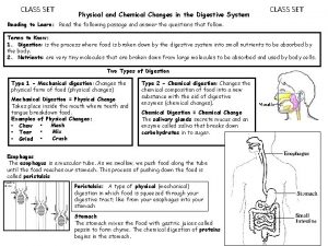
 Is the small intestine a physical or chemical change
 Http://www.innerbody.com/htm/body.html
 Overview of the digestive system
 Small intestine extends from
 Intestinal villus
 Another name for small intestine
 Digestive tract histology
 Small intestine peristalsis
 Anatomical directions frog
 Pearson
 Small bowel
 Small intestine mechanical digestion
 N
N Galt
Galt Brush border enzymes
 Peristalsis and segmentation
 Plicae circularis
 Digestive system prefixes and suffixes
 Lumen of small intestine
 Laparoyomy
 Pyloric stomach
 Small intestine mechanical digestion
 Parts of small intestine
 Cow eye retina
 Heart gross anatomy
 Optic disc cow eye
 Gross anatomy of skeletal system
 Gross anatomy of the lungs
 Gross anatomy of a long bone
 Gross anatomy of the gallbladder pancreas and bile passages
 Funiculus separans
 Sheep brain labeled
 Lời thề hippocrates
 Vẽ hình chiếu đứng bằng cạnh của vật thể
